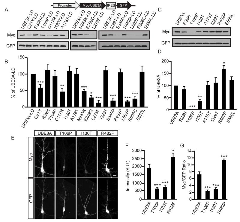
Figure 2. AS-linked missense mutations in UBE3A cause loss-of-function via distinct mechanisms.
(A and B) Representative western blot of protein levels for AS-linked mutations introduced into ligase-dead (LD) UBE3A and quantification (B). All UBE3A constructs were Myc-tagged, contained an IRES-GFP to normalize for expression and transfection efficiency and were transfected into HEK293T cells. Values are shown as the percent ± standard error of UBE3A-LD levels. n=3-6/condition; **p<0.005, ***p<0.0005. See Supplemental Information for detailed statistical methods.
(C and D) Representative western blot of UBE3A mutants that possess ubiquitin ligase activity (+Ligase) and quantification (C). Values are shown as the mean percent ± standard error of WT UBE3A levels, n=4, *p<0.005, ***p<0.0005.
(E - G) Immunofluorescence staining of Myc-tagged UBE3A and mutants in DIV 10 mouse cortical neurons, scale bar, 15 μm. Raw intensity values for Myc immunofluorescence (F) or Myc immunofluorescence normalized to GFP (G) are shown as the mean ± standard error. n=16-19 neurons/condition; *p<0.05, ***p<0.0005.

