Abstract
This study aimed to evaluate the beneficial effects of a traditional Chinese medicine named Gengnianchun (GNC) in ovariectomized Sprague-Dawley rats. The rats were randomly categorized into sham-operated group (Sham), saline-treated ovariectomized group (OVX), GNC-treated ovariectomized group (OVX+GNC), estradiol valerate-treated ovariectomized group (OVX+E). GNC and estradiol was administered for 1 month at dosages of 125 and 0.1 mg/day, respectively. Ovariectomy caused deterioration of learning and memory ability (P < 0.05), which was restored by treatment with GNC and estradiol (P < 0.05). Estrogen level and endometrial thickness significantly decreased in the OVX group (P < 0.05). These parameters significantly increased in the OVX+E group (P < 0.05) but did not change in the OVX+GNC group (P > 0.05). GNC and estradiol significantly increased the levels of norepinephrine (NE) and dopamine (DA) and decreased the levels of 5-hydroxytryptamine (5-HT) and 5-hydroxyindoleacetic acid (5-HIAA) in the hypothalamus (P < 0.05). The levels of interleukin-1 beta (IL-1β), interleukin-6 (IL-6), and tumor necrosis factor-alpha (TNF-α) significantly decreased and the levels of interleukin-2 (IL-2) and interferon-gamma (IFN-γ) increased in the OVX+GNC and OVX+E groups compared with those in the OVX group (P < 0.05). OVX rats treated with GNC and estradiol further exhibited reversed ovariectomy-induced weight gain and leptin resistance (P < 0.05). These results indicated that GNC demonstrated phytoestrogen-like properties without the side effects of estradiol valerate. Thus, GNC may confer protective and beneficial effects for management of menopausal syndrome.
Keywords: Gengnianchun, menopause, ovariectomy, estrogen, endometrium, cytokines, neurotransmitter, body weight, leptin
Introduction
Menopause is an important physiological event characterized by the cessation of ovarian function, resulting in low estrogen levels. Women with menopause syndrome experience a variety of symptoms, such as hot flashes, sweating, anxiety, depression, mood swings, sleep disorder, and learning and memory disorder; all of these symptoms are due to the cessation of ovarian estrogen production [1].
Approximately 40% of women suffering menopausal symptoms seek treatment to manage these conditions. The recommendations in the Global Consensus Statement on Menopausal Hormone Therapy in November 2012 [2] states that hormone replacement therapy (HRT) is the most effective treatment for menopausal symptoms; nevertheless, this treatment presents a complex pattern of risks and benefits. Studies have shown that women who undergo HRT demonstrated an increased risk of cardiovascular diseases, breast cancer, endometrial hyperplasia, and cancer [3]. Thus, many women refuse to use HRT and resort to alternative approaches for menopausal symptoms, with herbal mixtures being considered as an effective mode of complementary therapy.
Estrogen plays multiple biological roles and is required for the development of normal physiological function. Estrogen also regulates cognition, immune function, lipid metabolism, and neurotransmitters. This hormone exerts its effects via the interaction of its known estrogen receptors, namely, ERα and ERβ, with its target tissues [4].
Traditional Chinese medicine (TCM) has been used in Asian countries for over 5,000 years to prevent and treat diseases. According to TCM theory, menopausal syndrome is caused by kidney-liver weakness caused by essence deficiency accompanied with yin-yang imbalance and organ disharmony [5]. Herbal formulas classified as kidney/liver-tonifying mixtures are thus considered suitable for management of menopausal symptoms. As TCM consists of different herbs, the effects of the decoction are observed after the total and final reaction of the constituent compounds when administered to humans. Traditional Chinese herbs have long been used for improving menopausal symptoms because they exhibit minimal side effects [6]. Among different TCM types, Gengnianchun recipe (GNC) has been used for treatment of menopausal symptoms; GNC was originally developed based on many years of clinical experience to nourish kidney and liver.
In this study, we explored the effects of GNC in ovariectomized Sprague-Dawley rats. Estradiol level and endometrial thickness were quantified to confirm alleviation after ovariectomy. We used the Morris water maze to assess the effects of GNC on learning and memory ability. Dopaminergic and serotonergic indices, including norepinephrine (NE), dopamine (DA), 5-hydroxytryptamine (5-HT), and 5-hydroxyindoleacetic acid (5-HIAA), were determined to investigate the effects of GNC on neurotransmitters in the hypothalamus. The levels of cytokines, such as interleukin-1 beta (IL-1β), interleukin-6 (IL-6), tumor necrosis factor-alpha (TNF-α), interleukin-2 (IL-2), and interferon-gamma (IFN-γ), were measured to reveal the effects of GNC on the immune system. Serum leptin concentrations and body weight were evaluated to examine the effects of GNC on lipid metabolism. This study aimed to determine the action of GNC on dramatic alterations in the body caused by estrogen deficiency.
Materials and methods
Chinese medicinal formula
The GNC formula contained 12 crude herbs, which were prepared as shown in Table 1. The applied composition was referenced from TCM theory and our clinical experience. GNC was obtained from the Obstetrical and Gynecology Hospital of Fudan University. A total of 138 g of herbs was concocted into 25 g of concentrated power (Table 1).
Table 1.
Composition and preparation of GNC formula
| Crude herbs | Content |
|---|---|
| Radix Rehmanniae | 15 g |
| Rhizoma Coptidis | 3 g |
| Radix Paeoniae Alba | 12 g |
| Rhizoma Anemarrhenae | 15 g |
| Cistanche Salsa | 12 g |
| Radix Moridae Officinalls | 12 g |
| Poria | 9 g |
| Epimedium Brevicornums | 12 g |
| Cortex Phellodendri Amurensis | 9 g |
| Fructus Lycii | 12 g |
| Semen Cuscutae | 12 g |
| Carapax et plastrum Testudinis | 15 g |
Experimental animals
Ovariectomy is a standard surgical procedure used to induce menopause in experimental animals. We purchased 32 female Sprague-Dawley rats, with weights ranging from 220 g to 250 g and age of 3 months, from the Experimental Animal Center of the Chinese Academy of Sciences (Shanghai, China). The rats were kept under standard 12 h light and 12 h dark photoperiod with controlled temperature (23 ± 3°C) and 45%-60% humidity. Twenty-four rats were bilaterally ovariectomized to create menopause models, and the eight remaining rats were sham operated as non-ovariectomized controls (Sham). The ovariectomized rats were randomly divided into three groups containing eight rats each: saline-treated ovariectomized group (OVX), GNC-treated ovariectomized group (OVX+GNC), and estrogen-treated ovariectomized group (OVX+E). Treatments were started 1 week after bilateral ovariectomy under anesthesia with 10% chloral hydrate (0.7 ml/100 g). GNC and estrogen were administered once daily for 1 month at dosages of 5 and 0.4 mg/g, respectively. Physiological saline was given to rats in the untreated control and non-ovariectomized groups. One day after 1 month of treatment, the rats were tested in the Morris water maze. After the test, the rats were anesthetized with i.p. injection of chloral hydrate (0.7 ml/100 g) and then rapidly dissected. Cardiac blood, uteri, and hypothalami were collected for further analyses. Body weight was recorded on the first and last day of the experiment.
Morris water maze
The Morris water maze consisted of a circular pool with 1.5 m diameter and 0.8 m height, and the pool was filled to a level of 35 cm with water and maintained at approximately 25°C [7]. Pool water was made opaque by adding 150 ml of nontoxic paint. Maze performance was recorded by a video camera suspended above the maze and interfaced with a video tracking system. Extra-maze cues surrounding the maze were fixed at specific locations and made visible to the rats while in the maze. A clear plastic escape platform with 10 cm diameter was positioned 2 cm below the water surface. The first day of testing occurred 1 month after the treatment. The subjects were trained for 5 d with four trials per day (a total of 20 trials). The location of the submerged platform did not change throughout the experiment. For each trial, the rats were placed in the water facing the edge of the tank from one of the four start locations. The order of the start locations was randomized to ensure that the rats never started from the same location on any two consecutive trials. On each trial, the rats were allowed 90 s to locate the submerged platform. If they did not find the platform within the allotted time, the experimenter guided the subject to the platform. The rats were allowed to remain on the platform for 20 s before being removed from the maze and dried. We recorded the time required by the rats to locate the submerged platform on each trial as the escape latency period (ELP). When the rats could not locate the platform, their ELP was recorded as 90 s. At the end of testing on Day 5, the platform was removed from the tank and a probe trial was performed for 90 s. This trial is known as spatial probe performance (SPP) test, in which we recorded the time spent by the rats to search the platform.
Serum estradiol, cytokine, and leptin determination
Blood samples were extracted from the hearts of the rats after the last intragastric administration. The samples were allowed to clot at room temperature, and serum was separated by centrifugation at 1000 × g for 15 min and then stored at -80°C for biochemical analysis. Serum estradiol level was measured by radioimmunoassay with a commercial kit (Radim, Pomezia, Italy). The serum concentrations of IL-1β, IL-2, IL-6, IFN-γ, TNF-α, and leptin were determined through enzyme-linked immunosorbent assay with commercial kits (Abcam, USA). All measurements were carried out according to the manufacturer’s protocols.
Endometrial thickness determination
The uteri were removed from sacrificed rats, and the adhering fats were trimmed. The samples were fixed in 10% formalin for at least 24 h at room temperature. After fixation, the tissues were dehydrated in graded ethanol, cleared in xylene, and embedded in paraffin. The thin sections (4 μm thick) were mounted on glass slides, dewaxed, rehydrated with distilled water, and stained with hematoxylin and eosin. Endometrial thickness was observed under a light microscope.
Neurotransmitter (NE, DA, 5-HT, and 5-HIAA) determination in hypothalamus
After the rats were killed and their brains were rapidly removed, each hypothalamus was dissected on ice and then weighed. The hippocampi were homogenized and deproteinized with 5% perchloric acid solution. The homogenate was centrifuged at 13,200 rpm for 40 min at 4°C, and the supernatant was stored at -80°C. High-performance liquid chromatography (HPLC) was used to assay NE, DA, 5-HT, and 5-HIAA. The HPLC procedure was performed according to a previously described method with some minor modifications [8]. For monoamine analysis, an Agilent Eclipse XDB-C18 analytical column (150 mm × 4.6 mm, 5 μm; Agilent, USA) was used. The mobile phase consisted of 10% methanol and 90% aqueous solution containing 75 mM NaH2PO4, 1.7 mM orthosilicic acid, 25 μm ethylene diamine tetraacetic acid disodium salt, and 0.5 mM octanesulfonic acid sodium salt at a flow rate of 0.8 ml/min and pH of 3.5. The levels of NE, DA, 5-HT, and 5-HIAA were detected using an Antec Decard SDC electrochemical detector (Antec, Holland). The HPLC system was connected to a computer to quantify all compounds by comparing the area under the peaks with the area of reference standards by using HPLC software (Chromatography Station for Windows).
Statistical analysis
Statistical analysis of the experimental data was carried out using SPSS 13.0 software (SPSS, Chicago, IL, USA). A P-value of 0.05 or less was considered statistically significantly. The results of the evaluation were compared between OVX and the other groups with one-way analysis of variance.
Results
GNC improves learning and memory ability in Morris water maze test
ELP was measured in Morris water maze test, and the times that the rats across the platform was determined in SPP test. Ovariectomy significantly increased ELP and decreased the time spent by the rats across the platform (P < 0.05, Figure 1A and 1B). In the OVX+GNC and OVX+E groups, ELP decreased and the time spent by the rats across platform increased compared with those in the OVX group (Figure 1A and 1B). As shown in Figure 1C, the trails in the OVX group were randomly distributed. After treatment with GNC and estradiol, the trails concentrated near the platform (Figure 1C). These parameters, namely, ELP, time in SPP, and trail, showed that ovariectomy caused deterioration of learning and memory ability, which were restored after treatment with GNC and estradiol.
Figure 1.
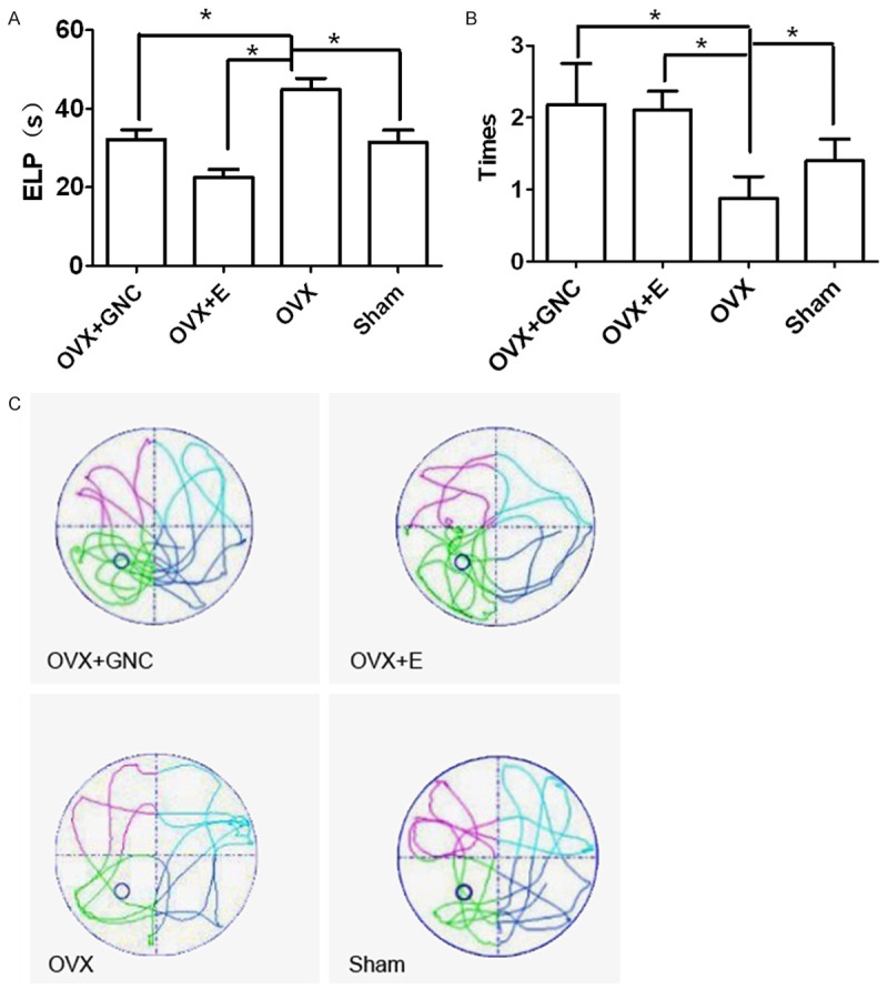
Effects of GNC on ovariectomized rats. A. ELP; B. Times that the rats across the platform; C. SPP trail of the OVX+GNC, OVX+E, OVX, and Sham groups. Data are expressed as mean values ± S.D. (n = 8). *P < 0.05, **P < 0.01.
GNC improves menopause syndrome without affecting estrogen concentration and endometrial thickness
Serum estrogen concentration in the OVX group significantly decreased compared with those in the Sham group (P < 0.01, Figure 2A). Ovariectomy also resulted in significantly decreased endometrial thickness (P < 0.01, Figure 2B). By contrast, GNC treatment did not affect serum estrogen level and endometrial thickness (P > 0.05, Figure 2A and 2B). The endometrium stained with hematoxylin and eosin was further observed under a light microscope as shown in Figure 2C. Serum estrogen concentration and endometrial thickness in the OVX+E group significantly increased compared with those in the OVX group (P < 0.01, Figure 2A and 2B).
Figure 2.
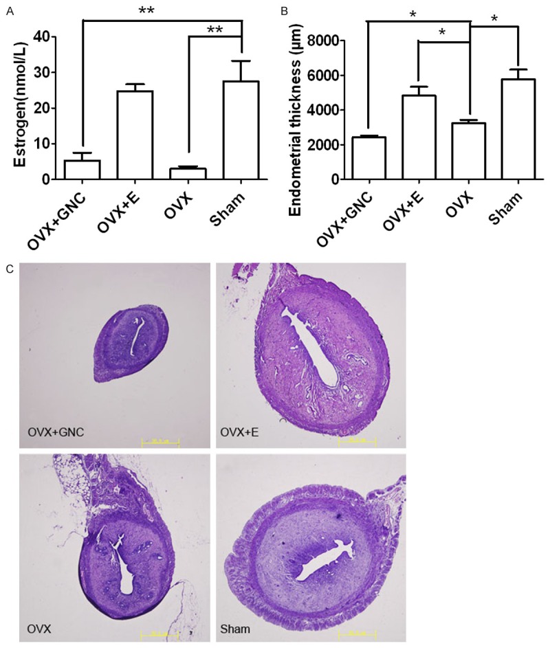
Effects of GNC on estrogen levels and endometrial thickness. A. Serum levels of estrogen; B. Endometrial thickness; C. endometrium stained with hematoxylin and eosin under a light microscope in the OVX+GNC, OVX+E, OVX, and Sham groups. Data are expressed as mean values ± S.D. (n = 8). *P < 0.05, **P < 0.01.
GNC decreases the levels of IL-1β, IL-6, and TNF-α and increases the levels of IL-2 and IFN-γ
As shown in Figure 3, the serum levels of IL-1β, IL-6, and TNF-α were significantly higher and the serum levels of IL-2 and IFN-γ were lower in the OVX group than those in the Sham group (P < 0.05, Figure 3A and 3B). In the OVX+GNC group, the serum levels of IL-1β, IL-6, and TNF-α were significantly lower and the serum levels of IL-2 and IFN-γ were higher than those in the Sham group (P < 0.05, Figure 3A and 3B). The serum levels of these cytokines did not significantly differ between the OVX+GNC and OVX+E groups (P > 0.05).
Figure 3.
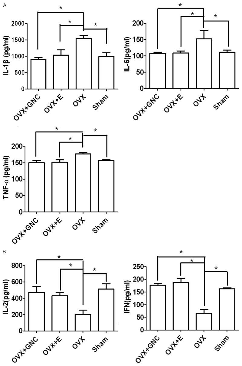
Effect of GNC on serum levels of cytokines. (A) Serum levels of IL-1β, IL-6, and TNF-α and (B) IL-2 and IFN-γ in the OVX+GNC, OVX+E, OVX, and Sham groups. Data are expressed as mean values ± S.D. (n = 8). *P < 0.05, **P < 0.01.
GNC upregulates the levels of NE and DA and downregulates the levels of 5-HT and 5-HIAA
As shown in Figure 4, ovariectomized rats demonstrated low levels of DA and NE (P < 0.05, Figure 4A) and high levels of 5-HT and 5-HIAA (P < 0.05, Figure 4B). The OVX+GNC and OVX+E groups presented significantly higher levels of DA and NE (P < 0.05, Figure 4A) and lower levels of 5-HT and 5-HIAA (P < 0.05, Figure 4B) than those in the OVX group. These results indicated that GNC treatment exhibited similar effect to estradiol treatment (P > 0.05).
Figure 4.
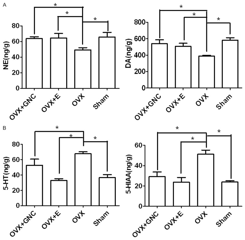
Effects of GNC on neurotransmitter levels. (A) Levels of NE and DA and (B) Levels of 5-HT and 5-HIAA in the hippocampus in the OVX+GNC, OVX+E, OVX, and Sham groups. Data are expressed as mean values ± S.D. (n = 8). *P < 0.05.
GNC ameliorates leptin resistance and reduces body weight
No significant differences were observed in the initial body weight among all groups (P < 0.05, Figure 5A). The final body weight significantly increased in the OVX group compared with that in the Sham group (P < 0.05, Figure 5A). Rats in the OVX+GNC and OVX+E group demonstrated significantly lower body weight than those in the OVX group (P < 0.05, Figure 5A). Leptin level was higher in the OVX group than that in the Sham group (P < 0.05, Figure 5B), but lower in the OVX+GNC group and OVX+E groups than that in the OVX group (P < 0.05, Figure 5B). No difference was observed between the OVX+GNC and OVX+E groups in terms of body weight and leptin level (P > 0.05, Figure 5B).
Figure 5.
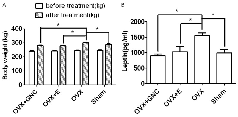
Effects of GNC on body weight and leptin levels. (A) Body weight before and after treatment and (B) Leptin level in the OVX+GNC, OVX+E, OVX, and Sham groups. Data are expressed as mean values ± S.D. (n = 8). *P < 0.05.
Discussion
Menopause leads to a wide range of symptoms, including hot flashes, night sweats, sleeping problems, and emotional and cognitive dysfunction. Studies on rodent and non-human primates rely on an ovariectomized model of surgical menopause, resulting in abrupt withdrawal of estrogen. Our study showed that ovariectomized rats showed a significant decrease in serum estrogen concentration and endometrial thickness compared with sham-operated rats. The reduction in endometrial thickness was caused by the lack of estrogen secreted by the ovaries. Administration of estradiol to ovariectomized rats for 1 month increased estrogen level and endometrial thickness. However, no significant difference in serum estrogen concentration and endometrial thickness was observed after GNC treatment of ovariectomized rats. Therefore, GNC could be considered a safe and effective complementary or alternative treatment for menopausal syndrome.
Complaints of memory loss are common during menopause in women [9]. Some studies on the relationship between menopause and learning and memory ability are impelled by pre-clinical evidence indicating that estrogen can help preserve cognitive function during aging. Estrogen deficiency in menopausal women causes memory decline and dementia in late life [10]. In the present study, the results of Morris water maze test revealed that ovariectomy impaired the learning and memory ability of the rats.
Hippocampus and prefrontal cortex, which serve as episodic and working memory, respectively, contain abundant estrogen receptors [11]. In animal and in vitro models, estrogen increases the levels of acetylcholine, promotes neuronal growth and synapse formation, acts as antioxidants, and regulates calcium homeostasis and second messenger system [12]. Therefore, estrogen fluctuation and withdrawal affects the central nervous system and potentially influences learning and memory ability. Healthy postmenopausal women who underwent HRT exhibited better performance on memory tests than those untreated with HRT [13]. Treatment of ovariectomized rats with estradiol and GNC significantly shortened the ELP and improved the trail in SPP test. No significant differences were observed between the OVX+GNC and OVX+E groups, suggesting that GNC and estradiol exhibited similar effects on cognitive function.
Some researchers have suggested that decreased estrogen level may lead to stress, anxiety, depression, sleep concerns, and hot flashes during menopausal transition [14]. Hot flashes and night sweats, which are the most common symptoms reported by menopausal women, severely affect their quality of life. Currently, HRT remains as the most effective treatment for menopause syndrome.
Emerging studies show that the effects of estrogen on depression, anxiety, and hot flashes are substantially mediated by the hypothalamic and pituitary function through the hypothalamic-pituitary-sexual gland axis [15]. This phenomenon is mainly due to estrogen receptors, which are widely present in cortex, pituitary, and hypothalamus [16]. Therefore, estrogen can affect the central nervous system to regulate neurotransmitter level. Changes in neurotransmitter levels are significant consequences of the deregulated gonadal hormone production during deterioration of many central nervous system activities, particularly those associated with hippocampal function [17]. DA and NE, which are important catecholaminergic monoamines, are involved in emotional disorders when their release is inhibited or their pertinent neurons are destroyed [18]. The serotonergic neurotransmitter 5-HT and its metabolite 5-HIAA are involved in regulation of diverse functions, such as hot flashes and night sweats, and have been proven to be decreased by estrogen in ovariectomized rats [19]. In addition, ovariectomized and male animals demonstrated reduced DA and NE functions compared with females with intact ovaries [20].
The present result showed that GNC significantly increased catecholaminergic neurotransmitter (NE and DA) levels and decreased serotonergic neurotransmitter (5-HT and 5-HIAA) levels, which were in accordance with the effects of estradiol. This finding suggested that neurotransmitter regulation could be another vital mechanism of GNC in ameliorating menopause syndrome.
Cytokines are key regulators of immune responses and are involved in neuroendocrine-immune interaction. Biological effects induced by cytokinesis include stimulation or inhibition of cell proliferation, apoptosis, antiviral activity, cell growth, cell differentiation, and inflammatory responses, as well as upregulation of the expression of surface membrane proteins [21]. Cytokines mainly include interleukins, interferons, tumor necrosis factors, and so on [22]. Some cytokines primarily induce inflammation, whereas other cytokines suppress inflammation. As cytokine function is fundamental to cytokinesis biology and clinical medicine, the balance between the effects of pro-inflammatory and anti-inflammatory cytokines must be considered in determination of disease outcomes.
Estrogen is a potential factor involved in regulation of immune function. Estrogen deprivation after menopause may upset the immunological balance in postmenopausal women, resulting in enhanced production of pro-inflammatory cytokines, including IL-1β, IL-6, and TNF-α [23], which are closely related to oxidative stress [24]. In addition, the serum concentrations of IL-2 and IFN-γ decreased in women with surgical menopause [25]. Studies have shown that HRT can improve the balance of cytokines through estrogen acting in immunocytes [26]. Under normal circumstances, immunoreaction is an elicited response to eliminate causes of initial cell injury and resultant tissues from the original insult [27]. After menopause, excessive inflammation and oxidative stress becomes a chronic condition, which continuously deteriorates the surrounding tissues and eventually results in severe chronic diseases, such as osteoporosis, tumors, cardiovascular diseases, and Alzheimer’s disease [28-32].
IL-1β is an important initiator of immune responses and plays a key role in the onset and development of a complex inflammatory cascade. IL-1β can induce IL-6 production, stimulate nitric oxide synthase activity, and increase the level of macrophage colony-stimulating factor [33]. IL-6 is a multifunctional cytokine important in host defense [34] and primarily regulates inflammatory responses [35]. TNF-α is a pleiotropic inflammatory cytokine involved in a variety of biological processes, including oxidative stress [36]. TNF-α initiates and regulates the cytokinesis cascade during an inflammatory response. The oxidative stress observed with aging and estrogen deficiency in postmenopausal women is partly related to TNF-α-mediated processes [32]. Many studies have reported that pro-inflammatory serum markers, particularly IL-6, increased after menopause in healthy women [37]. Other studies have also suggested that changes in the immune system of postmenopausal women are due to estrogen deprivation [38-40]. As IL-1β and TNF-α stimulate IL-6 production and IL-6 influences synthesis of IL-1β and TNF-α, scholars have suggested that the levels of these cytokinesis may be directly associated with one another [41]. IL-2 was discovered through its ability to induce the in vitro growth of activated T cells [42]. An age-dependent decrease in IL-2 production in T cells in senescent animals have also been well-documented [43]. IFN-γ further represents the pleiotropic nature of cytokinesis as it exhibits antiviral activity and activates cytotoxic T cells. Therefore, IL-2 and IFN-γ deficiency may lead to immunodeficiency.
The present study revealed that the immune system balance was upset in ovariectomized rats as evidenced by increased levels of IL-1β, IL-6, and TNF-α levels and decreased levels of IL-2 and IFN-γ. Moreover, treatments with GNC and estradiol were shown to decrease the serum levels of IL-1β, IL-6, and TNF-α and increase the serum levels of IL-2 and IFN-γ. Therefore, similar to estradiol, GNC may be used to regulate immunological balance and prevent severe tissue deterioration and chronic diseases.
A common clinical observation indicates that a large number of menopausal women experience weight gain, particularly central obesity. This observation suggests that the decline of estrogen not only increases body fat and weight, but also changes body fat distribution [44]. Obesity is a serious problem that heightens the risk of several chronic illnesses, including cancer development, cardiovascular diseases, and type 2 diabetes [45-47]. However, the exact mechanism of the menopausal effect on obesity has not been elucidated. Previous reports indicated that HRT inhibited the increase in fat mass in post-menopausal women by about 60% and concomitantly decreased cardiovascular risks [48,49].
Leptin is an obese gene product mainly expressed in adipose tissues [50]. Leptin is a key regulator of energy balance and functions in brain to decrease food intake and increase energy expenditure [51]. Serum leptin concentration is closely related to body fat and varies between males and females [52]. This sex difference may be associated with the stimulating role of estrogen or suppressing role of androgens on leptin production [53]. Previous studies suggested that that estradiol could be related to metabolism, production, and action of leptin in postmenopausal women [54].
Estrogen deficiency increases the accumulation of body fat in post-menopausal women; several studies found that ovariectomized rats presented low energy expenditure without corresponding decreases in energy intake, resulting in obesity [55]. Leptin functions as a feedback signal from the body energy storage to brain, and its circulating level diminishes during adiposis and increases during leanness [56]. Although obesity leads to increased leptin level, an excess of leptin does not lead to the phenotype of leanness as theoretically expected; this phenomenon is recognized as leptin resistance. Some studies have reported that OVX-induced weight gain is associated with increased circulating leptin level and leptin resistance [57], which is consistent with the results of the present study. The mechanism of leptin resistance remains unclear, but three possible related processes have been postulated, which include (1) failure of circulating leptin to reach its targets in the brain [58], (2) decrease in the expression of leptin receptor [59], and (3) inhibition of the signaling events within selected neurons in specific brain regions [60].
The present study revealed that GNC decreased body weight and leptin levels, which were similar between GNC- and estradiol-treated OVX rats. Therefore, GNC may be used to improve leptin resistance as evidenced by decreased serum leptin levels.
In conclusion, menopausal symptoms may become a problem not for only women, but also for their families and communities. Although HRT remains as the most effective treatment for vasomotor symptoms, recent evidence shows that many women no longer prefer HRT and are seeking alternatives. Therefore, in this period, GNC can be recommended as an effective replacement treatment.
As shown in our study, GNC can improve menopause syndrome as manifested by (1) increased learning and memory ability, (2) regulated neurotransmitter levels in the hypothalamus, (3) restored immune system balance, (4) ameliorated leptin resistance, and (5) reduced body weight. Moreover, GNC did not affect serum estrogen level and endometrial thickness. These results provided evidence for the potential benefits of GNC for menopause syndrome.
Acknowledgements
This study was supported by the New Chinese Medicine Drugs and Hospital Preparation Research of Shanghai Health Bureau (2011XY002) and the 3-year Action Plan on the TCM Plan of Shanghai-the Workshop Research Project of Famous Veteran Doctors of TCM.
Disclosure of conflict of interest
None.
References
- 1.Doyle BJ, Frasor J, Bellows LE, Locklear TD, Perez A, Gomez-Laurito J, Mahady GB. Estrogenic effects of herbal medicines from Costa Rica used for the management of menopausal symptoms. Menopause. 2009;16:748–755. doi: 10.1097/gme.0b013e3181a4c76a. [DOI] [PMC free article] [PubMed] [Google Scholar]
- 2.de Villiers TJ, Gass ML, Haines CJ, Hall JE, Lobo RA, Pierroz DD, Rees M. Global Consensus Statement on menopausal hormone therapy. Maturitas. 2013;74:391–392. doi: 10.1016/j.maturitas.2013.02.001. [DOI] [PubMed] [Google Scholar]
- 3.Tan MN, Kartal M, Guldal D. The effect of physical activity and body mass index on menopausal symptoms in Turkish women: a cross-sectional study in primary care. Bmc Womens Health. 2014;14:38. doi: 10.1186/1472-6874-14-38. [DOI] [PMC free article] [PubMed] [Google Scholar]
- 4.Roepke TA, Ronnekleiv OK, Kelly MJ. Physiological consequences of membrane-initiated estrogen signaling in the brain. Front Biosci (Landmark Ed) 2011;16:1560–1573. doi: 10.2741/3805. [DOI] [PMC free article] [PubMed] [Google Scholar]
- 5.Kim DI, Choi MS, Pak SC, Lee SB, Jeon S. The effects of Sutaehwan-Gami on menopausal symptoms induced by ovariectomy in rats. BMC Complement Altern Med. 2012;12:227. doi: 10.1186/1472-6882-12-227. [DOI] [PMC free article] [PubMed] [Google Scholar]
- 6.Su JY, Xie QF, Liu WJ, Lai P, Liu DD, Tang LH, Dong TT, Su ZR, Tsim KW, Lai XP, Li KY. Perimenopause Amelioration of a TCM Recipe Composed of Radix Astragali, Radix Angelicae Sinensis, and Folium Epimedii: An In Vivo Study on Natural Aging Rat Model. Evid Based Complement Alternat Med. 2013;2013:747240. doi: 10.1155/2013/747240. [DOI] [PMC free article] [PubMed] [Google Scholar]
- 7.Morris R. Developments of a water-maze procedure for studying spatial learning in the rat. J Neurosci Methods. 1984;11:47–60. doi: 10.1016/0165-0270(84)90007-4. [DOI] [PubMed] [Google Scholar]
- 8.Byers JP, Masters K, Sarver JG, Hassoun EA. Association between the levels of biogenic amines and superoxide anion production in brain regions of rats after subchronic exposure to TCDD. Toxicology. 2006;228:291–298. doi: 10.1016/j.tox.2006.09.009. [DOI] [PMC free article] [PubMed] [Google Scholar]
- 9.Weber MT, Mapstone M, Staskiewicz J, Maki PM. Reconciling subjective memory complaints with objective memory performance in the menopausal transition. Menopause. 2012;19:735–741. doi: 10.1097/gme.0b013e318241fd22. [DOI] [PMC free article] [PubMed] [Google Scholar]
- 10.McEwen B. Estrogen actions throughout the brain. Recent Prog Horm Res. 2002;57:357–384. doi: 10.1210/rp.57.1.357. [DOI] [PubMed] [Google Scholar]
- 11.Yamaguchi N, Yuri K. Estrogen-dependent changes in estrogen receptor-beta mRNA expression in middle-aged female rat brain. Brain Res. 2014;1543:49–57. doi: 10.1016/j.brainres.2013.11.010. [DOI] [PubMed] [Google Scholar]
- 12.Brann DW, Dhandapani K, Wakade C, Mahesh VB, Khan MM. Neurotrophic and neuroprotective actions of estrogen: basic mechanisms and clinical implications. Steroids. 2007;72:381–405. doi: 10.1016/j.steroids.2007.02.003. [DOI] [PMC free article] [PubMed] [Google Scholar]
- 13.Sherwin BB. Estrogen and/or androgen replacement therapy and cognitive functioning in surgically menopausal women. Psychoneuroendocrinology. 1988;13:345–357. doi: 10.1016/0306-4530(88)90060-1. [DOI] [PubMed] [Google Scholar]
- 14.Goveas JS, Hogan PE, Kotchen JM, Smoller JW, Denburg NL, Manson JE, Tummala A, Mysiw WJ, Ockene JK, Woods NF, Es peland MA, Wassertheil-Smoller S. Depressive symptoms, antidepressant use, and future cognitive health in postmenopausal women: the Women’s Health Initiative Memory Study. Int Psychogeriatr. 2012;24:1252–1264. doi: 10.1017/S1041610211002778. [DOI] [PMC free article] [PubMed] [Google Scholar]
- 15.Rubin BS. Hypothalamic alterations and reproductive aging in female rats: evidence of altered luteinizing hormone-releasing hormone neuronal function. Biol Reprod. 2000;63:968–976. doi: 10.1095/biolreprod63.4.968. [DOI] [PubMed] [Google Scholar]
- 16.Couse JF, Lindzey J, Grandien K, Gustafsson JA, Korach KS. Tissue distribution and quantitative analysis of estrogen receptor-alpha (ERalpha) and estrogen receptor-beta (ERbeta) messenger ribonucleic acid in the wild-type and ERalpha-knockout mouse. Endocrinology. 1997;138:4613–4621. doi: 10.1210/endo.138.11.5496. [DOI] [PubMed] [Google Scholar]
- 17.Genazzani AR, Bernardi F, Pluchino N, Begliuomini S, Lenzi E, Casarosa E, Luisi M. Endocrinology of menopausal transition and its brain implications. CNS Spectr. 2005;10:449–457. doi: 10.1017/s1092852900023142. [DOI] [PubMed] [Google Scholar]
- 18.Salgado-Pineda P, Delaveau P, Blin O, Nieoullon A. Dopaminergic contribution to the regulation of emotional perception. Clin Neuropharmacol. 2005;28:228–237. doi: 10.1097/01.wnf.0000185824.57690.f0. [DOI] [PubMed] [Google Scholar]
- 19.Pecins-Thompson M, Brown NA, Bethea CL. Regulation of serotonin re-uptake transporter mRNA expression by ovarian steroids in rhesus macaques. Brain Res Mol Brain Res. 1998;53:120–129. doi: 10.1016/s0169-328x(97)00286-6. [DOI] [PubMed] [Google Scholar]
- 20.Leranth C, Roth RH, Elsworth JD, Naftolin F, Horvath TL, Redmond DE Jr. Estrogen is essential for maintaining nigrostriatal dopamine neurons in primates: implications for Parkinson’s disease and memory. J Neurosci. 2000;20:8604–8609. doi: 10.1523/JNEUROSCI.20-23-08604.2000. [DOI] [PMC free article] [PubMed] [Google Scholar]
- 21.Rubio-Perez JM, Morillas-Ruiz JM. A review: inflammatory process in Alzheimer’s disease, role of cytokines. ScientificWorldJournal. 2012;2012:756357. doi: 10.1100/2012/756357. [DOI] [PMC free article] [PubMed] [Google Scholar]
- 22.Kovanen PE, Leonard WJ. Cytokines and immunodeficiency diseases: critical roles of the gamma(c)-dependent cytokines interleukins 2, 4, 7, 9, 15, and 21, and their signaling pathways. Immunol Rev. 2004;202:67–83. doi: 10.1111/j.0105-2896.2004.00203.x. [DOI] [PubMed] [Google Scholar]
- 23.Vural P, Akgul C, Canbaz M. Effects of hormone replacement therapy on plasma pro-inflammatory and anti-inflammatory cytokines and some bone turnover markers in postmenopausal women. Pharmacol Res. 2006;54:298–302. doi: 10.1016/j.phrs.2006.06.006. [DOI] [PubMed] [Google Scholar]
- 24.Reuter S, Gupta SC, Chaturvedi MM, Aggarwal BB. Oxidative stress, inflammation, and cancer: how are they linked? Free Radic Biol Med. 2010;49:1603–1616. doi: 10.1016/j.freeradbiomed.2010.09.006. [DOI] [PMC free article] [PubMed] [Google Scholar]
- 25.Kumru S, Godekmerdan A, Yilmaz B. Immune effects of surgical menopause and estrogen replacement therapy in peri-menopausal women. J Reprod Immunol. 2004;63:31–38. doi: 10.1016/j.jri.2004.02.001. [DOI] [PubMed] [Google Scholar]
- 26.Xia X, Zhang S, Yu Y, Zhao N, Liu R, Liu K, Chen X. Effects of estrogen replacement therapy on estrogen receptor expression and immunoregulatory cytokine secretion in surgically induced menopausal women. J Reprod Immunol. 2009;81:89–96. doi: 10.1016/j.jri.2009.02.008. [DOI] [PubMed] [Google Scholar]
- 27.Stopinska-Gluszak U, Waligora J, Grzela T, Gluszak M, Jozwiak J, Radomski D, Roszkowski PI, Malejczyk J. Effect of estrogen/progesterone hormone replacement therapy on natural killer cell cytotoxicity and immunoregulatory cytokine release by peripheral blood mononuclear cells of postmenopausal women. J Reprod Immunol. 2006;69:65–75. doi: 10.1016/j.jri.2005.07.006. [DOI] [PubMed] [Google Scholar]
- 28.Goldstein SL, Currier H, Watters L, Hempe JM, Sheth RD, Silverstein D. Acute and chronic inflammation in pediatric patients receiving hemodialysis. J Pediatr. 2003;143:653–657. doi: 10.1067/S0022-3476(03)00534-1. [DOI] [PubMed] [Google Scholar]
- 29.Heneka MT, O’Banion MK. Inflammatory processes in Alzheimer’s disease. J Neuroimmunol. 2007;184:69–91. doi: 10.1016/j.jneuroim.2006.11.017. [DOI] [PubMed] [Google Scholar]
- 30.Abrahamsen B, Bonnevie-Nielsen V, Ebbesen EN, Gram J, Beck-Nielsen H. Cytokines and bone loss in a 5-year longitudinal study--hormone replacement therapy suppresses serum soluble interleukin-6 receptor and increases interleukin-1-receptor antagonist: the Danish Osteoporosis Prevention Study. J Bone Miner Res. 2000;15:1545–1554. doi: 10.1359/jbmr.2000.15.8.1545. [DOI] [PubMed] [Google Scholar]
- 31.Kim JY, Hyun YJ, Jang Y, Lee BK, Chae JS, Kim SE, Yeo HY, Jeong TS, Jeon DW, Lee JH. Lipoprotein-associated phospholipase A2 activity is associated with coronary artery disease and markers of oxidative stress: a case-control study. Am J Clin Nutr. 2008;88:630–637. doi: 10.1093/ajcn/88.3.630. [DOI] [PubMed] [Google Scholar]
- 32.Moreau KL, Deane KD, Meditz AL, Kohrt WM. Tumor necrosis factor-alpha inhibition improves endothelial function and decreases arterial stiffness in estrogen-deficient postmenopausal women. Atherosclerosis. 2013;230:390–396. doi: 10.1016/j.atherosclerosis.2013.07.057. [DOI] [PMC free article] [PubMed] [Google Scholar]
- 33.Rossi F, Bianchini E. Synergistic induction of nitric oxide by beta-amyloid and cytokines in astrocytes. Biochem Biophys Res Commun. 1996;225:474–478. doi: 10.1006/bbrc.1996.1197. [DOI] [PubMed] [Google Scholar]
- 34.Hammacher A, Ward LD, Weinstock J, Treutlein H, Yasukawa K, Simpson RJ. Structure-function analysis of human IL-6: identification of two distinct regions that are important for receptor binding. Protein Sci. 1994;3:2280–2293. doi: 10.1002/pro.5560031213. [DOI] [PMC free article] [PubMed] [Google Scholar]
- 35.Raivich G, Bohatschek M, Kloss CU, Werner A, Jones LL, Kreutzberg GW. Neuroglial activation repertoire in the injured brain: graded response, molecular mechanisms and cues to physiological function. Brain Res Brain Res Rev. 1999;30:77–105. doi: 10.1016/s0165-0173(99)00007-7. [DOI] [PubMed] [Google Scholar]
- 36.Csiszar A, Labinskyy N, Smith K, Rivera A, Orosz Z, Ungvari Z. Vasculoprotective effects of anti-tumor necrosis factor-alpha treatment in aging. Am J Pathol. 2007;170:388–398. doi: 10.2353/ajpath.2007.060708. [DOI] [PMC free article] [PubMed] [Google Scholar]
- 37.Morley JE, Baumgartner RN. Cytokine-related aging process. J Gerontol A Biol Sci Med Sci. 2004;59:M924–929. doi: 10.1093/gerona/59.9.m924. [DOI] [PubMed] [Google Scholar]
- 38.Gameiro CM, Romao F, Castelo-Branco C. Menopause and aging: changes in the immune system--a review. Maturitas. 2010;67:316–320. doi: 10.1016/j.maturitas.2010.08.003. [DOI] [PubMed] [Google Scholar]
- 39.Giuliani N, Sansoni P, Girasole G, Vescovini R, Passeri G, Passeri M, Pedrazzoni M. Serum interleukin-6, soluble interleukin-6 receptor and soluble gp130 exhibit different patterns of age- and menopause-related changes. Exp Gerontol. 2001;36:547–557. doi: 10.1016/s0531-5565(00)00220-5. [DOI] [PubMed] [Google Scholar]
- 40.Yasui T, Maegawa M, Tomita J, Miyatani Y, Yamada M, Uemura H, Matsuzaki T, Kuwahara A, Kamada M, Tsuchiya N, Yuzurihara M, Takeda S, Irahara M. Changes in serum cytokine concentrations during the menopausal transition. Maturitas. 2007;56:396–403. doi: 10.1016/j.maturitas.2006.11.002. [DOI] [PubMed] [Google Scholar]
- 41.Bruunsgaard H, Pedersen BK. Age-related inflammatory cytokines and disease. Immunol Allergy Clin North Am. 2003;23:15–39. doi: 10.1016/s0889-8561(02)00056-5. [DOI] [PubMed] [Google Scholar]
- 42.Malek TR, Castro I. Interleukin-2 receptor signaling: at the interface between tolerance and immunity. Immunity. 2010;33:153–165. doi: 10.1016/j.immuni.2010.08.004. [DOI] [PMC free article] [PubMed] [Google Scholar]
- 43.Thoman ML, Weigle WO. Lymphokines and aging: interleukin-2 production and activity in aged animals. J Immunol. 1981;127:2102–2106. [PubMed] [Google Scholar]
- 44.Pasquali R, Casimirri F, Labate AM, Tortelli O, Pascal G, Anconetani B, Gatto MR, Flamia R, Capelli M, Barbara L. Body weight, fat distribution and the menopausal status in women. The VMH Collaborative Group. Int J Obes Relat Metab Disord. 1994;18:614–621. [PubMed] [Google Scholar]
- 45.Renehan AG, Tyson M, Egger M, Heller RF, Zwahlen M. Body-mass index and incidence of cancer: a systematic review and meta-analysis of prospective observational studies. Lancet. 2008;371:569–578. doi: 10.1016/S0140-6736(08)60269-X. [DOI] [PubMed] [Google Scholar]
- 46.Lavie CJ, Milani RV, Ventura HO. Obesity and cardiovascular disease: risk factor, paradox, and impact of weight loss. J Am Coll Cardiol. 2009;53:1925–1932. doi: 10.1016/j.jacc.2008.12.068. [DOI] [PubMed] [Google Scholar]
- 47.Kahn SE, Hull RL, Utzschneider KM. Mechanisms linking obesity to insulin resistance and type 2 diabetes. Nature. 2006;444:840–846. doi: 10.1038/nature05482. [DOI] [PubMed] [Google Scholar]
- 48.Abdulnour J, Doucet E, Brochu M, Lavoie JM, Strychar I, Rabasa-Lhoret R, Prud’homme D. The effect of the menopausal transition on body composition and cardiometabolic risk factors: a Montreal-Ottawa New Emerging Team group study. Menopause. 2012;19:760–767. doi: 10.1097/gme.0b013e318240f6f3. [DOI] [PubMed] [Google Scholar]
- 49.Ho SC, Wu S, Chan SG, Sham A. Menopausal transition and changes of body composition: a prospective study in Chinese perimenopausal women. Int J Obes (Lond) 2010;34:1265–1274. doi: 10.1038/ijo.2010.33. [DOI] [PubMed] [Google Scholar]
- 50.Zhang Y, Proenca R, Maffei M, Barone M, Leopold L, Friedman JM. Positional cloning of the mouse obese gene and its human homologue. Nature. 1994;372:425–432. doi: 10.1038/372425a0. [DOI] [PubMed] [Google Scholar]
- 51.Figlewicz DP, Benoit SC. Insulin, leptin, and food reward: update 2008. Am J Physiol Regul Integr Comp Physiol. 2009;296:R9–R19. doi: 10.1152/ajpregu.90725.2008. [DOI] [PMC free article] [PubMed] [Google Scholar]
- 52.Saad MF, Damani S, Gingerich RL, Riad-Gabriel MG, Khan A, Boyadjian R, Jinagouda SD, el-Tawil K, Rude RK, Kamdar V. Sexual dimorphism in plasma leptin concentration. J Clin Endocrinol Metab. 1997;82:579–584. doi: 10.1210/jcem.82.2.3739. [DOI] [PubMed] [Google Scholar]
- 53.Hube F, Lietz U, Igel M, Jensen PB, Tornqvist H, Joost HG, Hauner H. Difference in leptin mRNA levels between omental and subcutaneous abdominal adipose tissue from obese humans. Horm Metab Res. 1996;28:690–693. doi: 10.1055/s-2007-979879. [DOI] [PubMed] [Google Scholar]
- 54.Hadji P, Hars O, Bock K, Sturm G, Bauer T, Emons G, Schulz KD. The influence of menopause and body mass index on serum leptin concentrations. Eur J Endocrinol. 2000;143:55–60. doi: 10.1530/eje.0.1430055. [DOI] [PubMed] [Google Scholar]
- 55.Rogers NH, Perfield JW 2nd, Strissel KJ, Obin MS, Greenberg AS. Reduced energy expenditure and increased inflammation are early events in the development of ovariectomy-induced obesity. Endocrinology. 2009;150:2161–2168. doi: 10.1210/en.2008-1405. [DOI] [PMC free article] [PubMed] [Google Scholar]
- 56.Carter S, Caron A, Richard D, Picard F. Role of leptin resistance in the development of obesity in older patients. Clin Interv Aging. 2013;8:829–844. doi: 10.2147/CIA.S36367. [DOI] [PMC free article] [PubMed] [Google Scholar]
- 57.Ainslie DA, Morris MJ, Wittert G, Turnbull H, Proietto J, Thorburn AW. Estrogen deficiency causes central leptin insensitivity and increased hypothalamic neuropeptide Y. Int J Obes Relat Metab Disord. 2001;25:1680–1688. doi: 10.1038/sj.ijo.0801806. [DOI] [PubMed] [Google Scholar]
- 58.Levin BE, Dunn-Meynell AA, Banks WA. Obesity-prone rats have normal blood-brain barrier transport but defective central leptin signaling before obesity onset. Am J Physiol Regul Integr Comp Physiol. 2004;286:R143–150. doi: 10.1152/ajpregu.00393.2003. [DOI] [PubMed] [Google Scholar]
- 59.Scarpace PJ, Matheny M, Tumer N. Hypothalamic leptin resistance is associated with impaired leptin signal transduction in aged obese rats. Neuroscience. 2001;104:1111–1117. doi: 10.1016/s0306-4522(01)00142-7. [DOI] [PubMed] [Google Scholar]
- 60.Munzberg H, Myers MG Jr. Molecular and anatomical determinants of central leptin resistance. Nat Neurosci. 2005;8:566–570. doi: 10.1038/nn1454. [DOI] [PubMed] [Google Scholar]


