Sir,
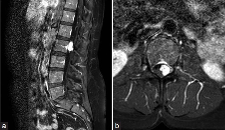
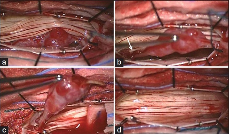
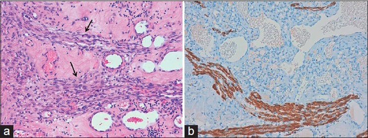
We read with great interest, the recent review about capillary hemangiomas of cauda equina published by Liu et al.[2] Reviewing this article and the pertinent literature we noted that no intraoperative picture of a capillary hemangioma of cauda equina has ever been published until now. A 45-year-old woman with 2 years history of low-back pain was recently admitted to our department. On admission, neurological exam was negative. Spinal magnetic resonance imaging showed a space occupying lesion at L2 level displacing the cauda equina roots [Figure 1]. She was then submitted, under neurophysiological monitoring, to surgery for tumor removal. After an L1–L2 laminotomy and opening of dura mater, the lesion was identified [Figure 2a and b]. The lesion appeared easily detachable from the healthy nerve roots. Thus, after dissection, the pathological nerve root was cut proximally and distally allowing the total removal of the lesion [Figure 2c and d]. Microscopically, the lesion was composed of many thin-walled vascular channels, lined by endothelial cells with interposed nerve fibers [Figure 3]. Thus, a diagnosis of nervous capillary hemangioma was made. Postoperatively the patient complained of mild hyposthenia in left leg, totally recovered at 3 months follow-up.
Figure 1.

Sagittal (a) and axial (b) postcontrast T1-weighted spinal magnetic resonance imaging showing a well-circumscribed lesion at L2 level with intense enhancement after gadolinium administration
Figure 2.

Intraoperative view of lesion. The lesion appeared as a white reddish elliptical-nodular lesion intimately involved with the nerve root displacing the cauda equina (a) Enlarged vessels were also evident along the nerve root (b, white arrow). After dissecting the lesion from cauda equina and cutting the originating nerve root distally (c) the lesion was totally removed (d)
Figure 3.

Histological picture (a) of the surgical specimen shows many, variably sized, thin-walled vascular channels lined by endothelial cells with interposed nerve fibers (black arrows) (H and E, original magnification ×20). Immunohistochemical staining (b) for neurofilament protein highlights the nerve fibers interspersed among the vascular spaces. The lesion was also positive at S-100 staining (not showed) (original magnification ×20)
To our knowledge, we report the first intraoperative picture of a capillary hemangioma of cauda equina. Only a few cases of capillary hemangiomas have been reported at cauda equina.[1,2] Histopathological examination is needed for diagnosis. Typical features are the presence of a myriad of small capillary sized vessels, reticularly arranged with normal nerve fascicles dispersed within the nodules of clustered capillaries.[3,4,5] Our picture confirms that, according with the microscopically examination, this tumor arises and develops intrinsically to the nerve root. Surgery is the therapy of choice, but a total removal is possible only cutting (cranially and caudally to the tumor) the pathological nerve root.
Footnotes
Contributor Information
Fabrizio Pignotti, Email: fabriziopignotti87@gmail.com.
Antonella Coli, Email: antonella.coli@rm.unicatt.it.
Eduardo Fernandez, Email: e.fernandez@rm.unicatt.it.
Nicola Montano, Email: nicolamontanomd@yahoo.it.
REFERENCES
- 1.Ganapathy S, Kleiner LI, Mirkin LD, Hall L. Intradural capillary hemangioma of the cauda equina. Pediatr Radiol. 2008;38:1235–8. doi: 10.1007/s00247-008-0947-1. [DOI] [PubMed] [Google Scholar]
- 2.Liu JJ, Lee DJ, Jin LW, Kim KD. Intradural extramedullary capillary hemangioma of the cauda equina: Case report and literature review. Surg Neurol Int. 2015;6:S127–31. doi: 10.4103/2152-7806.155701. [DOI] [PMC free article] [PubMed] [Google Scholar]
- 3.Mastronardi L, Guiducci A, Frondizi D, Carletti S, Spera C, Maira G. Intraneural capillary hemangioma of the cauda equina. Eur Spine J. 1997;6:278–80. doi: 10.1007/BF01322452. [DOI] [PMC free article] [PubMed] [Google Scholar]
- 4.Nowak DA, Gumprecht H, Stölzle A, Lumenta CB. Intraneural growth of a capillary haemangioma of the cauda equina. Acta Neurochir (Wien) 2000;142:463–7. doi: 10.1007/s007010050458. [DOI] [PubMed] [Google Scholar]
- 5.Nowak DA, Widenka DC. Spinal intradural capillary haemangioma: A review. Eur Spine J. 2001;10:464–72. doi: 10.1007/s005860100296. [DOI] [PMC free article] [PubMed] [Google Scholar]


