Abstract
Background and Objectives:
Fine needle aspiration cytology (FNAC) is an established out- patient procedure used in primary diagnosis of palpable thyroid lesions. A modified technique fine needle capillary sampling (FNCS) obviates the need of suction, is less painful, patient friendly and reported to overcome the problem of inadequate and bloody specimens. The aim of our study was to compare the efficacy and quality of FNCS with that of conventional FNAC in the lesions of thyroid.
Methods:
One hundred patients, presenting between January 2011 to December 2012 at Cytopathology Department of M M Institute of Medical Sciences and Research, Mullana, with diffuse and nodular thyroid lesions were enrolled with both the techniques being executed on the patients, beginning with FNA followed by FNCS. The smears were scored using five objective parameters i.e. background blood, cellular material, cellular degeneration, cellular trauma, and retention of appropriate architecture, in a single blind setting by a cyto-pathologist. The results were analyzed using Student’s test for paired data and chi- square analysis.
Results:
A highly significant differences (P<0.001) in favor of FNCS was observed for the background blood, cellular material and retention of architecture while total score favored FNA for cellular degeneration and degree of cellular trauma. Total scores and average score per case for FNCS was significantly better (P<0.001) than FNA. FNCS technique yielded more diagnostically superior and lesser number of unsatisfactory smears whereas greater number of diagnostically adequate samples was obtained by FNA technique.
Conclusion:
FNCS offers more number of diagnostically better quality smears. Both techniques could be supplementary on many occasions and substitutive on a few. Combination of the two techniques could offer better diagnostic accuracy.
Key Words: Fine Needle Aspiration, Fine Needle Capillary Sampling
Introduction
Fine needle aspiration cytology (FNAC) is firmly established as a first line investigative modality in the pretreatment evaluation of thyroid lesions. But this technique is not infrequently complicated by aspiration of significant quantities of blood, particularly in vascular organs like thyroid which compromise cellular preservation and interpretation. Furthermore, many diagnostic pitfalls exist in the interpretation of thyroid specimens making excellence of cellular material a prerequisite for diagnosis. In an attempt to overcome this problem, an alternative sampling method, non- aspiration fine needle cytology, was pioneered in France in 1980s and first described in the investigation of thyroid lesions by Santos and Leiman in 1988 (1, 2).
Several terms have been used to describe the technique; non-aspiration cytology, fine needle capillary sampling, fine needle non- aspiration, cytopuncture and fine needle sampling. It is reported to be easier to perform and most likely less painful. However, its efficacy compared with FNAC needs to be established.
The objective of this prospective study was to compare the efficacy and quality of FNCS with that of conventional technique of FNAC by using both the techniques in thyroid lesions to ascertain whether it could be chosen as a superior cytodiagnostic procedure in vascular organs like thyroid.
Materials and Methods
The present prospective study was conducted on 100 patients with diffuse and nodular thyroid lesions, attending the Cytopathology Department of M M Institute of Medical Sciences & Research, Mullana over a two year period i.e. from January 2011-December 2012. Both FNA and FNCS techniques were executed on the same patient in the same clinical session irrespective of consistency and size of lesions. FNA was done followed by FNC sampling in all the cases. All the procedures were performed by a single cytotechnician.
Every slide was evaluated in a single blind setting by a single cytopathologist without the prior knowledge of techniques utilized, thus prevented the observer bias. On an average, with the FNCS method 3 smears could be prepared for each case whereas with the FNA method 5 smears could be prepared for each case. FNA was performed by the conventional method as described in standard text books (3). FNCS was performed using a handheld 22-gauge needle without a syringe or handle, inserted into target lesion and moved back and forth in various directions. The cells were detached by cutting edge of the needle. After withdrawing the needle it was attached to a syringe filled with air and the materials was expressed on clean dry slides and smears were prepared in the usual manner. Half of the smears were immediately fixed in 95% ethyl alcohol for subsequent Papanicolaou staining, the remaining smears were air-dried and stained by May Grunwald Giemsa stain.
The smears were scored according to criteria using a predetermined scoring developed by Mair et al. (4). The two sampling techniques were compared using five objective parameters as background blood or clot; amount of cellular material; degree of cellular degeneration; degree of cellular trauma; and retention of appropriate architecture (Table 1). A cumulative score was obtained for each specimen which was then categorized into one of the three categories as unsuitable for cytodiagnosis (score 0-2), adequate for cytodiagnosis (score 3-6) and diagnostically superior (score 7-10). The scores were tabulated and results analyzed using categoric and Student’s test for paired data as well as chi- square analysis.
Table 1.
Point scoring system to classify quality of cytological aspirate
| Criterion | Quantitative description | Point score | |
|---|---|---|---|
| Background blood or clot | Large amount | Great compromise to diagnose Diagnosis possible |
0 |
| Moderate amount Minimal |
Diagnosis easy Specimen of textbook quality |
1 2 |
|
| Amount of cellular material | Minimal to absent Moderate Abundant |
Diagnosis not possible Sufficient for cytodiagnosis Diagnosis possible |
0 1 2 |
| Degree of cellular degeneration | Marked Moderate Minimal |
Diagnosis impossible Diagnosis possible Diagnosis easy |
0 1 2 |
| Degree of cellular trauma | Marked Moderate Minimal |
Diagnosis impossible Diagnosis possible Diagnosis obvious |
0 1 2 |
| Retention of appropriate architecture | Minimal to absent | Non diagnostic | 0 |
| Moderate | Some preservation of, e.g. follicle, papillae, acini, flat sheets, syncytia or single cell pattern | 1 | |
| Excellent architectural display closely reflecting histology; diagnosis obvious | 2 | ||
Results
For the five parameters studied objectively, a statistically highly significant differences (P<0.001) in favor of FNCS was observed for the parameters; background blood, amount of cellular material and retention of architecture. For the rest of the parameters- degree of cellular degeneration and degree of cellular trauma, the total score favored FNA with a statistically significant difference. On analyzing the total scores and average score per case of each sampling technique it was observed that the scores for FNCS were significantly better (P<0.001) than FNA as depicted in Table 2.
Table 2.
Comparison of FNAC and FNCS for various parameters
| Criterion |
FNAC
*
|
FNCS
**
|
P value | ||
|---|---|---|---|---|---|
| Mean±SD *** | Total | Mean±SD | Total | ||
| Background blood/clot | 0.56±0.499 | 56 | 1.74±0.485 | 174 | <0.001 |
| Amount of cellular material | 1.25±0.557 | 125 | 1.76±0.474 | 176 | <0.001 |
| Degree of cellular degeneration | 1.33±0.711 | 133 | 0.92±0.662 | 92 | <0.001 |
| Degree of cellular trauma | 1.32±0.584 | 132 | 0.85±0.479 | 85 | <0.001 |
| Retention of appropriate architecture | 0.96±0.530 | 96 | 1.83±0.428 | 183 | <0.001 |
| Total score | 542 | 710 | |||
| Average score per case | 5.42±2.113 | 7.1±1.761 | <0.001 | ||
| Diagnostically superior Diagnostically suitable |
40/100 84/100 |
55/100 95/100 |
<0.05 <0.05 |
||
Fine Needle Aspiration Cytology
Fine Needle Capillary Sampliry
Standard Deviation
Comparing the performance of both the techniques as shown in Table 3, it was observed that a total of 16 cases were unsuitable for diagnosis by FNA as compared to 5 cases by FNCS technique. The FNCS technique yielded more number of diagnostically superior (n=55) smears whereas FNA technique yielded more number of diagnostically adequate (n=44) smears. Table 4 depicts the frequencies of the various lesions encountered.
Table 3.
The Performance of FNAC and FNCS Techniques in Thyroid lesion
| Performance |
Techniques
|
|
|---|---|---|
| FNAC * | FNCS ** | |
| Diagnostically inadequate | 16(16.0) | 5(5.0) |
| Diagnostically adequate | 44(44.0) | 40(40.0) |
| Diagnostically superior | 40(40.0) | 55(55.0) |
Fine- Needle Aspiration Cytology
Fine- Needle Capillary Sampling; Figures In Parentheses Are In Percentage
Table 4.
Frequency of Various Thyroid Lesions
| Sample no. | Type of lesion | No. of cases | Percentage |
|---|---|---|---|
| 1 | Colloid goiter | 30 | 30 |
| 2 | Multinodular goiter | 23 | 23 |
| 3 | Thyroiditis | 21 | 21 |
| 4 | Follicular neoplasm | 19 | 19 |
| 5 | Papillary carcinoma | 6 | 6 |
| 6 | Medullary carcinoma | 1 | 1 |
| Total | 100 | 100 | |
Discussion
FNAC has been established as a valuable tool in the evaluation of thyroid nodules, since it is easy to perform, rapid, with a low complication rate and has a high degree of specificity and sensitivity (3). However, getting adequate cellularity is an inherent problem in thyroid FNA and is essentially due to increased vascularity of thyroid gland. FNA technique depends on suction and thus is at times painful, many times traumatic, as well as yield haemorrhagic material for cytologic study. In more recent time a modified technique called FNCS has come into vogue which obviates the need of suction. The principle depends solely on capillary action of the fine needle, where a fluid or semifluid substance ascends spontaneously into a narrow tube, in inverse proportion to the diameter of that tube (2). Therefore this allows the procedure to be much less painful, much less traumatic and more patient friendly (5). Kate et al, in an analysis of 670 patients, observed that FNCS allows greater ease of sampling with better control of hands during the procedure and a good perception of the lesion (6). FNCS technique provides smears of superb “text book quality”, without much blood in the background and with better cellularity.
A noteworthy finding in our study was that FNCS technique was well tolerated by the patients firstly because they were not apparently intimidated by the innocuous needle and handle and secondly because the method caused minimal pain.
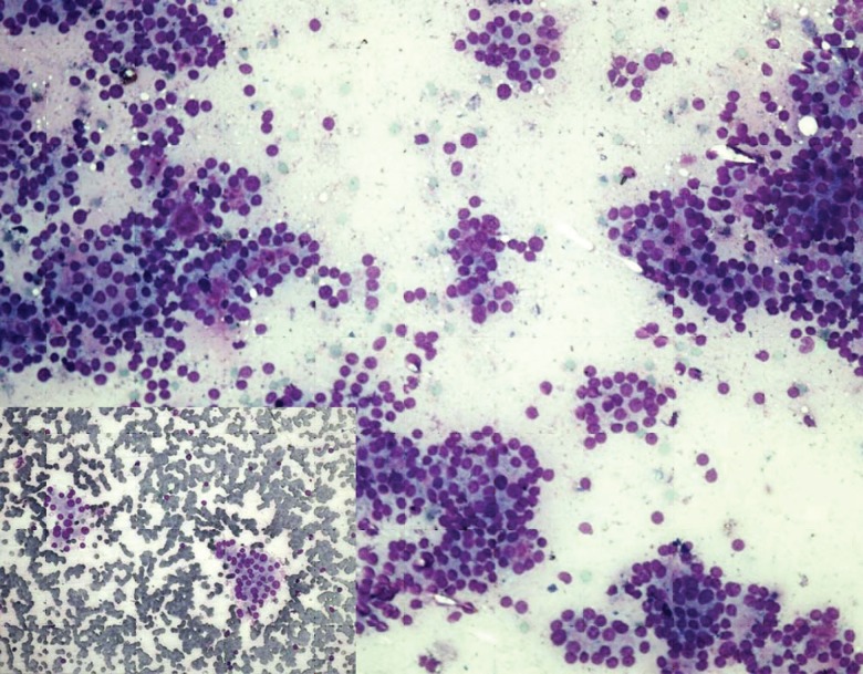
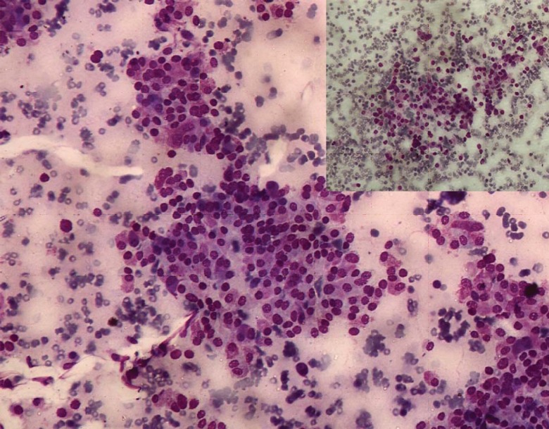
Results when compared for background blood contamination supported the FNCS and were statistically highly significant (Fig.1 and 2). FNCS was found to cause lesser degree of contamination because specimen was obtained by a spontaneous capillary action rather than suction due to negative pressure. In a previous study however, Akhtar et al. (7) did not notice a significant difference in the degree of dilution with blood. This might be because they continued the rotary passes until blood became visible in the hub of the needle. However, it should be remembered that whether it is FNCS or FNA, thyroid cytology samples are inevitably bloody.
Fig.1.
Photomicrograph of FNCS smear of follicular adenoma showing hypercellularity and minimal blood contamination in comparison to FNAC smear [inset] (May-Grunwald Giemsa, ×200
Fig. 2.
Photomicrograph of FNCS smear of medullary carcinoma showing better cellular yield, more preservation of cytoplasmic granules and less blood in the background in comparison to FNAC smear[inset] (May-Grunwald Giemsa, ×200)
Similar to the study by Akhtar et al. (7), the amount of cellular yield was found to be better by FNCS with a statistically highly significant difference. An observation similar to that of some previous workers but the difference was not statistically significant in these studies (8-10). In contrast, Jayaram and Gupta (11) observed that cellularity was higher in FNA smears than in FNCS smears in most of the goiters. Mair et al. (4) and Zajdela et al. (1) did not find any significant difference in the smears prepared by both the techniques.
Tumors of thyroid bleed easily and therefore hemodilution of the sample is common. Malignant cells being fragile are more prone to degeneration and trauma of suction. The application of suction to draw cells through a fine needle traumatizes fragile cells resulting in artifacts that can lead to diagnostic error. It was observed in our study that FNCS especially performed far better than FNA for malignant lesions i.e. yielded adequate material with less contamination of blood. Therefore the nature of lesion also has important bearing on the efficacy of the technique.
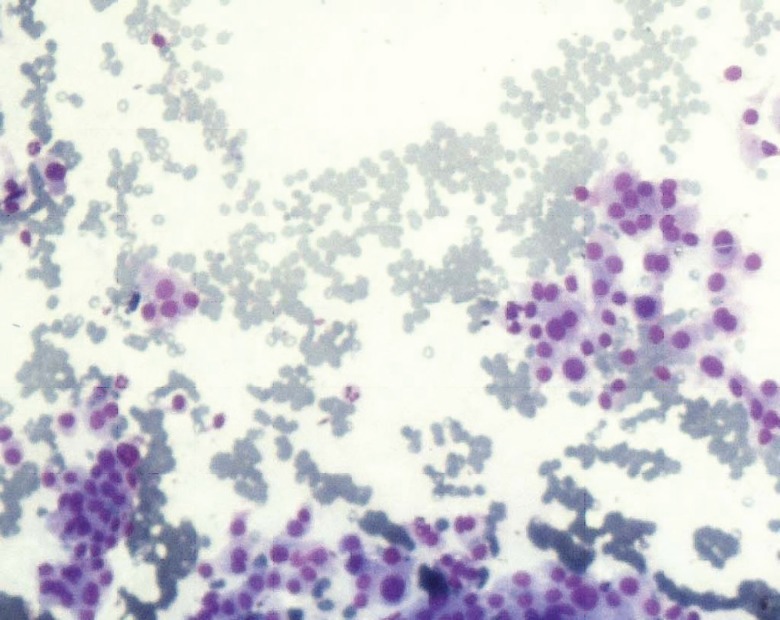
Unlike similar studies, the present study revealed a lesser degree of cellular degeneration and trauma in FNA as compared with FNCS smears. This was in agreement with the study by Baksh et al. (12); however the difference was statistically insignificant. Although the mean scores were more for FNCS as compared to FNA, but the difference was statistically insignificant in the studies by previous workers (8, 9, 13). We could not ascertain a specific reason for this, but possibly this could be because in the present study, FNA was executed first followed by FNC sampling. FNCS smears yielded better retention of architecture (Fig. 3) with similar findings reported by others (7- 9, 13).
Fig. 3.
Photomicrograph of FNCS smear of papillary carcinoma of thyroid showing high cellular yield with better retained architecture (May-Grunwald Giemsa, ×100)
When we analyzed the quality of specimens by comparing the performance of both the techniques with various studies, it was observed in the present study that the diagnosis was adequately possible in more number of cases by FNA technique but the FNCS yielded specimen of superior text book quality in more number of cases i.e. 55 as compared to 40 by FNAC and the difference was statistically significant (P<0.05; chi-square test), which was in agreement with the findings of other studies (2, 4, 8, 9, 14 ,15).
It has been emphasized in the literature that an experienced cytopathologist needed to interpret the aspirates. It would be wise to remember that the cytopathologist is only as good as the sample he or she obtains or that is provided to him or her. It is very difficult to diagnose/interpret if the smears contain only blood. An adequate specimen is the cornerstone for an accurate diagnosis and subsequent management of the patient. We agree that most of the diagnostic failures are due to unsatisfactory samples. Percentage of inadequate samples was less with FNCS (5%) than with FNA (16%) technique- an observation similar to that of Rizvi et al. (14), Santos and Leiman (2) and Ciatto et al. (16). This was at variance to the results obtained by Maurya et al. (9), Ghosh et al. (8) and Mahajan et al. (15), where the percentage of inadequate sampling was more with FNCS (38%, 10% and 20%; respectively). The dilution of cellular material with blood seemed to be main cause of higher rate of inadequacy by FNA technique. This is minimal with FNCS technique because of spontaneous ascent of material by capillary action. In FNA technique active and often high suction pressures, lead to immediate filling of needle with blood leading to withdrawal of needle, so less cellular yield while with FNCS there is more time to manipulate the needle.
Notwithstanding, when both the techniques were compared together for diagnostically suitable smears, it was observed that FNCS yielded more diagnostically suitable smears and the difference was statistically significant (P<0.05; chi-square test).
In the ultimate analysis when both the cumulative score as well as mean score for individual criteria were considered, it was observed that FNCS scored over FNA and the difference was statistically highly significant (P<0.001) i.e. amount of material was more with less contamination with blood and better preservation of cellular morphology and architecture.
Conclusion
It was observed that both the techniques have their own advantages and disadvantages. FNCS offers the distinct advantage of diagnostically better quality smears; FNA assures diagnostically adequate material quantitatively. Important advantage of FNCS is easy operation, better perception of tumor consistency, much more patient friendly and seemed to be better for diagnosing malignant lesions. This has been substantiated by a recent systematic review and meta-analysis which favors FNCS in terms of easy performance and better sample quality (17). On the whole FNCS is advantageous in some situations, whereas FNA scores over FNCS in some others. Both could be supplementary on many occasions but could be substitutive on a few. Though FNCS was diagnostic in a greater number of cases than FNA sampling, this study did not prove a clear superiority of FNCS over FNA. Also further studies by using two evaluators with comparison and randomization of sampling can give further insight into the role of FNCS. We strongly think that by combining both the techniques better diagnostic accuracy can be achieved and therefore both the techniques would be complimentary to each other.
Acknowledgment
We wish to acknowledge the immense help given by Mr Sanjeev Mittal, senior technician of our department. The authors declare that there is no conflict of interests.
References
- 1.Zajdela A, Zillhardt , Voillemet N. Cytological diagnosis by fine needle sampling without aspiration. Cancer. 1987;59(6):1201–5. doi: 10.1002/1097-0142(19870315)59:6<1201::aid-cncr2820590628>3.0.co;2-p. [DOI] [PubMed] [Google Scholar]
- 2.Santos JE, Leiman G. Non aspiration fine needle cytology: Application of a new technique to nodular thyroid diseases. Acta Cytol. 1988;32(3):353–6. [PubMed] [Google Scholar]
- 3.Orell SR, Sterrett GF, Walters MNI, Whitaker D. Manual and Atlas of fine needle aspiration cytology. 3rd ed. Philadelphia: Churchill Livingstone; 1999. [Google Scholar]
- 4.Mair S, Dunbar F, Becker PJ, Du Plessis W. Fine needle cytology- is aspiration suction necessary? A study of 100 masses in various sites. Acta Cytol. 1989;33(6):809–13. [PubMed] [Google Scholar]
- 5.Misra M, Chawla SC, Bandyopadhyay SP. Fine needle capillary sampling (Non aspiration technique) vs conventional FNAC in cytological diagnosis. Cytology. 2001;18(1):25–30. [Google Scholar]
- 6.Kate MS, Kamal MM, Bobhate SK, Kher AV. Evaluation of fine needle capillary sampling in superficial and deep- seated lesions An analysis of 670 cases. Acta Cytol. 1998;42(3):679–84. doi: 10.1159/000331826. [DOI] [PubMed] [Google Scholar]
- 7.Akhtar SS, Huq IU, Faiz-U-Din M, Reyes LM. Efficacy of fine needle capillary biopsy in the assessment of patients with superficial lymphadenopathy. Cancer Cytopathol. 1997;81(5):277–80. doi: 10.1002/(sici)1097-0142(19971025)81:5<277::aid-cncr4>3.0.co;2-k. [DOI] [PubMed] [Google Scholar]
- 8.Ghosh A, Mishra RK, Sharma SP, Singh HN, Chaturvedi AK. Aspiration vs non- aspiration technique of cytodiagnosis- A critical evaluation in 160 cases. Indian J Pathol Microbiol. 2000;43(2):107–12. [PubMed] [Google Scholar]
- 9.Maurya AK, Mehta A, Mani NS, Nijhawan VS, Batra R. Comparison of aspiration vs non-aspiration techniques in fine- needle cytology of thyroid lesions. J Cytol. 2010;27(2):51–4. doi: 10.4103/0970-9371.70737. [DOI] [PMC free article] [PubMed] [Google Scholar]
- 10.Kamal MM, Arjune DG, Kulkarni HR. Comparative study of fine needle aspiration and fine needle capillary sampling of thyroid lesions. Acta Cytol. 2002;46(1):30–4. doi: 10.1159/000326712. [DOI] [PubMed] [Google Scholar]
- 11.Jayaram G, Gupta B. Non aspiration fine needle cytology in diffuse and nodular thyroid lesion A study of 220 cases. Acta Cytol. 1991;35(6):789–90. [PubMed] [Google Scholar]
- 12.Baksh S, Masih K, Singh S, Das S. Diagnostic utility of fine needle non- aspiration cytology versus fine needle aspiration cytology in breast masses. Indian J Pathol Microbiol. 2004;47(3):319–21. [PubMed] [Google Scholar]
- 13.Raghuveer CV, Leekha I, Pai MR, Adhikari P. Fine needle aspiration cytology versus fine needle sampling without aspiration A prospective study of 200 cases. Indian J Med Sci. 2002;56(9):431–9. [PubMed] [Google Scholar]
- 14.Rizvi SA, Husain M, Khan S, Mohsin M. A comparative study of fine needle aspiration cytology versus non- aspiration technique in thyroid lesions. Surgeon. 2005;3(4):273–6. doi: 10.1016/s1479-666x(05)80091-5. [DOI] [PubMed] [Google Scholar]
- 15.Mahajan P, Sharma PR. Fine needle aspiration versus non- aspiration technique in cytodiagnosis in thyroid lesions. JK Science. 2010;12(3):120–2. [Google Scholar]
- 16.Ciatto S, Lossa A, Cicchi P, et al. Non aspiration fine needle cytology of thyroid tumors. Acta Cytol. 1983;33(5) [PubMed] [Google Scholar]
- 17.Pothier DD, Narula AA. Should we apply suction during fine needle cytology of thyroid lesions? A systematic review and meta-analysis. Ann R Coll Surg Engl. 2006;88(7):643–5. doi: 10.1308/003588406X149147. [DOI] [PMC free article] [PubMed] [Google Scholar]


