Abstract
Guinea pigs possess several biological similarities to humans and are validated experimental animal models1-3. However, the use of guinea pigs currently represents a relatively narrow area of research and descriptive data on specific methodology is correspondingly scarce. The anatomical features of guinea pigs are slightly different from other rodent models, hence modulation of sampling techniques to accommodate for species-specific differences, e.g., compared to mice and rats, are necessary to obtain sufficient and high quality samples. As both long and short term in vivo studies often require repeated blood sampling the choice of technique should be well considered in order to reduce stress and discomfort in the animals but also to ensure survival as well as compliance with requirements of sample size and accessibility. Venous blood samples can be obtained at a number of sites in guinea pigs e.g., the saphenous and jugular veins, each technique containing both advantages and disadvantages4,5. Here, we present four different blood sampling techniques for either conscious or anaesthetized guinea pigs. The procedures are all non-terminal procedures provided that sample volumes and number of samples do not exceed guidelines for blood collection in laboratory animals6. All the described methods have been thoroughly tested and applied for repeated in vivo blood sampling in studies within our research facility.
Keywords: Medicine, Issue 92, guinea pig, animal model, blood sampling, non-terminal, saphenous, tarsal, jugular
Introduction
The guinea pig is a valuable and validated experimental animal model due to a number of biological similarities to humans such as a requirement for a dietary supply of vitamin C, comparable plasma lipoprotein metabolizing enzymes and lipoprotein profiles, as well as shared similarities with human placentation and prenatal development1-3,7,8. This makes the guinea pig an attractive and suitable model for studying effects of diet-associated diseases such as non-alcoholic fatty liver disease, cardiovascular diseases and putative effects of vitamin C deficiency but also the effect of e.g., maternal dietary intervention on the developing offspring.
Repeated blood sampling is often required in both long and short term in vivo studies. Due to species-specific anatomical differences modulation of the sampling technique is necessary in order to obtain blood samples in guinea pigs compared to other rodent models4,5. This includes variation in the course of veins and arteries, and also that guinea pigs do not have a tail, thus have different requirements and options for both sampling and handling. Sampling from the saphenous or tarsal vein, induces minimal discomfort and stress in the animal and does not require anaesthesia3,9. This technique allows repeated samples of small amounts of blood (100-400 µl), e.g., to determine biochemical markers in plasma. If an even smaller sample size is sufficient (50-100 µl), this can quite easily be obtained by puncturing the ear vein. This technique can also be carried out in conscious animals and is useful for example when measuring blood glucose10,11. Sampling from the jugular vein allows for collection of larger amounts of blood (1-2 ml) but requires anaesthesia.
Here, we present four different blood sampling techniques for either conscious or anaesthetized guinea pigs. The procedures are all non-terminal procedures provided that sample volumes and number of samples do not exceed guidelines for blood collection in laboratory animals6. It is in the interests of good science as well as of animal welfare that stress should be kept to a minimum. Consequently, only well-trained and competent personal should perform the animal procedures.
Protocol
The shown procedures were performed according to protocols approved by the Danish Animal Experimentation Inspectorate under the Ministry of Food, Agriculture and Fisheries.
1. Blood Sampling from the Lateral Saphenous or Tarsal Vein
Perform this procedure without anaesthesia and the help of one investigator, one handling the guinea pig, and one collecting the sample.
Shave the tarsal region of the hind leg with an electric shaver until the veins are visible. Ensure the area is large enough to avoid fur interfering with the puncture site.
Place the hind leg in a lukewarm water bath and gently rub the leg, making the veins more dilated and evident.
Wipe the leg with a clean towel and disinfect the puncture site with gauze or a cotton swab dampened with 70% alcohol.
Restrain the guinea pig and extend the hind leg downward while applying gentle pressure immediately above the knee joint to provide stasis.
Puncture the vein using a 21 G needle or lancet. Collect blood e.g., by using a collection or capillary tube. Gently manipulate the leg to aid the blood flow.
Release stasis, once the sample has been obtained. Apply a piece of gauze or cotton wool with gentle pressure to the puncture site until bleeding has stopped.
Return the animal to its cage and monitor for 5-10 min to ensure hemostasis.
2. Blood Sampling from the Ear Vein
Perform this procedure without anaesthesia either alone or preferably with the help of one colleague.
Disinfect the dorsal surface of the ear using gauze or a cotton swab dampened with 70% alcohol.
Apply light pressure to the base of the ear to provide stasis.
Puncture the vein using a 21 G needle or lancet, and collect blood.
Release stasis, once the sample has been obtained, Apply a piece of gauze or cotton wool with gentle pressure to the puncture site until bleeding has stopped.
Return the animal to its cage and monitor for 5-10 min to ensure hemostasis.
3. Blood Sampling from the Jugular Vein
Carry out this procedure using the help of one additional investigator; one handling the guinea pig and one collecting the sample.
Anaesthetize the guinea pig with a suitable anaesthetic for the entire duration of this procedure. A mixture of tiletamine (0.93 mg/kg), zolazepam (0.93 mg/kg), xylazine (1.49 mg/kg) and butorphanol (0.06 mg/kg) can be used.
Place the anaesthetized guinea pig on its back and gently retract the forelegs caudally.
Shave and disinfect (e.g., with 70% alcohol) the ventral part of the neck.
Coat the syringe with either heparin or EDTA prior to sampling in order to reduce risk of clotting.
Palpate the clavicle and insert a 25 G (or 23 G) needle mounted on a 1 ml syringe approximately 1 cm lateral to the midline at the level of the shoulder joint. Keep the syringe in a slight upward and medial angle while applying a slight negative pressure. Avoid damaging the underlying structures as the jugular vein is situated quite superficial. Tilt the neck of the guinea pig slightly backwards to provide increased stasis of the vein.
Release the front legs when the blood sample is obtained, and alleviate stasis before the needle is retracted. Use a dry piece of gauze or cotton wool to apply gentle pressure to the puncture site.
Return the animal to a recovery cage and monitor to ensure hemostasis and recovery from anaesthesia.
Representative Results
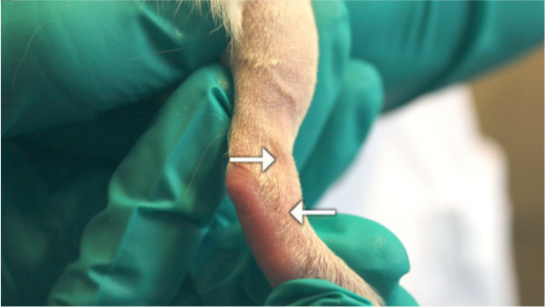
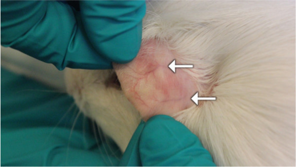
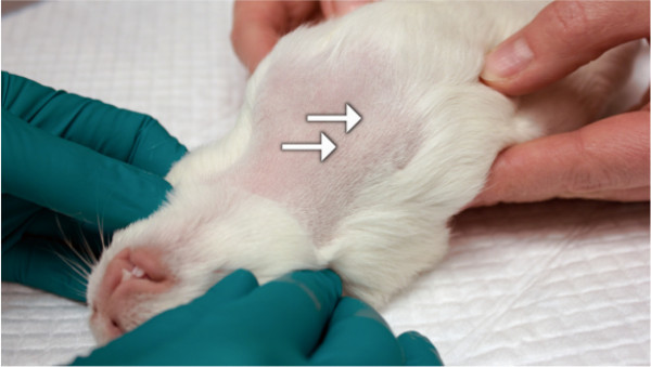
Four different approaches to non-terminal in vivo blood sampling techniques in either conscious or anaesthetized guinea pigs have been presented. Figure 1 illustrates the course of the lateral saphenous and tarsal vein in the guinea pig and possible puncture sites. Ear veins visible on the dorsal surface of a guinea pig ear are illustrated in Figure 2. These veins can be used for blood sampling when only a very small volume is required. The position of the guinea pig for jugular blood sampling is show in Figure 3. The course of the jugular vein is illustrated by the arrows with the uppermost arrow pointing at the recommended puncture site.
 Figure 1. The lateral saphenous and tarsal vein in the guinea pig. The image illustrates the course and the puncture site for blood sampling from the lateral saphenous (upper arrow) or the tarsal vein (bottom arrow) in the guinea pig.
 Figure 2. Ear veins. This image illustrates ear veins (arrows) visible on the dorsal surface of a guinea pig ear. These veins can be used for blood sampling when only a very small volume is required.
 Figure 3. Position of the guinea pig for jugular vein blood sampling. This illustrates the position of the guinea pig and the puncture site for jugular vein blood sampling. The arrows indicate the course of the jugular vein with the uppermost arrow pointing towards the recommended puncture site.
Discussion
Compared to mice and rats, guinea pigs are fearful by nature and must be approached calmly and handled correctly in order to reduce stress. From an animal welfare point of view, acting with care is naturally a priority; however, this will also reduce the risk of anxiety-associated effects on the collected data e.g., increased blood levels of corticosteroids and glucose12. Likewise, daily handling will habituate the animals to normal procedures and may serve to reduce stress even further. Practical expertise in a particular blood sampling technique should be gained by first observing an experienced operator and thereafter conducting the technique under supervision until competence is achieved. The chosen technique should ideally both reduce stress and discomfort to a minimum but also to ensure survival as well as compliance with requirements of sample size and accessibility. For all presented blood sampling methods aseptic techniques should be used to minimize risk of infection and ensure collection of uncontaminated samples.
The illustrated approaches to blood sampling ensure survival of the animal provided that sample volume and frequency is achieved according to guidelines and can all be applied for repeated sampling in both short and long term studies6. This enables consistent monitoring of individual animals over time hereby reducing variance and number of included animals in compliance with the 3Rs’(reduction)13,14. The techniques presented here have different ranges of obtainable sample volume (a few µl up to 1-2 ml) which should be considered prior to commencing an experiment. Though procedures for collecting blood from the jugular vein in un-anaesthetized guinea pigs have been described12, we strongly recommend the use of anaesthesia to reduce stress and discomfort as much as possible. When doing so, the choice of anaesthesia should be carefully addressed, noting that guinea pigs are notoriously difficult to anaesthetize and display considerable variation effectiveness of the anaesthetic as well as a high sensitivity towards induced respiration depression15,16. As the jugular vein in guinea pigs is neither visible nor palpable any reduction in stasis/venous dilation may increase sampling difficulties, thus it is generally recommended to avoid vaso-constricting agents in anaesthesia-mixtures. Furthermore, known effects of specific anaesthetizing agents which may interfere with the desired data collection e.g., cardiovascular effects and/or increased blood glucose levels following α2- agonist actions in rodents should be assessed16,17. The use of inhalation anaesthesia such as isoflurane may also be an option, however as intubation in guinea pigs is a very difficult task care must be taken to avoid interference by the inhalation-mask with the head/neck position as this may complicate the procedure unnecessarily. Generalized anaesthesia e.g., by inhalation is also required for cardiac puncture which may provide a larger sample volume. However, cardiac puncture is a terminal procedure and therefore not applicable for repeated sampling and will not be described further.
Potential adverse effects such as hemorrhage, bruising, thrombosis, infection and scaring may occur following puncture of a vein. In our experience, the guinea pig does not exhibit an increased risk of such undesired complications compared to other rodent models provided that the procedure is done as described in the current manuscript. As the jugular vein cannot be visualized from the outer surface, it is very important to relieve stasis prior to withdrawing the needle and to provide adequate hemostasis in order to minimize risk of peri-venous hemorrhage and bruising. In the event of adverse effects, treatment should always be sought from the responsible veterinarian. For reasons of good animal welfare and science, serious considerations must be given to the combined effect of sample volume and frequency and choice of sampling technique. All the described blood sampling techniques have been thoroughly tested and applied for repeated in vivo blood sampling in studies within our research facility.
Disclosures
The authors have nothing to disclose. All procedures were approved by the Animal Experimentation Act of Denmark, which is in accordance with the Council of Europe Convention ETS 123.
Acknowledgments
We acknowledge the skilful assistance of Annie B. Kristensen, Elisabeth V. Andersen, Belinda B. Bringtoft and the animal caretakers at the Department of Experimental Medicine, Frederiksberg Campus, Faculty of Health and Medical Sciences, University of Copenhagen, Denmark. Jacob Lønholdt is thanked for his excellent technical assistance in filming the procedures. This study was supported in part by the LIFEPHARM Centre for in vivo Pharmacology and University of Copenhagen.
References
- DeOgburn R, et al. Effects of increased dietary cholesterol with carbohydrate restriction on hepatic lipid metabolism in Guinea pigs. Comp.Med. 2012;62(2):109–115. [PMC free article] [PubMed] [Google Scholar]
- Fernandez M, Volek JS. Guinea pigs a suitable animal model to study lipoprotein metabolism, atherosclerosis and inflammation. Nutr Metab (Lond) 2006;3(17) doi: 10.1186/1743-7075-3-17. [DOI] [PMC free article] [PubMed] [Google Scholar]
- Frikke-Schmidt H, Tveden-Nyborg P, Birck MM, Lykkesfeldt J. High dietary fat and cholesterol exacerbates chronic vitamin C deficiency in guinea pigs. Br J Nutr. 2011;105(1):54–61. doi: 10.1017/S0007114510003077. [DOI] [PubMed] [Google Scholar]
- Hem A, Smith AJ, Solberg P. Saphenous vein puncture for blood sampling of the mouse, rat, hamster, gerbil, guinea pig, ferret and mink. Lab Anim. 1998;32(4):364–368. doi: 10.1258/002367798780599866. [DOI] [PubMed] [Google Scholar]
- Parasuraman S, Raveendran R, Kesavan R. Blood sample collection in small laboratory animals. J Pharmacol Pharmacother. 2010;1(2):87–93. doi: 10.4103/0976-500X.72350. [DOI] [PMC free article] [PubMed] [Google Scholar]
- Guillen J. FELASA guidelines and recommendations. J Am Assoc Lab Anim Sci. 2012;51(3):311–321. [PMC free article] [PubMed] [Google Scholar]
- Ye P, Cheah IK, Halliwell B. High fat diets and pathology in the guinea pig Atherosclerosis or liver damage. Biochim Biophys Acta. 2013;1832(2):355–364. doi: 10.1016/j.bbadis.2012.11.008. [DOI] [PubMed] [Google Scholar]
- Tveden-Nyborg P, et al. Maternal vitamin C deficiency during pregnancy persistently impairs hippocampal neurogenesis in offspring of guinea pigs. PLoS One. 2012;7(10):12–18391. doi: 10.1371/journal.pone.0048488. [DOI] [PMC free article] [PubMed] [Google Scholar]
- Schjoldager JG, Tveden-Nyborg P, Lykkesfeldt J. Prolonged maternal vitamin C deficiency overrides preferential fetal ascorbate transport but does not influence perinatal survival in guinea pigs. Br J Nutr. 2013;110(9):1573–1579. doi: 10.1017/S0007114513000913. [DOI] [PubMed] [Google Scholar]
- Greulich S, et al. Secretory products of guinea pig epicardial fat induce insulin resistance and impair primary adult rat cardiomyocyte function. J Cell Mol Med. 2011;15(11):2399–2410. doi: 10.1111/j.1582-4934.2010.01232.x. [DOI] [PMC free article] [PubMed] [Google Scholar]
- Swifka J, Weiss J, Addicks K, Eckel J, Rosen P. Epicardial fat from guinea pig a model to study the paracrine network of interactions between epicardial fat and myocardium. Cardiovasc Drugs Ther. 2008;22(2):107–114. doi: 10.1007/s10557-008-6085-z. [DOI] [PubMed] [Google Scholar]
- Pilny AA. Clinical hematology of rodent species. Vet.Clin North Am Exot Anim Pract. 2008;11(3):523–vii. doi: 10.1016/j.cvex.2008.04.001. [DOI] [PubMed] [Google Scholar]
- Franco NH, Olsson IA. Scientists and the 3Rs: attitudes to animal use in biomedical research and the effect of mandatory training in laboratory animal science. Lab Anim. 2014;48(1):50–60. doi: 10.1177/0023677213498717. [DOI] [PubMed] [Google Scholar]
- Van LJ, et al. eAssessing the application of the 3Rs a survey among animal welfare officers in The Netherlands. Lab Anim. 2013;47(3):210–219. doi: 10.1177/0023677213483724. [DOI] [PMC free article] [PubMed] [Google Scholar]
- Sloan RC, et al. High doses of ketamine-xylazine anesthesia reduce cardiac ischemia-reperfusion injury in guinea pigs. J Am Assoc Lab Anim Sci. 2011;50(3):349–354. [PMC free article] [PubMed] [Google Scholar]
- Jacobson CA. A novel anaesthetic regimen for surgical procedures in guinea pigs. Lab Anim. 2001;35(3):271–276. doi: 10.1258/0023677011911598. [DOI] [PubMed] [Google Scholar]
- Saha JK, Xia J, Grondin JM, Engle SK, Jakubowski JA. Acute hyperglycemia induced by ketamine xylazine anesthesia in rats mechanisms and implications for preclinical models. Exp Biol Med(Maywood) 2005;230(10):777–784. doi: 10.1177/153537020523001012. [DOI] [PubMed] [Google Scholar]


