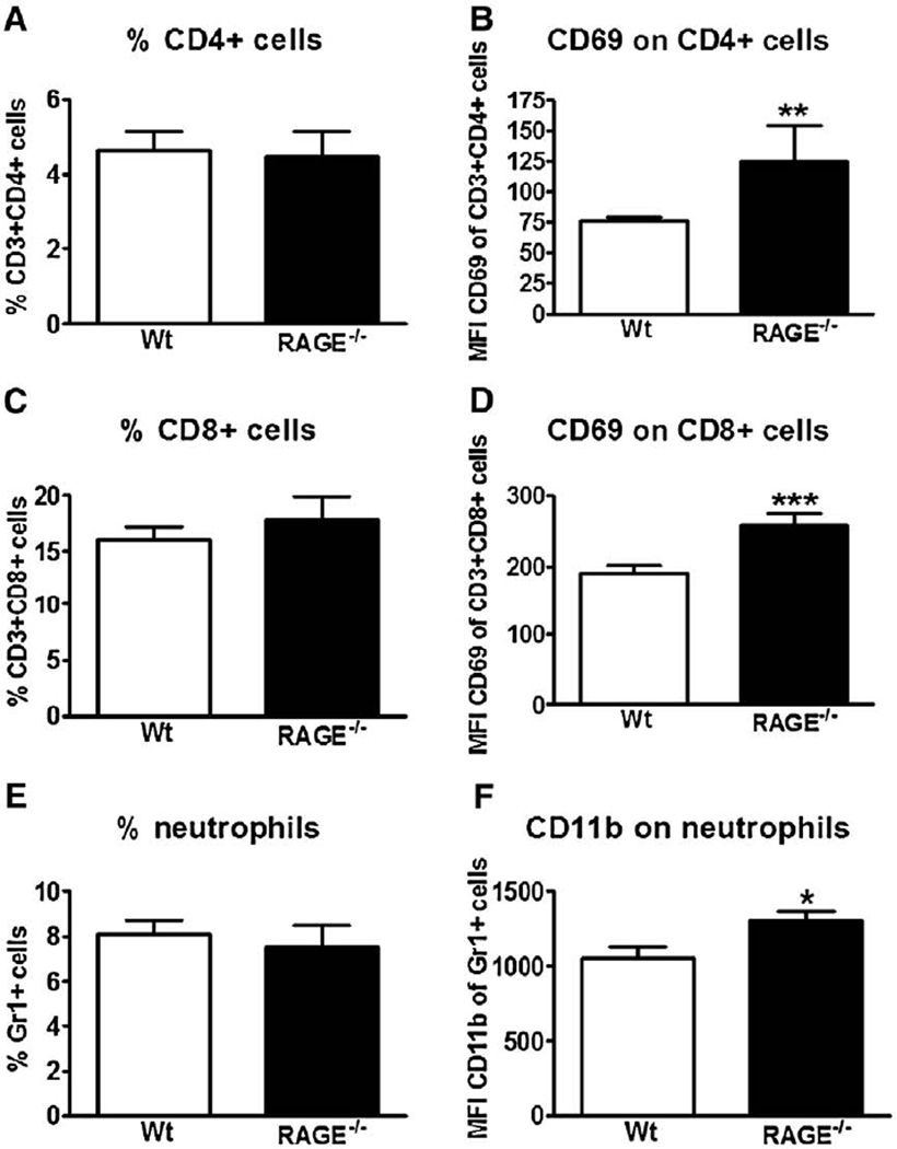
Fig. 5.
Receptor for advanced glycation end product deficient (RAGE−/−) mice show enhanced activation of pulmonary T lymphocytes and neutrophils. Wild-type (wt) and RAGE−/− mice were intranasally inoculated with influenza A virus (IAV). After 8 days, lung cell suspensions were collected and flow cytometry was performed as described in the Materials and methods section. Results are represented as percentage of CD4+ (A), CD8+ (C) cells and neutrophils (E) in the lungs and as the mean fluorescence intensity of CD69 surface expression within the CD4+ (B) and CD8+ (D) population and of CD11b surface expression within the Gr1+ population (F). Data are means ± SEs of 8–9 mice/genotype. *P<0.05, vs. wt mice. **P<0.01, vs. wt mice. ***P<0.005, vs. wt mice (Mann–Whitney U test).

