Figure 4.
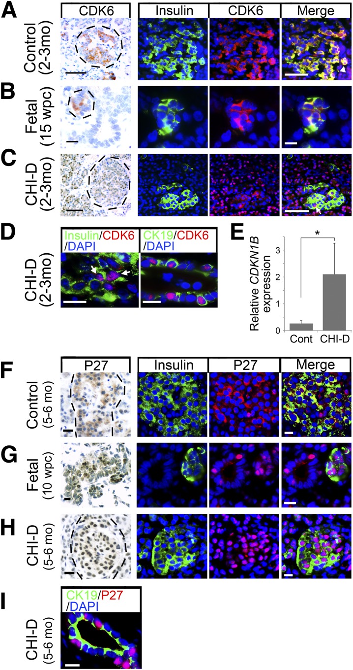
Increased nuclear localization of CDK6 and P27 in CHI-D. Bright-field immunohistochemistry counterstained with toluidine blue and dual immunofluorescence counterstained with DAPI for CDK6 (A–D) or P27 (F–I) in postnatal control pancreas, samples of fetal pancreas, and CHI-D. Costaining is with insulin except for samples with CK19 in D and I. Hatched lines in the bright-field images encircle islets. A–D: Arrowhead in the merged panel of A points to insulin-positive cells, in which CDK6 localizes to both cytoplasm and nucleus. In C, the cytoplasmic CDK6 is very much reduced in CHI-D compared with control (Cont) or fetal β-cells, whereas the arrow in the merged panel (and arrows in D) points to clear nuclear CDK6 in insulin-positive cells. E: Quantitative RT-PCR showing increased CDKN1B expression in CHI-D (mean ± SE from five cases across the first 13 months of age) compared with age-matched controls (n = 3). *P < 0.05, Mann-Whitney U test. F–I: P27 is almost exclusively cytoplasmic in postnatal control β-cells, whereas it is almost exclusively nuclear in fetal and CHI-D β-cells and in CK-19-positive duct cells. Scale bars = 50 µm (A and C), 20 µm (B), and 10 µm (D and F–I). mo, months.

