Abstract
The coincidence of alopecia and a tumour may indicate the paraneoplastic nature of alopecia. Paraneoplastic alopecia is not uncommon in animals, feline paraneoplastic alopecia being the best example known. We present a case of alopecia coinciding with the presentation of a cholangiocarcinoma in a woman. Following surgical resection of the tumour, alopecia resolved spontaneously and it reappeared on local recurrence, 2 years later. As far as pathogenesis is concerned, the coincidence of alopecia and cholangiocarcinoma may indicate the paraneoplastic nature of alopecia as a rare complication of this rare tumour in humans. This also implies that common interspecies mechanism(s) must exist as far as this paraneoplastic complication is concerned.
Background
Alopecia associated with internal tumours is not very common in humans but feline paraneoplastic alopecia is well described.1 It usually coincides with diagnosis of a tumour and it may resolve following complete excision of the tumour. Recurrence of hair loss following recurrence of the tumour may be expected, but this is rarely observed in animals owing to termination of euthanasia. Accordingly, a complete documentation of the paraneoplastic nature of alopecia in veterinary oncology is not always achieved. Cholangiocarcinoma is rare in western populations; paraneoplastic manifestations of this rare malignancy are rare as well. We report a case of alopecia developing as a presenting sign of cholangiocarcinoma in a Caucasian woman which resolved following resection of the tumour and reappeared following recurrence of the tumour. This case points to common interspecies mechanisms complicating this unusual malignancy with alopecia.
Case presentation
A 48-year-old Greek woman, married, had no children, currently living in the Athens area, with a free medical history and free of exposure to drugs, chemicals, tobacco or radiation, was admitted for investigation of a liver mass detected by ultrasonography. The mass was 6cm in diameter and it was located in the right liver lobe (segment V).
Investigations
On examination, the patient was found to be free of any sign apart from a round hair loss area just on the top of the skull, some 7 cm2 in surface and final sharp edges; this hair loss was noticed by her some weeks before ultrasound investigation. Clinical diagnosis was that of an alopecia areata. Local cultures proved negative for microorganisms but no skin biopsy was conducted as per the patient's decision. Serum protein electrophoresis was normal. Serology testing for viruses, tumour markers and the Venereal Disease Research Laboratory test were negative; anti-nuclear antibodies, antibodies to DNA, smooth-muscle antibodies and mitochondrial antibodies were negative. Thyroid hormonal measurements were normal; anti-thyroid peroxidase and anti-thyroglobulin antibodies were also negative.
Treatment
Following a further biochemical, haematological and imaging work-up that was otherwise negative, the patient was subjected to a partial liver resection, resulting in a complete removal of the tumour. Its histology revealed a biliary carcinoma (cholangiocarcinoma); a single portal lymph node was infiltrated by the tumour. No other indication of any residual mass or any distal metastasis existed. Shortly after operation the head hair loss resolved and new hair appeared, resulting in complete remission of alopecia.
Outcome and follow-up
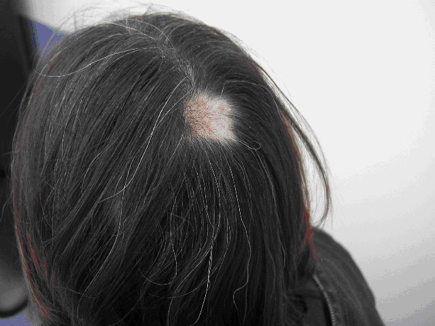
A regular follow-up MRI after operation was negative for any sign of local or systemic recurrence for the next 20 months. In February 2011, the patient became jaundiced and showed signs of bile duct obstruction both in blood chemistry and in imaging. In parallel, a relapse of the alopecia lesion was noted on the same top of head, as indicated in figure 1. No other alopecia plaques were found anywhere else. No evidence for any distal metastasis existed.
Figure 1.

Alopecia lesion relapsed on the same top of head.
A second operation was conducted by the same surgeons in March 2011. During this operation, two small tumours, some 2 cm in diameter each, were found at the middle of the hepatoduodenal ligament, pressing the common bile duct from outside and resulting in obstruction. The initial part of the duodenum was also adherent to them. These round tissues were resected and proved to be the recurrence of cholangiocarcinoma, but there was no bile tree infiltration. Although the cut surface of the liver from the previous resection looked suspicious and resected, it did not show any sign of recurrence.
The patient was reluctant to undergo any further investigation and decided not to be given chemotherapy.
Discussion
Lessons from animal oncology sometimes prove useful to human medicine and vice versa. This may apply in cases of paraneoplastic syndromes, that is, manifestations accompanying an internal tumour.2 The spectrum of paraneoplastic manifestations is broad. Skin paraneoplastic syndromes are of importance in terms of early detection or early recognition of tumour relapse.
Our case presented with alopecia areata. Speculations on the aetiology of this condition seem endless, stretching over many years; putative factors include focal infections, endocrine or nervous system disturbances, toxic factors and dental problems; emotional factors also play a significant role, but in most cases the cause remains undetermined.3 4
This alopecia coincided with the diagnosis of intrahepatic cholangiocarcinoma. It resolved spontaneously without any other intervention following surgical resection and it reappeared on relapse.
In our case, this temporal association points to the paraneoplastic nature of skin manifestation. It is clear that the role of emotional stress in all alopecia cases,4 whether paraneoplastic or not, cannot be excluded in cancer sufferers. Obviously, this role in animals is more difficult to establish.
The mechanisms underlying paraneoplastic alopecia remain obscure; a criterion that must be satisfied before considering an alopecia as paraneoplastic is that it must follow a parallel course with the relevant tumour; that is, remission of cancer results in clearing of alopecia and relapse of cancer results in recurrence. This requirement may not be fulfilled in animals as following a diagnosis of cancer the animal is usually subjected to euthanasia.
In cats, tumours potentially accompanied by alopecia are pancreatic carcinoma and cholangiocarcinoma.5
In humans, several tumours are complicated by particular paraneoplastic syndromes. Skin paraneoplastic dermatomyositis is not uncommon in gut tumours.6 Paraneoplastic myasthenia may complicate small-cell lung carcinoma. Paraneoplastic alopecia may develop in several human cancers, including malignant lymphomas; the deranged cellular immunity in this disease is proposed to be causative factor.7 Alopecia may also complicate gastrointestinal stromal tumours.8 In evaluating the paraneoplastic nature of alopecia, one must exclude the effects of anti-tumour agents on hair growth.
Cholangiocarcinoma in humans is rare, being more common in the Far East. The aetiology remains unclear, although some authors underline the role of parasites and dietary carcinogens. In Caucasians, it is currently far less frequent but a rising frequency has been recently noted in the USA.9 Data on specific paraneoplastic syndromes accompanying this tumour are not available; this applies not only to the West where the tumour is sporadic but also to the Far East where it prevails. Interestingly, another very unusual paraneoplastic syndrome, acquired porpurea cutanea tarda, was reported in the course of a cholangiocarcinoma in a Caucasian.10
Feline paraneoplastic alopecia manifesting as multiple lesions in many areas of the body appears late in the course of a malignant tumour, namely pancreatic adenocarcinoma and bile duct carcinoma.1 5
For a dermatosis to be considered as paraneoplastic the so-called Curth postulates should be satisfied, namely: concurrent onset, parallel course, regression of skin disease after treatment of the tumour, return of cutaneous signs after recurrence of the tumour, uniform relation between skin disease and malignancy (ie, a specific tumour cell type or site is associated with a characteristic cutaneous eruption), significant association between malignancy and cutaneous disease, and genetic association between them.11 Notably, not all criteria must be met to postulate a relation between skin manifestation and underlying malignancy.12
Our case points to a possibility for an interspecies mechanism on the interaction of tumour and skin. As in humans paraneoplastic alopecia remains a rather rare manifestation, better recording may lead to better understanding of common aspects of it in humans and animals. Preliminary data indicate that genome-wide association studies may reveal molecular genetic link(s) between cancer and alopecia.13 14
Learning points.
Manifestations of rare tumours must be properly recorded and reported.
Skin symptoms may indicate an underlying malignancy.
Early detection of relapse of a tumour may be preceded by skin manifestations.
Manifestations reminiscent of animal pathology may be useful to human medicine from practical and theoretical points of view.
Footnotes
Competing interests: None.
Patient consent: Obtained.
References
- 1.Marconato L, Albanese F, Viacava P, et al. Paraneoplastic alopecia associated with hepatocellular carcinoma in a cat. Vet Dermatol 2007;18:267–71. [DOI] [PubMed] [Google Scholar]
- 2.McLean DI. Toward a definition of cutaneous paraneoplastic syndrome. Clin Dermatol 1993;11:11–13. [DOI] [PubMed] [Google Scholar]
- 3.Muller SA, Winkelmann RK. Alopecia areata: an evaluation of 736 patients. Arch Dermatol 1963;88:106–13. [DOI] [PubMed] [Google Scholar]
- 4.Cohen IH, Lichtenberg JD. Alopecia areata. Arch Gen Psychiat 1967;17:608–14. [DOI] [PubMed] [Google Scholar]
- 5.Turek MM. Cutaneous paraneoplastic syndromes in dogs and cats: a review of the literature. Vet Dermatol 2003;14:279–96. [DOI] [PubMed] [Google Scholar]
- 6.Vayopoulos G, Constantopoulos C, Fotiou C, et al. Dermatomyositis / polymyositis and carcinoma of the ampulla of vater. Ann Rheum Dis 1987;46:945–6. [DOI] [PMC free article] [PubMed] [Google Scholar]
- 7.Mlczoch L, Attarbaschi A, Dworzak M, et al. Alopecia areata and multifocal bone involvement in a young adult with Hodgkin's disease. Leuk Lymphoma 2005;46:623–7. [DOI] [PubMed] [Google Scholar]
- 8.Axel J, Weickert U, Dancygier H. Gastrointestinal tumor (GIST) of the esophagus in a 34-year-old man: clubbed fingers and alopecia arealis as an early paraneoplastic phenomenon. [Article in German]. Dtsch Med Wochenschr 2005;130:2380–3. [DOI] [PubMed] [Google Scholar]
- 9.El-Serag HB, Mason AC. Rising incidence of hepatocellular carcinoma in the United States. N Engl J Med 1999;340:745–50. [DOI] [PubMed] [Google Scholar]
- 10.Sökmen M, Demirsoy H, Ersoy O, et al. Paraneoplastic porphyria cutanea tarda associated with cholangiocarcinoma: case report. Turk J Gastroenterol 2007;18:200–5. [PubMed] [Google Scholar]
- 11.Curth HO. Skin lesions and internal carcinoma. In: Andrade R, Gumport SL, Popkin GL, Rees TD, eds. Cancer of the skin. Philadelphia: WB Saunders, 1976:1308–9. [Google Scholar]
- 12.Thiers BH, Sahn RE, Callen JP. Cutaneous manifestations of internal malignancy. CA Cancer J Clin 2009;59:73–98. [DOI] [PubMed] [Google Scholar]
- 13.Forstbauer LM, Brockschmidt FF, Moskvina V, et al. Genome-wide pooling approach identifies SPATA5 as a new susceptibility locus for alopecia areata. Eur J Hum Genet 2012;20:326–32. [DOI] [PMC free article] [PubMed] [Google Scholar]
- 14.Kumar M, Zhao X, Wang X-W. Molecular carcinogenesis of hepatocellular carcinoma and intrahepatic cholangiocarcinoma: one step closer to personalized medicine? Cell Biosci. 2011;1:5 Published Online 24 January 2011. doi: 10.1186/2045-3701-1-5. [DOI] [PMC free article] [PubMed] [Google Scholar]


