Abstract
Cat scratch disease causes the majority of cases of neuroretinitis. Neuroretinitis is characterised by clinical features of papillitis, macular oedema and macular star. We report a case study of infection with Bartonella henselae most likely transmitted by a bull ant sting. The patient presented with blurred vision and reduced visual acuity after being stung by an ant in her garden some 7 days earlier. Further testing revealed positive serology to B henselae and the patient improved with appropriate treatment.
Background
Neuroretinitis is characterised by clinical features of papillitis, macular oedema and macular star.
The majority of cases are seen in cat scratch disease. Other causes include syphilis, lyme disease, mumps, leptospirosis and idiopathic.
Cat scratch disease is a systemic condition caused by infection with Bartonella henselae, a Gram-negative rod. Ocular features include Parinaud oculoglandular syndrome, retinal vascular occlusions, panuveitis, intermediate uveitis and focal choroiditis. Neuroretinitis occurs in approximately 1–2% of cases.1 Systemic features can include fever, malaise and generalised lymphadenopathy.
As the name suggests, the infection is typically transmitted by a bite or scratch from a cat or flea. Other suspected sources include dogs2 and monkey bites,3 thorns and splinters.4
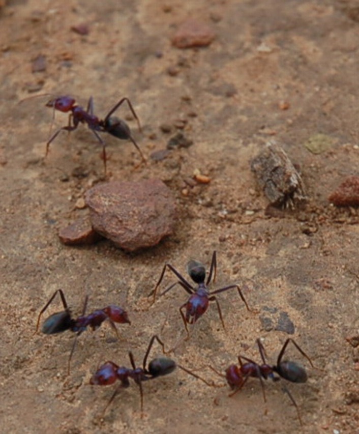
We describe a case of neuroretinitis and probable B henselae infection in a patient who was bitten by a bull ant (genus Myrmecia) in South Australia (figure 1).
Figure 1.

Bull ants (myrmecia species).
Case presentation
A 52-year-old Caucasian woman presented with a 3-day history of blurred vision. The patient at this stage did not mention anything else of note in the history.
On examination, best-corrected visual acuity (BCVA) was hand movements OS and 6/4.8 OD.
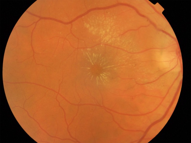
Examination revealed a quiet eye with normal anterior segment. Posterior segment examination revealed papillitis, macular oedema and macular star in the right eye (figure 2). Examination of the left eye was unremarkable.
Figure 2.
Right fundus showing neuroretinitis and macular star.
On further enquiry, the patient denied any contact with cats or recent trauma. She did, however, describe being bitten and stung by a ‘Bull ant’ in the garden, 7 days prior to developing visual symptoms. She brought in an example for identification. Shortly after receiving the sting, the patient described having rigours and sweats, and pain at the sting site. The site was visible on the right inner thigh when the patient presented. There was no lymphadenopathy.
Full blood count, urea and electrolytes and blood serology for Bartonella, Toxocara, Toxoplasma, Herpes viruses and Borellia were requested. The only positive findings were titres of 256 for B henselae on paired sera 4 weeks apart, the first sample being taken 10 days following the sting. This is consistent with B henselae infection.
Treatment was started with oral erythromycin 500 mg four times a day for 3 months and was well tolerated. BCVA improved to 6/10 OD 3 months after original presentation with residual red colour desaturation and a paracentral scotoma.
Discussion
Bull ants are aggressive creatures ranging in size from 15 to 25 mm. They usually nest underground in dry, sandy soils with the entrance surrounded by fine gravel. The ants attack any animal which threatens the nest. The ants deliver a painful sting by first biting the attacker with their mandibles, and then curling their abdomens to deliver one or multiple venomous stings.
Bull ant stings are an important cause of life-threatening anaphylaxis in Australia,5 although the venom itself is not lethal in humans. Acute renal failure with widespread internal organ necrosis and haemorrhage with eventual death (euthanasia) has been reported in a dog following massive envenoma.6
The venom contains histamine-like activity, a heat-sensitive haemolytic factor and causes the release of cyclooxygenase products.7 We felt it was unlikely that the venom was responsible for this patient's ocular presentation because it was unilateral, presented some time after the sting and has not been reported previously despite bull ant stings being relatively common. Discussions with an infectious disease physician and an entomologist specialising in ants revealed that B henselae was probably introduced via the stinger or mandibles by providing an entry point into the skin. Bartonella species have been shown to have remarkable strategies to adapt to their hosts and arthropod vectors;8 and they can survive for several days in faeces of vectors.8 9 It is therefore likely that the bacteria were introduced into the skin by the bull ant. This case represents a previously unreported mode of transmission for B henselae causing neuroretinitis. On the basis of this case and other known causes, we suggest that the term cat scratch disease is no longer used for infection with B henselae.
Learning points.
Bartonella henselae can be transmitted by a sting of the South Australian bull ant.
Neuroretinitis should trigger the investigation of a variety of causes, even if some appear unlikely.
Footnotes
Competing interests: None.
Patient consent: Obtained.
References
- 1.Ormerod LD, Dailey JP. Ocular manifestations of cat scratch disease. Curr Opin Ophthalmol 1999;10:209–16. [DOI] [PubMed] [Google Scholar]
- 2.Tsukahara M, Tsuneoka H, Iino H, et al. Bartonella henselae infection from a dog. Lancet 1998;352:1682. [DOI] [PubMed] [Google Scholar]
- 3.O'Rourke LG, Pitulle C, Hegarty BC, et al. Bartonella quintana in cynomolgus monkey (Macaca fascicularis). Emerg Infect Dis 2005;11:1931–4. [DOI] [PMC free article] [PubMed] [Google Scholar]
- 4.Kalogeropoulos C, Koumpoulis I, Mentis A, et al. Bartonella and intraocular inflammation: a series of cases and review of literature. Clin Ophthalmol 2011;5:817–29. [DOI] [PMC free article] [PubMed] [Google Scholar]
- 5.Douglas RG, Weiner JM, Abramson MJ, et al. Prevalence of severe ant-venom allergy in southeastern Australia. J Allergy Clin Immunol 1998;101:129–31. [DOI] [PubMed] [Google Scholar]
- 6.Abraham LA, Hinkley CJ, Tatarczuch L, et al. Acute renal failure following Bull Ant mass envenomation in two dogs. Aust Vet J 2004;82:43–7. [DOI] [PubMed] [Google Scholar]
- 7.Matuszek MA, Hodgson WC, Sutherland SK, et al. Pharmacological studies of the venom of an Australian bulldog ant (Myrmecia pyriformis). Natural Toxins 1994;2:36–43. [DOI] [PubMed] [Google Scholar]
- 8.Chomel BB, Boulouis HJ, Breitschwerdt EB, et al. Ecological fitness and strategies of adaptation of Bartonella species to their hosts and vectors. Vet Res 2009;40:29. [DOI] [PMC free article] [PubMed] [Google Scholar]
- 9.Cunningham ET, Koehler JE. Ocular bartonellosis. Am J Ophthalmol 2000;130:340–9. [DOI] [PubMed] [Google Scholar]


