Abstract
We describe a rare case report of unscarred uterus rupture (UR) diagnosed in the puerperium after a vacuum extraction (VE) delivery of a healthy newborn. In this instance, no risk factors were found apart from the use of VE in the setting of prolonged deceleration. The suspicion of the diagnosis was made because of the patient's constant distressing abdominal pain with peritoneal signs as well as a drop in haemoglobin. In the exploratory laparotomy, a 2000 ml haemoperitoneum and a complete transverse tear of the uterine fundus 10 cm long was found in a structurally normal uterus. Peritoneal lavage was effected and the tear was repaired. A very high index of suspicion is needed and the longer the delay in making the diagnosis, and starting treatment, the greater the clinical risk. Since the risk of UR in subsequent pregnancies is very high, caesarean delivery is recommended in any future pregnancy, after fetal pulmonary maturity is confirmed.
Background
A prior uterine scar is the major risk factor for uterine rupture. Rupture of an unscarred uterus (UU) is a much rarer obstetric event than the one that occurs in a scarred uterus. Its incidence is estimated to range from 1 in 5700 to 1 in 20 000 pregnancies.1
Rupture of a pregnant uterus may be associated with maternal mortality (especially in developing countries), maternal morbidity, particularly peripartum hysterectomy and a high incidence of perinatal mortality and morbidity.2
Risk factors for UU rupture described in the literature include advanced maternal age, grand multiparity, macrossomy, multiple gestation, longer labour, lack of access to emergency obstetrical services, uterine anomalies (eg, bicornuate or didelphys uterus and those secondary to a diethylstilbestrol exposure), maternal connective tissue disease, in particular Ehlers-Danlos syndrome, abnormal placentation, trauma, obstetric manoeuvres (eg, internal version and breech extraction, instrumental delivery), labour induction and augmentation and manual removal of the placenta.1 3
Classical clinical manifestations of uterine rupture are fetal bradycardia (the most common), which may be preceded by variable or late decelerations, maternal manifestations of constant abdominal pain (although it can be masked by regional analgesia) and signs of intra-abdominal haemorrhage. Vaginal bleeding is not a cardinal symptom and may be modest, despite a major haemoperitoneum. Other common signs are maternal tachycardia and hypotension that can progress to hypovolaemic shock, cessation of uterine contractions, loss of station of the fetal presenting part, uterine tenderness and change in uterine shape.
The differential diagnosis includes obstetric conditions such as placental abruption, intra-amniotic infection, liver rupture in severe pre-eclampsia and all the other non-obstetric causes of acute abdomen.1 4
The confirmation of the diagnosis is typically made at laparotomy.
Case presentation
A 38-year-old female non-smoker, without any relevant medical or surgical history, with obstetric antecedents of two spontaneous miscarriages, without curettage, and one eutocic delivery in 2001 with a newborn weighing 3428 g, without complications.
Her actual pregnancy underwent uneventfully besides a risk of preterm labour at 27 weeks. At 39+5 weeks, she was admitted to our emergency room in spontaneous labour (4 cm dilatation, intact membranes, at 04:30) and cephalic presentation.
No oxytocin augmentation was administrated. Continuous external electronic fetal monitoring was used. There was spontaneous membrane rupture at 06:00, with clear amniotic fluid. The delivery occurred at 07.51 with vacuum extraction because of a prolonged deceleration. The newborn weighed 4030 g and the Apgar index was 4/8. The episiotomy was sutured. The third station of labour underwent uneventfully. In subsequent hours, she started to be distressed, referring to continuous abdominal pain.
Investigations
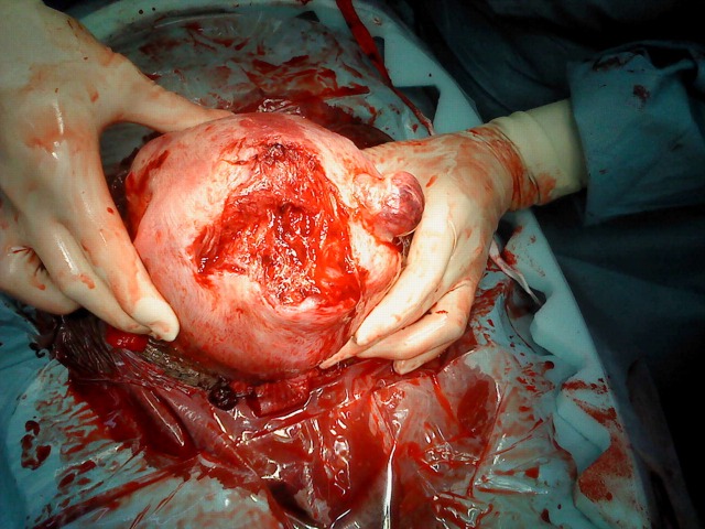
On examination, she was pale, hypotense, taquicardic and the abdomen was tender and with peritoneal signs. Her haemoglobin concentration dropped from 12.4 to 7.2 g/dl and the abdominopelvic ultrasound suggested haemoperitoneum. An exploratory laparotomy was decided upon and we found a 2000 ml haemoperitoneum and a complete transverse tear of the uterine fundus 10 cm long in a structurally normal uterus (figure 1).
Figure 1.
Uterine tear found in exploratory laparotomy.
Treatment
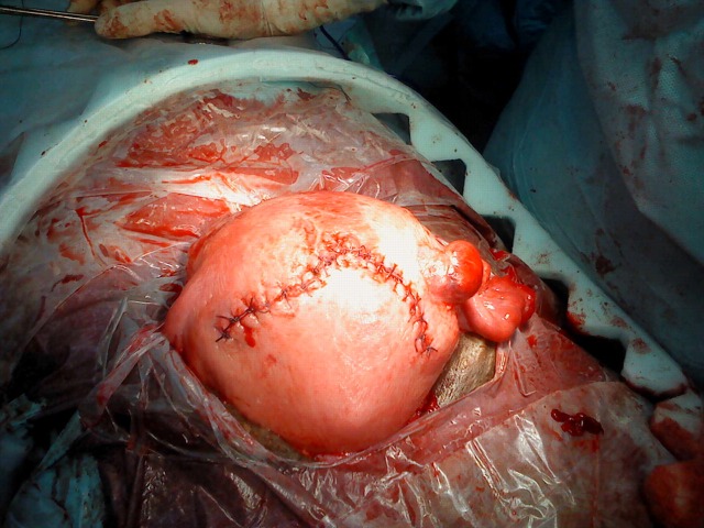
Peritoneal lavage was effected and the tear repaired (figure 2). She received fluids, four units of packed red blood cells and one unit of fresh frozen and empirical antibiotherapy (cefoxitin and metronidazole).
Figure 2.
Uterine tear repaired.
Outcome and follow-up
The patient's postoperative course was uneventful and she was discharged on her fifth postoperative day. The patient was advised on the need for her to book early in subsequent pregnancies and to be delivered by elective caesarean section.
Discussion
There are very few reports of UU rupture. Although a rare event, the UU is not immune to rupture as demonstrated in this report. A very high index of suspicion is needed and the longer the delay in making the diagnosis and starting treatment, the greater the clinical risk. The only risk factor encountered in this case was the delivery with vacuum extraction in the setting of a prolonged deceleration and not prolonged or obstructed labour. The rupture probably occurred in this intrapartum phase.
The risk of recurrent rupture ranges from 22% to 100% and appears to be highest when the uterine fundus was involved.1 Whether the use of imaging (ultrasound or magnetic resonance) improves the outcome in future pregnancies remains to be validated, but it seems prudent nonetheless. Currently, there are no methods available to accurately predict uterine rupture.
Learning points.
A high index of suspicion is needed because the rupture of an unscarred uterus is a rare and unexpected obstetric event.
It may be associated with high maternal and neonatal morbimortality.
Urgent delivery is imperative.
The risk of recurrence is very high.
Caesarean delivery is recommended in future pregnancies, after fetal pulmonary maturity is confirmed.
Footnotes
Competing interests: None.
Patient consent: Obtained.
References
- 1.Smith JF, Wax JR. Rupture of the unscarred uterus. In: UpToDate, Lockwood, CJ (Ed), UpToDate. Waltham, MA, 2012.
- 2.Turner MJ. Uterine rupture. Best Pract Res Clin Obstet Gynaecol 2002;16:69–79. [DOI] [PubMed] [Google Scholar]
- 3.Smith JG, Mertz HL, Merril DC. Identifying risk factors for uterine rupture. Clin Perinatol 2008;35:85–99. [DOI] [PubMed] [Google Scholar]
- 4.Chigbu B, Onwere S, Kamanu C, et al. Rupture of the uterus in a primigravida: a case report. Niger J Clin Pract 2010;13:233–4. [PubMed] [Google Scholar]


