Abstract
Flexor tenosynovitis is an aggressive closed-space infection of the digital flexor tendon sheaths of the hand. We present a case of pyogenic flexor tenosynovitis in an immunocompromised patient and discuss the importance of early diagnosis and referral to a specialist hand surgery unit. A 61-year-old man visited his general practitioner because of swelling and tenderness of his left index finger. The patient was discharged on oral antibiotics but returned 4 days after because of deterioration of his symptoms and was referred to a plastic surgery unit. A diagnosis of flexor tenosynovitis was made and the patient required multiple debridements in theatre, resulting in the amputation of the infected finger. Pyogenic flexor tenosynovitis is a relatively common but often misdiagnosed hand infection. Patients with suspected flexor tenosynovitis should be referred and treated early to avoid significant morbidity, especially when risk factors for poor prognosis are present.
Background
Pyogenic flexor tenosynovitis is a closed-space infection of the flexor tendon sheath of the fingers.1 Although relatively common, pyogenic flexor tenosynovitis is often misdiagnosed, leading to significant morbidity. We report a case of late presentation of flexor tenosynovitis in a high-risk patient and discuss the importance of early diagnosis and referral of flexor tenosynovitis to a specialist hand surgery unit.
Case presentation
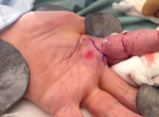
A 61-year-old gentleman visited his general practitioner (GP) because of swelling and tenderness of his left index finger following an injury the day before. His medical history included Crohn's disease for which the patient was on immunosuppressant agents. The patient was diagnosed to have superficial skin infection and was sent home on oral antibiotics. Four days following the onset of his symptoms the patient returned to his GP because of deterioration of his symptoms and was referred to a plastic surgery unit. When seen by the plastic surgery team he was generally unwell, pyrexial and had associated nausea and vomiting. On examination, his left index finger was grossly swollen, had a fixed flexion deformity, mottled skin with blistering, pain on passive movement and was extremely tender (figure 1). A clinical diagnosis of flexor tenosynovitis was made and the patient was started immediately on intravenous antibiotics.
Figure 1.
Preoperative findings show severe swelling and erythema of the left index finger with signs of ischaemia.
Investigations
His wound swabs grew β-haemolytic streptococcus group A and antibiotic treatment was changed according to microbiology advice.
Differential diagnosis
The differential diagnosis included subcutaneous abscess, felon, herpetic whitlow and flexor tenosynovitis.
Treatment
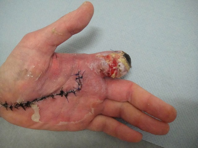
He was taken to theatre for an open flexor sheath washout. During the operation, there was frank pus in the flexor sheath and evidence of infection extending up to the carpal tunnel. The patient required further washouts and debridements in theatre because of continuous evidence of infection. Nine days following the initial presentation and following multiple visits to theatre he developed significant necrosis of the skin of his index finger, therefore a decision was made to proceed with an amputation of his finger (figure 2).
Figure 2.
Resulting amputation of the left index finger following multiple debridements.
Outcome and follow-up
He made an uneventful recovery and was discharged 7 days following his amputation.
Discussion
Flexor tenosynovitis is a relatively common and aggressive closed space infection of the digital flexor sheaths, accounting for 2.5–9.4% of hand infections.2 Early diagnosis and treatment of pyogenic flexor tenosynovitis are necessary to prevent tendon necrosis, adhesion formation and spread of infection to the deep fascial spaces. Because of the poor vascularisation of synovial sheaths, bacterial proliferation is rapid, the host defend mechanisms and antibiotic penetration are limited, therefore the infection can spread rapidly within the sheath.1 3
Patients usually present with a history of rapid development and progression of pain, swelling and erythema following a penetrating injury, typically on the volar aspect of the proximal or distal interphalangeal joint. A useful indicator of flexor tenosynovitis during our examination are the four cardinal signs as described by Kanavel:4
Uniform, symmetric digit swelling.
Digit is held in partial flexion at rest.
Excessive tenderness along the entire flexor tendon sheath.
Pain along the tendon sheath with passive digit extension.
Not all Kanavel signs are present in all cases. The most reliable signs of flexor tenosynovitis are excessive tenderness along the tendon sheath and pain on passive extension.5 6
The differential diagnosis of flexor tenosynovitis includes:1
Local abscess: no tenderness along entire tendon and no pain on passive extension.
Inflammatory diseases (ie, rheumatoid arthritis, gout and aseptic tenosynovitis): sometimes difficult to differentiate septic from non-septic causes of flexor tenosynovitis, may require aspiration of the synovial sheath.
Herpetic whitlow: more distal findings and small skin vesicles.
Felon: more distal findings.
Pyarthrosis: swelling localised around the joint, traumatic injury on dorsal aspect of finger and no tenderness along the entire tendon.
The commonest causative agents in flexor tenosynovitis are Staphylococcus and Streptococcus, therefore the suggested empirical treatment is either penicillin with B-lactamase inhibitor or a combination of penicillin and first generation cephalosporin.6 7 Haematogenous spread of Neisseria gonorrhoea has been described and should be suspected if there is no history of trauma.3
Pang et al identified five risk factors that were associated with poor prognosis:2
An age of more than 43 years.
The presence of diabetes mellitus, peripheral vascular disease or renal failure.
The presence of subcutaneous purulence.
Digital ischaemia.
Polymicrobial infection.
Conservative treatment with intravenous antibiotics should only be considered when patients present within the first 24 h of the onset of symptoms and have mild symptoms and signs. Surgical treatment should be considered if the clinical symptoms do not improve in the first 12 h. Conservative treatment should very rarely be considered for immunocompromised or diabetic patients.1 6
A popular approach for the surgical treatment of flexor tenosynovitis, initially described by Neviaser,5 is closed continuous irrigation for 48 h through a catheter inserted through two small incisions placed just proximally and distally from the tendon sheath. Lille et al8 have demonstrated that there is no statistically significant benefit of continuous irrigation when compared with intraoperative debridement and irrigation alone. Hee-Nee et al2 recommended a treatment plan according to the clinical signs of severity:
Early sheath irrigation in patients with no subcutaneous purulence or digital ischaemia.
Open debridement for patients with subcutaneous purulence but no digital ischaemia
Patients with subcutaneous purulence and digital ischaemia may require an amputation.
Learning points.
Flexor tenosynovitis is a relatively common and aggressive hand infection.
Flexor tenosynovitis is often misdiagnosed.
Early diagnosis and treatment are necessary in order to avoid significant morbidity.
Footnotes
Competing interests: None.
Patient consent: Obtained.
References
- 1.Green DP, Hotchkiss RN, Pederson WC. Green's operative hand surgery. Vol 1 Philadelphia, USA: Churchill Livingstone, 1999. [Google Scholar]
- 2.Pang HN, Teoh LC, Yam AK, et al. Factors affecting the prognosis of pyogenic flexor tenosynovitis. J Bone Joint Surg Am 2007;89:1742–8.. [DOI] [PubMed] [Google Scholar]
- 3.Clark DC. Common acute hand infections. Am Fam Physician 2003;68:2167–76.. [PubMed] [Google Scholar]
- 4.Kanavel AB. Infections of the hand: a guide to the surgical treatment of acute and chronic suppurative processes in the fingers, hand and forearm. 7th edn Philadelphia: Lea & Febiger, 1939. [Google Scholar]
- 5.Neviaser RJ. Closed tendon sheath irrigation for pyogenic flexor tenosynovitis. J Hand Surg 1978;3:462–6.. [DOI] [PubMed] [Google Scholar]
- 6.Boles SD, Schmidt CC. Pyogenic flexor tenosynovitis. Hand Clin 1998;14:567–78.. [PubMed] [Google Scholar]
- 7.Moran G, Talan D. Hand infections. Emerg Med Clin North Am 1993;11:601–19.. [PubMed] [Google Scholar]
- 8.Lille S, Hayakawa T, Neumeister NM, et al. Continuous postoperative catheter irrigation is not necessary for the treatment of suppurative flexor tenosynovitis. J Hand Surg Eur Vol 2000;25:304–7.. [DOI] [PubMed] [Google Scholar]


