Abstract
An inherited or acquired deficiency of protein S leads to a prothrombotic state, with predisposition to venous thrombosis. We describe a case of cerebral venous sinus thrombosis (CVST) associated with acquired protein S deficiency and recanalisation within 15 days of anticoagulation. A 38-year-old man presented with recurrent headache, vomiting, altered sensorium and one episode of transient left hemiparesis. Magnetic resonance venography showed poor flow in the deep cerebral venous sinuses with extensive collateral venous channel formation, which resolved after 15 days of anticoagulation, along with clinical improvement. Serum protein S activity was found to be markedly low (16% of biological reference). CVST should be suspected in a patient with acute features of raised intracranial pressure or focal neurological deficit, and a patient without obvious clinical predisposition for a prothrombotic state should be evaluated for underlying thrombophilic states like protein S deficiency.
Background
Protein S is an endogenous vitamin K-dependent anticoagulant, which prevents clotting by acting as a cofactor for activated protein C in the degradation of clotting factors Va and VIIa.1 An inherited or acquired deficiency of protein S leads to a prothrombotic state, with predisposition to thrombosis. It is a cause of cerebral venous sinus thrombosis (CVST), with only a few sporadic reports in the literature.2 3 Furthermore, with anticoagulation, recanalisation occurs within 4 months of therapy in most cases of cerebral venous thrombosis.4 We describe a case of acquired protein S deficiency-associated CVST with recanalisation within 15 days of anticoagulation therapy.
Case presentation
A 38-year-old man presented to our hospital with a history of headache and vomiting about 2weeks back followed by altered sensorium. He developed weakness of the left half of the body a few minutes after the onset of headache. His weakness improved on the next day. He remained asymptomatic for 4 days after this event, following which he again had an episode of headache, vomiting and altered sensorium for which he was brought to our hospital. He was a non-smoker and had no features suggestive of systemic or haematologic malignancy. He had no history suggestive of thrombotic episodes in any family members. Clinical examination revealed a young disoriented man. Pulse rate was 84/min, blood pressure 130/80 mm Hg and respiratory rate was 18/min. Cranial nerve examination was normal. He was moving all four limbs and maintaining posture but was not cooperative for a detailed motor system examination. His deep tendon reflexes were brisk and plantars were bilateral flexors. Sensory and cerebellar examination could not be performed.
Investigations
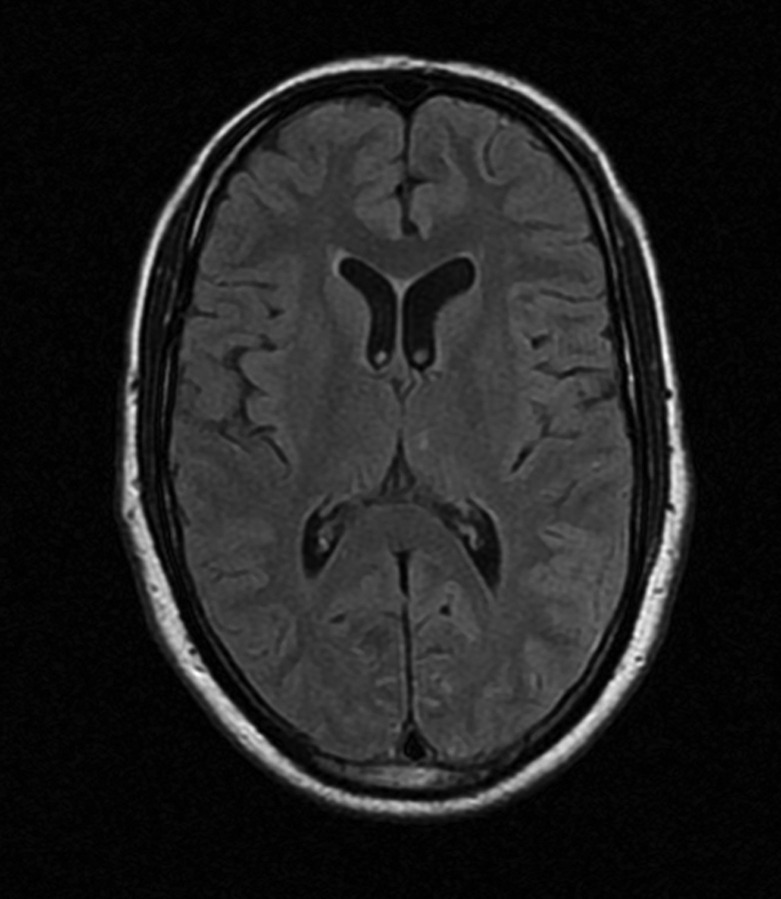
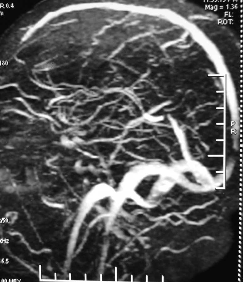
On laboratory investigations, haemoglobin level was 12 g/dl, total leucocyte count was 8900/cumm with normal differentials. Platelet count was 240 000/cumm. Chest x-ray did not reveal any abnormality. The patient was negative for HIV and hepatitis B serology. Cerebrospinal fluid (CSF) examination showed 5 cells/cumm with normal CSF proteins and sugar levels. CSF ELISA for antibodies against endemic viruses: Japanese encephalitis, dengue, herpes simplex and varicella zoster were negative. Considering history of altered sensorium with transient focal neurodeficit, MRI with MR angiography of the brain was requested. MRI cranium and arteriograhy did not reveal any abnormality (figure 1). However MR venography was showing poor flow in the deep cerebral venous sinuses with extensive collateral venous channel formation (figure 2). A diagnosis of CVST was thus considered and the patient was investigated for a possible hypercoagulable state. Serum protein S activity was found to be markedly low (16% of biological reference) (normal range: 77–143%). Other factors that were tested included protein C activity, homocysetine, lupus anticoagulant, antinuclear antibody antiphospholipid antibodies, antithrombin III which were found to be unremarkable.
Figure 1.
MRI, T2 fluid-attenuated inversion-recovery image revealed normal study.
Figure 2.
Magnetic resonance venography showed poor flow in deep venous sinuses with the presence of collaterals.
Treatment
Patient was started on anticoagulation therapy with low-molecular-weight heparin, enoxaparin 60 mg twice daily, after withdrawing blood sample for above procoagulant factors estimation. The patient received decongestive therapy and other supportive measures. Patient's sensorium improved within 2 days of starting therapy.
Outcome and follow-up
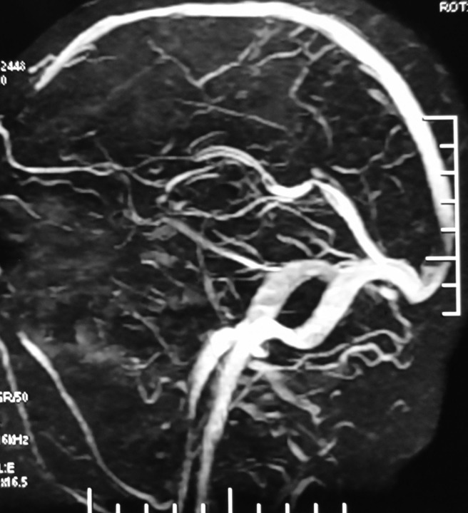
A repeat MR venography was performed 15 days later, which revealed recanalisation in the form of striking improvement in flow with a marked decrease in collaterals (figure 3). The patient was shifted on oral anticoagulation with warfarin and discharged. He was asymptomatic on further follow-up. Serum protein S estimation was repeated 8 weeks after the initial presentation and it was found to be persistently low (18%), further confirming the protein S deficiency.
Figure 3.
MR venogram on repeat study demonstrated recanalisation of deep venous sinuses with disappearance of collaterals.
Discussion
This young patient had come to us with recurrent headache, vomiting, altered sensorium and a transient hemiparesis episode, which led us to consider possibility of either a cerebrovacular episode or viral encephalitis. Viral encephalitis was refuted by a normal CSF examination. Further, in view of a normal MRI and MR ateriography, we considered the diagnosis of CVST, which was confirmed by poor flow and extensive collaterals in MR venography. The rich collaterals and absence of an obvious occlusive thrombus suggests a recurrent, chronic thrombotic process in cerebral venous sinuses. Thus, transient focal neurodeficit is an unusual presentation of CVST and the diagnosis can be made in such cases only with a high index of suspicion for CVST.
Protein S (named in reference to its isolation and characterisation in Seattle in 1979) is a vitamin K-dependent protein synthesised in the liver, vascular endothelium and megakaryocytes. It helps in cleaving activated clotting factors Va and VIIa on vascular endothelium by acting in association with protein C. Protein S deficiency can be inherited or acquired. Bertina classified protein S deficiency into three clinical subtypes based on laboratory findings. Type I refers to deficiency of both free and total protein S as well as decreased protein S activity; type II shows normal plasma values, but decreased protein S activity; and type III shows decreased free protein S levels and activity, but normal total protein S levels.5 Of 71 protein S-deficient members from 12 Dutch pedigrees, 74%, 72% and 38% of the individuals sustained deep vein thrombosis (DVT), superficial thrombophlebitis and pulmonary emboli, respectively.6 However, CVST has been described in only a few case reports. Acquired protein S deficiency secondary to HIV and pregnancy leading to CVST has been described in a previous case report. As per our knowledge, ours is the first case report of an idiopathic, acquired protein S deficiency causing CVST. Few case reports have also implicated protein S deficiency in recurrent ischaemic strokes in the young.7–9 Similarly, progressive intracranial occlusive disease has been reported in association with protein S deficiency.10 However, current data do not support an association between hereditary protein S deficiency and an increased risk of arterial thrombosis.11 The recommended therapy for CVST is anticoagulation with heparin followed by oral anticoagulation, which should be continued indefinitely for patients with underlying thrombophilia.12 Despite similar treatment as that for DVT, CVST management poses a unique management issue, with respect to discontinuation of anticoagulation. Because DVT in the limbs is precipitated by immobilisation, it is assumed that after a sufficient period of anticoagulation and achieving mobility, anticoagulation can be discontinued, which cannot be the case with CVST. Hence, assessment of recanalisation is important in CVST. A study showed that most of the recanalisation occurs during initial 4 months of anticoagulation therapy.4 Another study demonstrated a recanalisation rate of 60% at 4 weeks after CVST.13 Recanalisation of CVST has been described as early as 24 h in a previous case report.14 Recanalisation is affected most importantly by endogenous fibrinolysis. Recanalisation in CVST can have potential therapeutic implications in framing future guidelines of therapy, particularly with regard to discontinuation of anticoagulation. Thus, the scarcity of data regarding recanalisation in CVST and its implications in outcome of patients mandates a major study addressing these issues.
Learning points.
Cerebral venous sinus thrombosis (CVST) should be suspected in a patient with acute features of raised intracranial pressure or focal neurological deficit.
A transient ischaemic attack-like presentation is also possible in CVST, and in such cases if work-up for an arterial stroke is negative, then a venography should be considered.
Patients of CVST should be evaluated for underlying thrombophilic states like protein S deficiency in the absence of well-defined causes/risk factors.
The early institution of anticoagulants in cerebral venous thrombosis is helpful in achieving early recanalisation in CVST.
Footnotes
Competing interests: None.
Patient consent: Obtained.
References
- 1.Oktar N, Dalbasti T. Protein S deficiency, epileptic seizures, sagittal sinus thrombosis and hemorrhagic infarction after ingestion of dimenhydrinate. Turk Neurosurg 2008;18:85–8. [PubMed] [Google Scholar]
- 2.Cros D, Comp PC, Beltran G, et al. Superior sagittal sinus thrombosis in a patient with protein S deficiency. Stroke 1990;21:633–6. [DOI] [PubMed] [Google Scholar]
- 3.Burneo JG, Elias SB, Barkley GL. Cerebral venous thrombosis due to protein S deficiency in pregnancy. Lancet 2002;359:892. [DOI] [PubMed] [Google Scholar]
- 4.Baumgartner RW, Studer A, Arnold M, Georgiadis D. Recanalisation of cerebral venous thrombosis. J Neurol Neurosurg Psychiatry 2003;74:459–61. [DOI] [PMC free article] [PubMed] [Google Scholar]
- 5.Bertina RM. Nomenclature proposal for protein S deficiency. XXXVI Annual Meeting of the Scientific and Standardization Committee of the ISTH; Barcelona, Spain, 1990. [Google Scholar]
- 6.Engesser L, Broekmans AW, Briet E, et al. Hereditary protein S deficiency: clinical manifestations. Ann Intern Med 1987;106:677. [DOI] [PubMed] [Google Scholar]
- 7.Hooda A, Khandelwal PD, Saxena P. Protein S deficiency: recurrent ischemic stroke in young. Ann Indian Acad Neurol 2009;12:183–4. [DOI] [PMC free article] [PubMed] [Google Scholar]
- 8.Girolami A, Simioni P, Lazzaro AR, et al. Severe arterial thrombosis in a patient with protein S cerebral deficiency (moderately reduced total and markedly reduced free protein S): a family study. Thromb Haemost 1989;61:144–7. [PubMed] [Google Scholar]
- 9.Sie P, Boneu B, Bierme R, et al. Arterial thrombosis and protein S deficiency. Thromb Haemost 1989;62:1040. [PubMed] [Google Scholar]
- 10.Barinagarrementeria F, Cantú Brito C, Izaguirre R, et al. Progressive intracranial occlusive disease associated with deficiency of protein S. Report of two cases. Stroke 1993;24:1752–6. [DOI] [PubMed] [Google Scholar]
- 11.Brouwer JP, Veeger NJGM, van der Schaaf W, et al. Difference in absolute risk of venous and arterial thrombosis between familial protein S deficiency type I and type III. Results from a family cohort study to assess the clinical impact of a laboratory test-based classification. Br J Haematol 2005;128:703. [DOI] [PubMed] [Google Scholar]
- 12.Einhäupl K, Stam J, Bousser MG, et al. European Federation of Neurological Societies. EFNS guideline on the treatment of cerebral venous and sinus thrombosis in adult patients. Eur J Neurol 2010;17:1229.–. [DOI] [PubMed] [Google Scholar]
- 13.Stolz E, Trittmacher S, Rahimi A, et al. Influence of recanalization on outcome in dural sinus thrombosis: a prospective study. Stroke 2004;35:544–7. [DOI] [PubMed] [Google Scholar]
- 14.Vanden Abeele B, Saliou G, Lehmann P, et al. Recanalization of cerebral venous thrombosis within 24 h: a case report. Neurology 2007;69:221. [DOI] [PubMed] [Google Scholar]


