



Abstract
The immune reconstitution inflammatory syndrome (IRIS) after starting antiretroviral treatment for HIV infection can be caused by a great variety of pathogens. Among these are non-tuberculous mycobacteria (NTM), with Mycobacterium avium complex being the most commonly described finding. Antimycobacterial treatment of NTM in cases of IRIS is controversial. We report the case of a 39-year-old man diagnosed with HIV-1 infection during admission to hospital with Pneumocystis jirovecii pneumonia (PCP) and a CD4 cell count of 60/μl. The patient started antiretroviral treatment and made an uneventful recovery from the PCP diagnosis, but was readmitted after 2.5 months with a purulent infrapatellar bursitis on the left knee. A surgical prodedure was performed and Mycobacterium malmoense was grown from the pus from the bursa. The patient recovered without supplemental antimycobacterial treament. To our knowledge, this is the first report on IRIS caused by M malmoense.
Background
Infection with non-tuberculous mycobacteria (NTM) occurs with an increasing incidence not only in immunocompromised patients, but also in immunocompetent individuals.1 2 Although the vast majority of NTM disease in patients with HIV infection is caused by Mycobacterium avium complex (MAC), other NTM are also important pathogens.3 4 Mycobacterium malmoense, first described in Sweden in 1977 by Schröder and Juhlin, is the second leading cause of NTM disease in Sweden and has with increasing frequency been isolated in Northern Europe where it is primarily presenting with pulmonary manifestations; disseminated disease has also been described.5–9
We present a case history with isolation of M malmoense from an infrapatellar purulent bursitis in a patient with HIV after starting antiretroviral treatment.
Case presentation
A 39-year-old man presented to the emergency department at a tertiary hospital with a 6-month history of unintended weight loss (22 kg), severe fatigue, dry irritative cough and severe shortness of breath during physical activity. The patient was febrile without focal symptoms and underwent an extensive examination including a transthoracic echocardiography, chest film, spiral CT, ultrasound of the testis and CT of thorax and abdomen. The latter revealed a single lymph node enlargement in the right hilus region, otherwise no abnormal findings were revealed. Biochemical tests showed an elevated erythrocyte sedimentation rate of 34 mm/h, a normal leucocyte count with mild neutropaenia (1.56×10⁹/l) and lymphopaenia (0.83×10⁹/l), and slight anaemia with an Hb of 7.7 mmol/l.
The patient had a medical history with arthroscopic surgery for a meniscus injury in his left knee in 2000 and 2003, an episode of unexplained urine retention in 2005 and an operation for a perianal abscess in 2007. Further, he had a travel history with a stationing in the Balkans in 1994 as a soldier, a vacation trip to Egypt in 2001 and to Croatia in 2008. He had no history of drug abuse, and he had been in a stable monogameous relationship for 15 years, apart from a sexual contact with a man 6–8 years previously. His wife and children were all healthy. Finally, in 1995 his mother had a recurrence of pulmonary tuberculosis, for which she was successfully treated. The entire family was examined for tuberculosis—none were found latently infected.
The patient was referred to the Department of Infectious Diseases at our hospital and was found to be alert and oriented with fever, progression in respiratory symptoms and seborrhoeic dermatitis. The only other subjective complaint was intermittent pain from the left knee, which was normal at inspection. The patient tested positive for HIV-1 with a CD4 count of 60/μl and a plasma HIV-RNA of 1 500 000 copies/ml. Because of the respiratory symptoms, Pneumocystis pneumonia (PCP) was suspected and empirical treatment with sulphamethoxazole/trimethoprim at a standard dose of 1600 mg/320 mg four times a day was started. The PCP diagnosis was subsequently confirmed with a positive fluorescence microscopy for Pneumocystis jirovecii on broncho-alveolar lavage fluid.
Three weeks after the patient tested positive for HIV, antiretroviral treatment was started in the outpatient clinic with Efavirenz 600 mg once a day emtricitabine 200 mg and tenofovir disoproxil 245 mg od. Five weeks after starting treatment, the CD4 cell count had risen to 240/μl with a decrease in plasma HIV-RNA to 2600 copies/ml.
Two-and-a-half months after the HIV diagnosis, the patient was hospitalised with increasing pain for 1 week in his left knee as the only subjective symptom. On arrival, the patient was afebrile with a clinically suspected prepatellar bursitis anterolateral at the left knee, which was not, at that time, aspirated. The patient was treated with a short course of 2 days with intravenous dicloxacillin 1 g four times a day and discharged without antibiotics.
After 14 days, the patient was re-admitted due to increasing pain from his clinically suspected prepatellar bursitis. An ultrasound was performed finding a deep infrapatellar bursitis (figure 1). An aspiration was performed with purulent matter and an incision was done; the cavity was rinsed and was left to heal. There was not sent material for pathology. Antibiotic treatment with intravenous dicloxacillin tid was commenced, but stopped after 3 days because direct microscopy of pus showed many acid-fast bacilli and the pus was without bacterial growth. No steroids were used. The Mycobacterium tuberculosis gamma-interferon releasing assay (QuantiFERON-TB) was negative.An x-ray of the left knee showed no sign of osteomyelitis. As localised atypical mycobacteriosis was suspected as part of immune reconstitution inflammatory syndrome after initiation of antiretroviral treatment, it was decided to observe the patient without extended antibiotic treatment. Two weeks after the surgical procedure, the CD4 cell count was stable at 250/µl with a further decrease in plasma HIV-RNA to 380 copies/ml.
Figure 1.
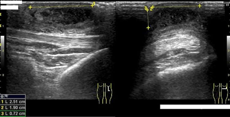
Ultrasound of the patients left knee with the finding of a soft tissue process anterolateral measuring 25 cm×19 cm×0.7 cm.
Three weeks after the incision of the infrapatellar purulent bursitis was performed, the final microbiology results showed no PCR amplification of atypical mycobacterium, and a negative PCR for M tuberculosis. However, growth of M malmoense MGIT in the culture medium was identified by means of InnoLipa species (from Innogenetics, Technologiepark 6, 9052 Gent, Belgium) with clear hybridisation to M malmoense specific probes (1, 2, 13 and 18) (16–23 S amplification followed by hybridisation to the species-specific probes).10 At follow-up in our outpatient clinic after 2, 11 and 26 weeks, the wound healed and left only slight scarring. The patient suffered from a depression and a post-traumatic stress disorder, but was somatically well without signs of ongoing infection with atypical mycobacteriosis.
Discussion
M malmoense has been isolated from environmental sources such as water and soil.3 Over the past three decades, the incidence of M malmoense infections, compared with other NTM, has increased especially in Northern Europe. This could largely be attributed to the increased awareness of mycobacteria as potential pathogens, the improved laboratory methods and perhaps the discontinuation of BCG vaccination of newborns as suggested in other studies.5 11 12
M malmoense infection is rarely associated with invasive disease in immunocompetent hosts, but is well described in patients with underlying pulmonary disease or in otherwise immunocompromised patients.6 7 13 14 Patients frequently present with pulmonary disease, as the most common site of infection and the presentation is much similar to infection with other non-tuberculous mycobacteria and M tuberculosis.15 Uncommon sites of infection have been reported, such as the skin, bones and joints.5 7 16–23 Furthermore, infection with M malmoense has a tendency to distribute in two peaks: in children with cervical lymphadenitis and in adults with predominant pulmonary infections.5
The risk of disease from NTM increases with the severity of immunodeficiency. In HIV-infected individuals, a CD4 cell count below 50 cells/mm3 is a risk factor for NTM disease.24 The patient in our case had a nadir CD4 count of 60 cells/mm3. A few weeks before the clinical presentation of the infrapatellar purulent bursitis, the patient started antiretroviral treatment leading to a risk of immune reconstitution inflammatory syndrome (IRIS), the paradoxical worsening of pre-existing infectious processes after the start of therapy. IRIS with known or previously undiagnosed mycobacterial infection has been frequently described among HIV-infected patients. The vast majority of NTM infections resulting in IRIS are due to MAC, but cases of IRIS related to infection with a variety of NTM have been described (table 1).5 25–30
Table 1.
Reported cases of immune reconstitution inflammatory syndrome associated with NTM
| Reference | Number of patients | NTM | IRIS presentation |
| Phillips et al25 | 1 | Mycobacterium simiae | Fever Hepatosplenomegaly |
| Phillips et al26* | 51 |
MAC (N=43) Mycobacterium genavense (N=2) Smear positive and culture negative (N=6) |
Peripheral lymphadenitis Pulmonary-thoracic disease Intra-abdominal disease Miscellaneous presentations |
| Lawn et al24* | 80 |
MAC (N=64) Mycobacterium xenopi (N=4) M genavense (N=1) Mycobacterium scrofulaceum (N=1) Mycobacterium kansasii (N=2) Mycobacterium leprae (N=6) BCG (N=2). |
Peripheral lymphadenitis leprosy and reversal reaction Pulmonary-thoracic disease |
| Puthanakit et al27 | 14 |
MAC (N=4) Mycobacterium Scrofulaceum (N=3) Mycobacterium kansasii (N=1) Mycobacterium simiae (N=1) Mycobacterium bovis (N=3) BCG (N=2) |
Peripheral lymphadenitis Pulmonary-thoracic disease Intra-abdominal disease Subcutaneous abscesses |
| Leone et al28 | 1 | M xenopi | Pulmonary infection |
| Shelburne et al29 | 11 | MAC | Localised lymphadenitis hepatosplenomegaly |
*Some of the references are overlapping.
BCG, Bacille Calmette-Guerin; IRIS, immune reconstitution inflammatory syndrome; MAC, Mycobacterium avium complex; NTM, non-tuberculous mycobacteria.
The infection was localised with no sign of joint or bone involvement, no supplementary antimycobacterial treatment was thus given following the surgical incision of the infrapatellar purulent bursitis. Currently, there is no consensus for treatment of M malmoense infection. In vitro susceptibility testing does not necessarily correlate with the in vivo activity of the antimicrobial agent or the clinical outcome, but a combination including clarithromycin, rifampicin and ethambutol have been used.3 31
To our knowledge, the isolation of M malmoense as part of an IRIS in an HIV infected patient has not been reported previously. Given the rise in incidence of infection with M malmoense this case highlights the importance of considering atypical infection as an important differential diagnosis in particularly immunocompromised patients.
Learning points.
In HIV-infection, the immune reconstitution inflammatory syndrome secondary to starting antiretroviral therapy can be caused by Mycobacterium malmoense.
Localised infection with M malmoense can be treated with surgery alone without supplementary antimycobacterial treatment, even in HIV-infected patients with low CD4 cell counts.
PCR for atypical mycobacteria is optimised for microscopy-positive sputum, which must be borne in mind when the method is used on other materials, for example, pus, in which it does not work satisfactory.
Atypical infection with atypical clinical presentation, for example, infrapatellar bursitis caused by Mycobacterium malmoense, should be considered as a differential diagnosis in severily immunocompromised patients.
Acknowledgments
We would like to thank MSc PhD Vibeke Østergaard Thomsen for critically reading the manuscript and assisting with the diagnostic procedures.
Footnotes
Competing interests: None.
Patient consent: Obtained.
References
- 1.Wayne LG, Sramek HA. Agents of newly recognized or infrequently encountered mycobacterial diseases. Clin Microbiol Rev 1992;5:1–25. [DOI] [PMC free article] [PubMed] [Google Scholar]
- 2.Moore JE, Kruijshaar ME, Ormerod LP, et al. Increasing reports of non-tuberculous mycobacteria in England, Wales and Northern Ireland, 1995–2006. BMC Public Health 2010;10:612. [DOI] [PMC free article] [PubMed] [Google Scholar]
- 3.Griffith DE, Aksamit T, Brown-Elliott BA, et al. An official ATS/IDSA statement: diagnosis, treatment, and prevention of nontuberculous mycobacterial diseases. Am J Respir Crit Care Med 2007;175:367–416. [DOI] [PubMed] [Google Scholar]
- 4.Lan R, Yang C, Lan L, et al. Mycobacterium tuberculosis and non-tuberculous mycobacteria isolates from HIV-infected patients in Guangxi, China. Int J Tuberc Lung Dis 2011;15:1669–75. [DOI] [PubMed] [Google Scholar]
- 5.Henriques B, Hoffner SE, Petrini B, et al. Infection with Mycobacterium malmoense in Sweden: report of 221 cases. Clin Infect Dis 1994;18:596–600. [DOI] [PubMed] [Google Scholar]
- 6.Hoefsloot W, Boeree MJ, van Ingen J, et al. The rising incidence and clinical relevance of Mycobacterium malmoense: a review of the literature. Int J Tuberc Lung Dis 2008;12:987–93. [PubMed] [Google Scholar]
- 7.Zaugg M, Salfinger M, Opravil M, et al. Extrapulmonary and disseminated infections due to Mycobacterium malmoense: case report and review. Clin Infect Dis 1993;16:540–9. [DOI] [PubMed] [Google Scholar]
- 8.Chocarra A, Gonzalez Lopez A, Breznes MF, et al. Disseminated infection due to Mycobacterium malmoense in a patient infected with human immunodeficiency virus. Clin Infect Dis 1994;19:203–4. [DOI] [PubMed] [Google Scholar]
- 9.Schröder KH, Juhlin I. Mycobacterium malmoense sp. nov. Int J Syst Bacteriol 1977;27:241–6. [Google Scholar]
- 10.Johansen IS, Lundgren BH, Thyssen JP, et al. Rapid differentiation between clinically relevant mycobacteria in microscopy positive clinical specimens and mycobacterial isolates by line probe assay. Diagn Microbiol Infect Dis 2002;43:297–302. [DOI] [PubMed] [Google Scholar]
- 11.Katila ML, Brander E, Backman A. Neonatal BCG vaccination and mycobacterial cervical adenitis in childhood. Tubercle 1987;68:291–6. [DOI] [PubMed] [Google Scholar]
- 12.Romanus V, Hallander HO, Wahlen P, et al. Atypical mycobacteria in extrapulmonary disease among children. Incidence in Sweden from 1969 to 1990, related to changing BCG-vaccination coverage. Tuber Lung Dis 1995;76:300–10. [DOI] [PubMed] [Google Scholar]
- 13.Willocks L, Leen CL, Brettle RP, et al. Isolation of Mycobacterium malmoense from HIV-positive patients. J Infect 1993;26:345–6. [DOI] [PubMed] [Google Scholar]
- 14.Haq I, Isenberg DA. Myositis and swollen knees: disease or treatment complication? Ann Rheum Dis 2002;61:544–6. [DOI] [PMC free article] [PubMed] [Google Scholar]
- 15.Hoefsloot W, van Ingen J, de Lange WC, et al. Clinical relevance of Mycobacterium malmoense isolation in The Netherlands. Eur Respir J 2009;34:926–31. [DOI] [PubMed] [Google Scholar]
- 16.Castor B, Juhlin I, Henriques B. Septic cutaneous lesions caused by Mycobacterium malmoense in a patient with hairy cell leukemia. Eur J Clin Microbiol Infect Dis 1994;13:145–8. [DOI] [PubMed] [Google Scholar]
- 17.France AJ, McLeod DT, Calder MA, et al. Mycobacterium malmoense infections in Scotland: an increasing problem. Thorax 1987;42:593–5. [DOI] [PMC free article] [PubMed] [Google Scholar]
- 18.Krusche-Mandl I, Decramer A, Boltuch-Sherif J, et al. (Non-tuberculous mycobacterial infections of the hand and wrist: a retrospective review of five cases from a single centre). Handchir Mikrochir Plast Chir 2009;41:283–7. [DOI] [PubMed] [Google Scholar]
- 19.Piersimoni C, Scarparo C. Extrapulmonary infections associated with nontuberculous mycobacteria in immunocompetent persons. Emerg Infect Dis 2009;15:1351–8; quiz 544. [DOI] [PMC free article] [PubMed] [Google Scholar]
- 20.Prince H, Ispahani P, Baker M. A Mycobacterium malmoense infection of the hand presenting as carpal tunnel syndrome. J Hand Surg Br 1988;13:328–30. [DOI] [PubMed] [Google Scholar]
- 21.Syed AA, O'Flanagan J. Mycobacterium malmoense infection. An unusual cause of wrist swelling. J Hand Surg Br 1998;23:811–12. [DOI] [PubMed] [Google Scholar]
- 22.Whitehead SE, Allen KD, Abernethy VE, et al. Mycobacterium malmoense septic arthritis. J Infect 2003;46:60–1. [DOI] [PubMed] [Google Scholar]
- 23.Callaghan R, Allen M. Mycobacterium malmoense infection of the knee. Ann Rheum Dis 2003;62:1047–8. [DOI] [PMC free article] [PubMed] [Google Scholar]
- 24.Benator DA, Gordin FM. Nontuberculous mycobacteria in patients with human immunodeficiency virus infection. Semin Respir Infect 1996;11:285–300. [PubMed] [Google Scholar]
- 25.Lawn SD, Bekker LG, Miller RF. Immune reconstitution disease associated with mycobacterial infections in HIV-infected individuals receiving antiretrovirals. Lancet Infect Dis 2005;5:361–73. [DOI] [PubMed] [Google Scholar]
- 26.Phillips DR, Krishnan H, Watson J. First UK report of successful treatment of Mycobacterium simiae and immune reconstitution inflammatory syndrome in an HIV-seropositive patient. Sex Transm Infect 2008;84:271–2. [DOI] [PubMed] [Google Scholar]
- 27.Phillips P, Bonner S, Gataric N, et al. Nontuberculous mycobacterial immune reconstitution syndrome in HIV-infected patients: spectrum of disease and long-term follow-up. Clin Infect Dis 2005;41:1483–97. [DOI] [PubMed] [Google Scholar]
- 28.Puthanakit T, Oberdorfer P, Akarathum N, et al. Immune reconstitution syndrome after highly active antiretroviral therapy in human immunodeficiency virus-infected thai children. Pediatr Infect Dis J 2006;25:53–8. [DOI] [PMC free article] [PubMed] [Google Scholar]
- 29.Leone S, Giglio S, Maio P, et al. Mycobacterium xenopi pulmonary infection resulting in self-limited immune reconstitution inflammatory syndrome in an HIV-1 infected patient. New Microbiol 2009;32:415–17. [PubMed] [Google Scholar]
- 30.Shelburne SA, Visnegarwala F, Darcourt J, et al. Incidence and risk factors for immune reconstitution inflammatory syndrome during highly active antiretroviral therapy. AIDS 2005;19:399–406. [DOI] [PubMed] [Google Scholar]
- 31.Banks J, Jenkins PA, Smith AP. Pulmonary infection with Mycobacterium malmoense–a review of treatment and response. Tubercle 1985;66:197–203. [DOI] [PubMed] [Google Scholar]