Description
A 40-year-old female patient presented with a progressive hearing loss in her right ear during the last 3 months. She did not have otorrhea or otalgia. She did not report any attack of vertigo or tinnitus. On physical examination, the right external auditory canal (EAC) was completely occluded by a hard mass, which was fixed and covered with wax. Pure tone audiometry revealed a severe conductive hearing loss in the right ear with normal hearing in the left ear.
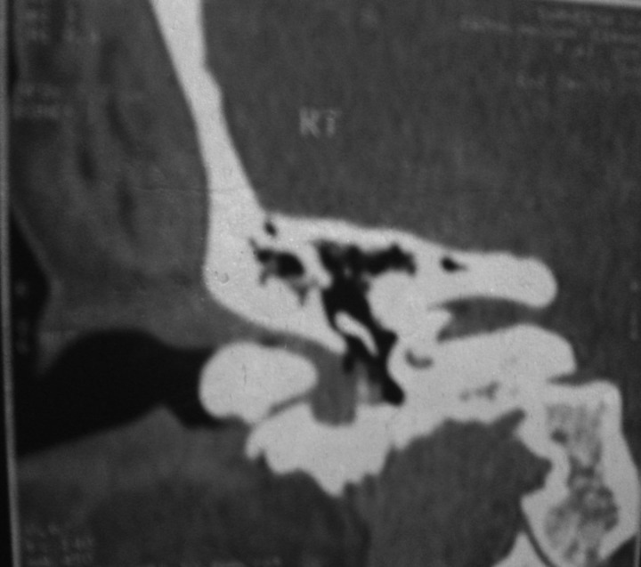
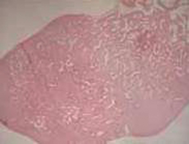
CT of the temporal bone revealed a 1.7 cm×1.5 cm-sized, ovoid bony mass completely filling and occluding the inner part of the right bony EAC (figure 1). Preliminary diagnosis of the right EAC osteoma was made. The decision was taken to remove the osteoma surgically. Through the postauricular approach, it was confirmed that the origin of the osteoma was the posterior wall of the EAC. Removal of the osteoma was performed through its pedicle to avoid recurrences. Histopathological examination of the removed mass was made to rule out other similar conditions. It showed fibrovascular canals, surrounded by bone lamellae and the diagnosis of right external auditory canal osteoma was confirmed (figure 2).
Figure 1.
CT of the temporal bone showing 1.7 cm×1.5 cm-sized, ovoid bony mass completely filling and occluding the inner part of the right bony external auditory canal.
Figure 2.
Histopathology of the removed mass showing fibrovascular canals, surrounded by bone lamellae. The diagnosis of osteoma was confirmed.
Osteoma in the EAC is an uncommon benign lesion, which presents as a solitary, unilateral and slow-growing pedunculated mass in the bony canal.1 2 It is usually asymptomatic, but symptoms can arise if a canal obstruction occurs.2 Treatment of EAC osteoma is surgical removal through its pedicle to avoid recurrences.2
Learning points.
External auditory canal (EAC) osteoma is an uncommon benign lesion which is usually asymptomatic, but symptoms can arise if a canal obstruction occurs resulting in hearing loss.
Diagnosis can be made by physical examination, which reveals the EAC completely occluded by a hard mass that may be covered by wax. Pure tone audiometry reveals a conductive hearing loss in the affected ear. CT of the temporal bone reveals a bony mass completely filling the bony EAC.
Treatment of EAC osteoma is surgical removal through its pedicle to avoid recurrences. Histopathological examination of the removed mass is essential to rule out other similar conditions.
Footnotes
Competing interests: None.
Patient consent: Obtained.
References
- 1.Ebelhar AJ, Gadre AK. Osteoma of the external auditory canal. Ear Nose Throat J 2012;91:96–100. [DOI] [PubMed] [Google Scholar]
- 2.Carbone PN, Nelson BL. External auditory osteoma. Head Neck Pathol 2012;6:244–6. [DOI] [PMC free article] [PubMed] [Google Scholar]


