Abstract
A 60-year-old woman, after a femoral neck fracture and joint replacement, underwent a Girdlestone's procedure and received aggressive antimicrobial therapy in order to completely eradicate the fungal infection Candida glabrata. In the majority of such cases, a revised hip arthroplasty would be considered following debridement. However, due to the recurrence of this infection and a key associated risk factor, radical removal with concurrent drug therapy was the only option.
Background
Infection is an adverse and potentially disastrous consequence of any interventional procedure. Hip prostheses can be particularly problematic. When investigating causative infectious agents it is rare to find the presence of fungal infections (such as Candida albicans). It is even rare for Candida glabrata to infect hip prostheses. This case illustrates how infrequently occurring infections can result in life-altering circumstances for patients. It also demonstrates how important it is to utilise specialist centres in order to manage complex clinical scenarios.
Case presentation
A 60-year-old woman presented to hospital with an excruciatingly painful left hip, following a fall. She had a medical history of rheumatoid arthritis, a cerebrovascular accident, previous deep vein thrombosis and vasculitis resulting in amputation of several fingers on the right hand and small bowel ischaemia with consequential fitting of an ileostomy bag. She is a non-smoker and non-drinker. On x-ray, the patient was found to have a femoral neck fracture and underwent an internal screw fixation of the left femur.
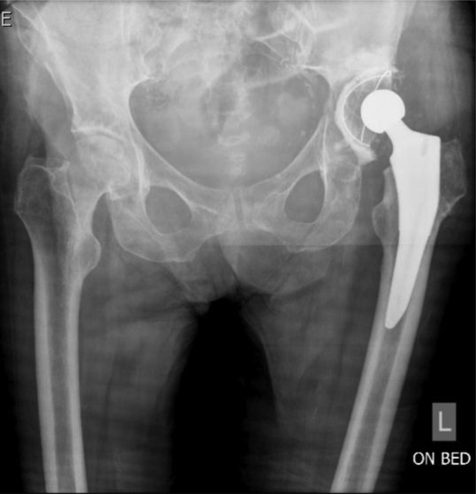
She was later re-admitted with severe left hip pain. x-Rays revealed avascular necrosis of the femoral head and the patient had a left total hip arthroplasty (figure 1). Subsequently, an infection ensued (symptoms included fever, vomiting and groin pain) and the patient underwent a number of arthroscopic washouts in an attempt to eliminate the infection from the hip joint. The patient was tested positive for Escherichia coli and Pseudomonas aerogenosa.
Figure 1.
Anterio-posterior (AP) radiograph of pelvis showing left hip total arthroplasty.
Treatment
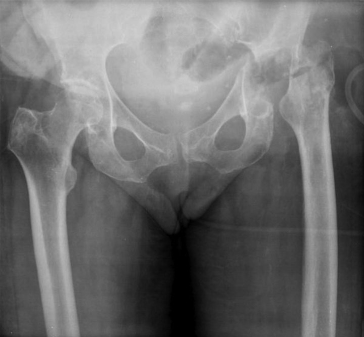
A Girdlestone's procedure was later performed, as previous arthroscopic washouts had been unsuccessful (figure 2). Despite this aggressive procedure, a preoperative MRI scan showed infection within the acetabulum, which extended down the shaft of the femur. Skin involvement was also present in this case. A debridement of the left hip with deep tissue sampling and a vastus muscle flap was performed in a bone specialist unit. Postoperatively the patient was given vancomycin and tazocin while awaiting a microbiology report. The results showed that there was in fact C glabrata present with P aeroginosa (sensitive to ceftazadime, meropenem, piptaz and ciprofloxacin) and E coli (sensitive to ceftazadime, ceftriaxone, meropenem and ciprofloxacin). The decision was made to start the patient on intravenous ceftazadime (2 g thrice a day) and caspofungin (50 mg once a day). This treatment will be continued for 6 weeks after which oral ciprofloxacin (750 mg twice a day) will be introduced.
Figure 2.
AP radiograph post-Girdlestone's procedure (note leg shortening due to the removal of prosthesis).
Outcome and follow-up
The outcome for a patient without associated risk factors for further infection and complications would be a revised hip prosthesis/reconstruction. Owing to this patient's associated comorbidity (vasculitis) which could result in further future infections; it would not be advisable to undertake such an invasive procedure in this instance. Instead, conservative management to aid daily living has been incorporated into her care plan. This includes physiotherapy to improve her range of movement and strength in her left leg and referral for orthotic shoes for expected postoperative shortened leg length.
Discussion
Joint infection has been a concern of orthopaedic specialists for decades. Whether it is the original joint or a prosthetic one, the management plan is complex and far from straightforward. Our case has proven to be challenging and has resulted in long-term hospitalisation of the patient. One aspect of this case that contributed to the difficulty in appropriate management planning was the rarity of this hip joint infection. C glabrata is the least common Candida species found in hip joint infections. In 2001, Ramamohan et al had documented in their research only two published cases of this particular infection.1 Candida accounts for less than 1% of all cases of prosthetic joint infections. The most commonly infecting Candida species are C albicans and Candida parapsilosis.2 In the absence of common practice it was difficult to fully identify the infection and establish an effective treatment plan.
Overall, the risk of infection in general in knee and hip replacement surgery is comparatively higher than that of smaller joints, due to the longer operation time, low blood flow to cortical bone and larger dead-space surrounding these larger prostheses. Haematoma formation can occur within this dead-space and can disrupt blood supply to the surrounding tissue and prevent antibiotic entry.2 In addition, prosthetic joints are ideal for microbes to adhere to and form extracellular polymers providing a matrix that allows further adhesion. Organisms contained within biofilms are somewhat intractable to antimicrobial therapy. Biofilm-associated infections of prostheses may reoccur once antibiotic therapy has been stopped. 2–4
Risk factors for infection with a Candida species include: immunocompromised patients, diabetes mellitus, antibiotic use and prosthetic devices. Infection of prosthetic joint with Candida species can occur for up to 4 years postsurgery. Specific risk factors for infection with C glabrata include: surgery, prophylaxis with azoles and catheters (urinary and vascular).2
This patient had many of these risk factors present. Mucosal and systemic infections with C glabrata have become more prevalent recently due to the increased usage of immunosuppressive treatments. It is important to highlight that immunosuppressive treatment would have rendered her more susceptible to this particular fungus.2–4
Carbon et al carried out a study to retrospectively look at 69 cases of hip and knee prosthetic joint infections and discovered that you would only consider leaving the prostheses in the patient and use debridement to remove the infection where the symptom period for the patient was a short-time span. In the majority of cases this did not ultimately control the infection in the prosthetic joint.5 Prenzel et al document a case where a Candida infection resulted in the removal of the prostheses and a combination antifungal therapy was administered to leave the patient infection free. After this therapy the hip prosthesis was revised and no infection has returned to date.6 Azam et al reported a case of a patient with associated comorbidities who had developed a Candida tropicalis infection and after unsuccessful debridement; a regime of antifungals and removal of the prostheses enabled the patient to return to a preinfective state and the prosthesis was revised.7 These publications illustrate how, in most cases, the patient can return to a state of normality similar to that before the infective/infected period. This is not possible with our patient as, in her case, the likelihood of a returning infection after revision is considered too high and therefore a permanent removal of her left hip joint is the best solution. Definitive treatment of an infected joint prosthesis with a Candida species is removal due to the production of a biofilm.2 8
Learning points.
Rare infections of joints still occur in everyday clinical settings and there are particular risk factors for developing and prolonging these infections.
Isolation of the causative organism is vital to ensure effective eradication of the infection and improves prognosis.
Removal of the joint prosthesis remains the only definitive method alongside appropriate antifungal therapy to eliminate the organism.
Comorbidities (such as vasculitis) can complicate clinical diagnosis and result in difficult management decisions for the healthcare team involved.
Footnotes
Competing interests: None.
Patient consent: Obtained.
References
- 1.Ramamohana N, Zeineha N, Grigorisa P, et al. Candida glabrata infection after total hip arthroplasty. J Infect 2001;42:74–6. [DOI] [PubMed] [Google Scholar]
- 2.Kojic E, Darouiche R. Candida infections of medical devices. Clin Microbiol Rev 2004;17:255–67. [DOI] [PMC free article] [PubMed] [Google Scholar]
- 3.Eukaryotes Genomes – CANDIDA GLABRATA. http://www.ebi.ac.uk/2can/genomes/eukaryotes/Candida_glabrata.html (accessed 19 Oct 2011).
- 4.Verstrepen KJ, Klis FM. Flocculation, adhesion and biofilm formation in yeasts. Mol Microbiol 2006;60:5-5–15. [DOI] [PubMed] [Google Scholar]
- 5.Tattevin P, Crémieux A, Pottier P, et al. Prosthetic joint infection: when can prosthesis salvage be considered? Clin Infect Dis 1999;29: 292–5. [DOI] [PubMed] [Google Scholar]
- 6.Prenzel KL, Isenberg J, Helling HJ, et al. [Candida infection in hip alloarthroplasty]. Unfallchirurg 2003;106: 70–2. [DOI] [PubMed] [Google Scholar]
- 7.Azam A, Singh PK, Singh VK, et al. A rare case of candida tropicalis infection of a total hip arthroplasty: a case report and review of literature. Malays Orthop J 2008;2(2): 43–6. [Google Scholar]
- 8.Phelan DM, Osmon DR, Keating MR, et al. Delayed reimplantation arthroplasty for candidal prosthetic joint infection: a report of 4 cases and review of the literature. Clin Infec Dis 2002;34: 930–8. [DOI] [PubMed] [Google Scholar]


