Abstract
A 47-year-old man is investigated in the ear, nose and throat department for a 6-month history of left submandibular swelling and pain. He underwent several investigations, before a large styloid process was found on CT imaging of the neck. The patient underwent surgical excision of the enlarged styloid process and stylohyoid ligament. He recovered well with no long-term complications and his symptoms were fully resolved.
Background
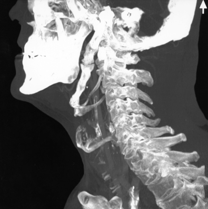
This is the case of a young man who has a long history of symptoms of a neck lump and pain. Several investigations are required to reach a diagnosis and make a plan about the definitive management of the patient. The diagnosis of Eagle's syndrome was made after a CT scan demonstrated an enlarged styloid process and a thickened, ossified stylohyoid ligament. There was also found to be an articulation between the styloid process and the ossified ligament; a feature which was also described by Eagle in his original case reports.1
This was a difficult diagnosis to make but once it was reached a definitive plan for treatment could be made. In this case, treatment was surgical excision via an external approach. This gave resolution in symptoms and a good result for the patient.
Case presentation
A 47-year-old man presented to the general ear nose and throat (ENT) clinic with a 6-month history of a swelling in the left submandibular triangle. He had discomfort in that area on swallowing which radiated to his ear but the lump had not changed in size and he was otherwise well with no other symptoms. On examination, he was found to have a 3 cm submandibular lump that was non-tender and the rest of the examination was unremarkable.
Investigations
The initial investigations were ultrasound and barium swallow. The barium swallow was normal and the ultrasound showed only an enlarged left submandibular gland with no other definite abnormality. This was followed by an MRI of his neck, which showed a lesion that appeared to be arising from the deep lobe of the left parotid gland, related to the carotid sheath and parapharyngeal fat. A CT scan was then performed which showed an enlarged left styloid process with an ossified, enlarged stylohyoid ligament (figure 1).
Figure 1.
CT neck with contrast showing gross enlargement of the styloid process, and enlargement and ossification of the stylohyoid ligament.
Differential diagnosis
The differential diagnosis in any case of a neck lump and pain is malignancy. In this case, both submandibular and parotid gland lesions were suspected on imaging.
Treatment
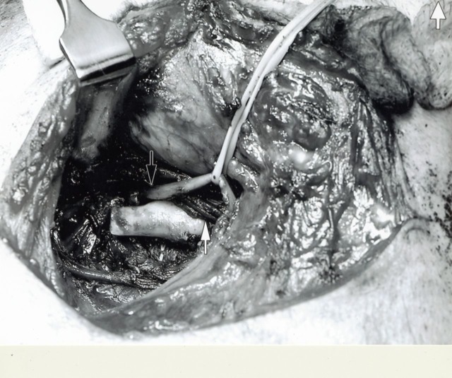
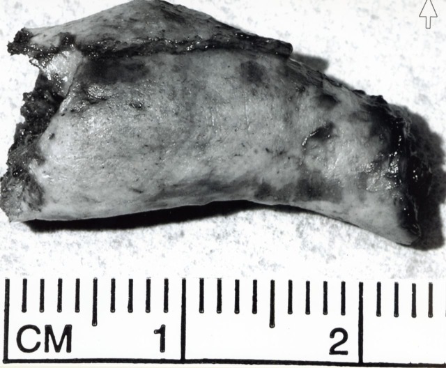
An external surgical approach was undertaken. The styloid process as well as the stylohyoid ligament were exposed and found to be grossly enlarged. The ossified stylohyoid ligament which was articulating with the hyoid bone was then excised (figure 2). The excised segment measured 2.4 cm (figure 3). The wound was closed in layers and a drain was inserted. The patient went home the following day.
Figure 2.
Exposure, via an external approach, of the ossified stylohyoid ligament. Note the posterior belly of the digastric muscle (white arrow) and the hypoglossal nerve (Black arrow).
Figure 3.
Surgically resected portion of the stylohyoid ligament.
Outcome and follow-up
During the follow-up 2 weeks later, he had a hypoglossal neuropraxia. This was managed conservatively, and 4 weeks later, the tongue movements returned to normal and the patient was free of his original symptoms.
Discussion
Eagle's syndrome is the term given to symptomatic elongation of the styloid process or mineralisation of the styloid or stylohyoid ligament. Eagle first reported two cases in 1937; since then, there has been debate over the criteria for diagnosis and different management options.1
Accepted symptoms include throat pain and odynophagia, globus and difficulty swallowing as well as ear, facial and neck pain.2 The history of symptoms has been given following trauma, classically tonsillectomy and as a chronic pain problem. Diagnosis requires a precise history from the patient and appropriate imaging to demonstrate the styloid process. Imaging would show the abnormal anatomy of either an elongated styloid process or a calcified stylohyoid ligament. The normal length of a styloid process is approximately 2.5 cm, with a length over 3 cm being considered abnormal.3 The diagnosis of Eagle's syndrome can be supported by relief of the symptoms after local anaesthetic infiltration around a palpable styloid process in the tonsillar fossa.4 5 Treatment options are surgical and non-surgical. Surgical excision can be performed either via intraoral or extraoral approach.
Although non surgical management of Eagle's syndrome has been used, the only treatment shown to produce long-term symptom control is excision of the elongated styloid. This can be done by either an intraoral or extraoral approach. Each has advantages and limitations and both have been advocated by previous authors.6
The intraoral technique starts with tonsillectomy, if not already performed. The elongated styloid process can then be palpated. The overlying mucosa is incised so the styloid can be dissected to its origin on the temporal bone and removed. Ligamentous attachments are separated from the tip. The muscles and mucosa can be closed in layers. This technique affords a quick postoperative recovery and a short hospital stay. It spares the scar, fascial dissection and risk of paraesthesia associated with the external approach. However, its disadvantages are that it gives limited access and visualisation to the surgical field, increasing the risk of injury to nearby structures. Also, opening deep cervical spaces through the oropharynx increases infection risk.4–6
There are variations in the external approach. Once the submandibular gland and sternocleidomastoid are exposed, the posterior belly of digastric is retracted to expose the styloid process which can then be excised. Because this approach gives better access and visualisation, it is preferable to the intraoral one. It has, however, a longer postoperative recovery and hospital stay, and can also result in greater auricular paraesthesia.7–9 The overall success rate for treatment, whether it be surgical or not, is said to be in the range of 80%
Learning points.
Eagle's syndrome can present with vague symptoms and the diagnosis can be difficult.
Patients with vague head and neck pain can go undiagnosed for some time and often have multiple investigations.
It is an uncommon but important diagnosis to make as it is effectively treated.
Non-surgical options are available but surgery is well tolerated and produces good results.
Footnotes
Competing interests: None.
Patient consent: Obtained.
References
- 1.Watt W, Eagle MD. Elongated styloid process: report of two cases. Arch Otolaryngol 1937;25:584–7. [Google Scholar]
- 2.Baugh RF, Stocks RM. Eagle's syndrome: a reappraisal. Ear Nose Throat J 1993;72:341–4. [PubMed] [Google Scholar]
- 3.Murtagh RD, Caracciolo JT, Fernandez G. CT findings associated with Eagle syndrome. Am J Neuroradiol 2001;22:1401–2. [PMC free article] [PubMed] [Google Scholar]
- 4.Beder E, Ozgursory OB, Ozgursory SK. Current diagnosis and transoral surgical treatment of Eagle's syndrome. J Oral Maxillofac Surg 2005;63:1742–5. [DOI] [PubMed] [Google Scholar]
- 5.Chrcanovic BR, Custodio ALN, de Oliveira DRF. An intraoral surgical approach to the styloid process in Eagle's syndrome. J Oral Maxillofac Surg 2009;13:145–51. [DOI] [PubMed] [Google Scholar]
- 6.Chase DC, Zarmen A, Bigelow WC, et al. Eagle's syndrome: a comparison of intraoral versus extraoral surgical approaches. Oral Surg Oral Med Oral Pathol 1986;62:625–9. [DOI] [PubMed] [Google Scholar]
- 7.Buono U, Mangone GM, Michelotti A, et al. Surgical approach to the stylohyoid process in Eagle's syndrome. J Oral Maxillofac Surg 2005;63:714–16. [DOI] [PubMed] [Google Scholar]
- 8.Ceylan A, Köybaşioğlu A, Çelenk F, et al. Surgical treatment of elongated styloid process: Experience of 61 cases. Skullbase 2008;18:289–95. [DOI] [PMC free article] [PubMed] [Google Scholar]
- 9.Diamond LH, Cottrell DA, Hunter MJ, et al. Eagle's syndrome: a report of 4 patients treated using a modified extraoral approach. J Oral Maxillofac Surg 2001;59:1420–6. [DOI] [PubMed] [Google Scholar]


