

Abstract
Patient: Female, 70
Final Diagnosis: Metal hypersensitivity
Symptoms: Joint pain • swelling • instability
Medication: —
Clinical Procedure: Revision total knee arthroplasty
Specialty: Orthopedics and Traumatology
Objective:
Unusual clinical course
Background:
Metal hypersensitivity is an uncommon complication after total knee arthroplasty (TKA) that can lead to significant functional impairment and aseptic prosthesis failure.
Case Report:
We describe a 70-year-old patient who presented with persistent pain, swelling, and instability 2 years after a primary TKA. The patient had a history of metal hypersensitivity following bilateral metal-on-metal total hip arthroplasty (THA) that was revised to ceramic-on-polyethylene implants. Knee radiographs showed severe osteolysis with implant loosening. Serum cobalt was elevated and serum chromium was significantly elevated, while joint aspiration and inflammatory marker levels ruled out a periprosthetic infection. Revision TKA was performed, with intraoperative tissue pathology and postoperative leukocyte transformation testing confirming metal hypersensitivity as the cause for aseptic implant failure.
Conclusions:
This case report demonstrates the clinical and laboratory signs that suggest metal hypersensitivity in total knee arthroplasty and the potential for joint function restoration with revision surgery.
MeSH Keywords: Arthroplasty, Replacement, Knee; Metal-on-Metal Joint Prostheses; Reoperation
Background
Total knee arthroplasty (TKA) is a effective treatment option in patients with debilitating pain and limited knee function from joint arthritis [1]. However, the procedure can result in perioperative complications, which most commonly include infection, instability, and implant failure, that often require revision arthroplasty to restore patient function and mobility [2]. Metal hypersensitivity, or an allergic reaction to metal ions, is another potential complication of knee arthroplasty. It occurs rarely and unpredictably, but can cause significant joint dysfunction and implant failure [3–5]. Hypersensitivity can be distinguished from infection based on joint aspiration analysis and inflammatory marker levels; however it is otherwise a diagnosis of exclusion due to the lack of a sensitive confirmatory test as well as unique clinical symptoms [3,4].
In this report we present a patient with a history of failed bilateral Metal-on-Metal (MoM) bearing total hip arthroplasty (THA) due to hypersensitivity who went on to develop a similar reaction after her subsequent TKA. The aim of this work is to draw attention to diagnostic techniques and surgical principles to treat TKA aseptic failure induced by metal hypersensitivity.
Case Report
A 70-year-old woman presenting with osteoarthritis and medial joint space loss (Figure 1) underwent left TKA at an outside institution. The primary surgeon used a posterior-stabilized implant with cobalt-chromium components (Attune Knee System, Depuy Synthes, Warsaw, Indiana) (Figure 2). There were no significant complications during the perioperative period. Subsequently the patient reported new onset pain, swelling, and joint instability of her left knee. The pain started several months after the surgery, was associated with standing from a seated position and prolonged weight-bearing. Physical therapy, assistive ambulatory devices, anti-inflammatory medications, and pain management evaluation were all utilized; however, the symptoms persisted. Periprosthetic fracture and joint infection were ruled out, with records reporting normal CRP and ESR levels with a joint aspiration resulting in a leukocyte count of 1650 cells/ml, 85% macrophages, and a low polymorphonucleocyte percentage. The recommendation was made for revision TKA surgery due to aseptic failure, implant loosening and instability. Of note, the patient previously had bilateral metal-on-metal bearing THA done by an outside surgeon three years prior that were revised to ceramic heads and polyethylene acetabular liners; although there were no formal laboratory studies performed by this surgeon, the presumptive diagnosis was metal hypersensitivity. Her femoral stems and acetabular cups were retained.
Figure 1.

Preoperatively, there is evidence of severe osteoarthritis at the left knee with elimination of the medial joint space, subchondral sclerosis, and marginal osteophyte formation.
Figure 2.

Postoperatively after the initial total knee arthroplasty, there are well-aligned components with a neutral tibial slope. There is no evidence of osteolysis or loosening.
On initial examination at our clinic, 14 months after the index TKA procedure, the surgical incision over the knee was well healed without significant swelling, erythema, drainage, or other evidence of infection. The patient exhibited tenderness at the medial and lateral joint lines and had a limited and painful passive range of motion between 10 and 110 degrees. Mild to moderate effusion was present. She had full strength with active knee flexion and extension. There was moderate varus and valgus laxity on extension, mid flexion, and full flexion. The patient was neurovascular intact and had full strength in all distributions distally. Imaging showed significant osteolysis, loss of tibial posterior slope, and settling of the tibial base-plate into varus as compared to her previous images taken after surgery (Figure 3).
Figure 3.

At 14 months postoperatively, there is now evidence of osteolysis, especially at the medial and posterior tibia, with shifting of tibial baseplate into varus and a negative tibial slope.
The patient elected to pursue revision TKA. Because of her history of sensitivity to cobalt and chromium after her metal on metal bearing THAs as well as her current TKA failure, the decision was made to use an oxidized zirconium femoral component and a titanium based tibial baseplate (Legion Oxinium, Smith & Nephew, Memphis, Tennessee) in the revision surgery. The procedure was undertaken 17 months after the primary TKA.
An incision was made through the previous scar, and a medial parapatellar approach was used to enter the joint. Upon arthrotomy through the previous incision, aggressive and hypertrophic fibrous synovitis was immediately identified around the joint (Figure 4). The synovial fluid appeared hazy and yellow. A complete synovectomy was performed and samples sent for histology analysis. Intraoperative joint fluid analysis resulted in 10% polymorphonucleocytes, 9% lymphocyte, 81% macrophage, and a leukocyte cell count of 533 cells/ml, with a very low likelihood of infection as the cause of failure. The tibial liner showed signs of mild wear. The femoral component was removed revealing severe bone loss and erosion, especially at the anterior femur and posterior femoral condyles, which were nearly completely resorbed. The lateral femoral epicondyle had a stress fracture line, most likely due to the weakened and resorbed bone leading to valgus collapse; this was seen after implant removal and was reduced and secured using a locking plate. The tibial component was completely loose and removed by hand, revealing again severe bone erosion and cyst formation. After the femur and tibia had been debrided to yield adequate bone stock, the revision TKA procedure was then completed using a varus-valgus constrained implant (Legion Total Knee System, Smith & Nephew, Memphis, Tennessee). Augments were utilized to reconstitute the anatomical joint line, achieve equal flexion and extension gaps, and ensure overall knee stability (Figure 5).
Figure 4.

Intraoperatively at the revision surgery, there is significant synovitis surrounding the components, with the excised synovium appearing fibrous and inflamed.
Figure 5.
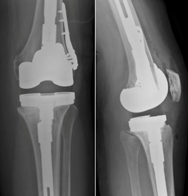
Postoperatively after the revision total knee arthroplasty, there are well-aligned long-stemmed components. There is no evidence of osteolysis or loosening.
The synovium, which was sent to pathology, was found to have granulation tissue, fibrosis, and focal giant cell reaction and calcification (Figure 6). IgE and eosinophil levels were both high, indicating a systemic allergic response. The patient’s serum was sent for analysis (Associated Regional and University Pathologists, Salt Lake City, Utah) with results showing elevated levels of serum cobalt at 2.4 ug/L (0.08–0.5 ug/L reference) and serum chromium at >1000 ug/L (0.06–0.93 ug/L reference). Leukocyte sensitivity tests (Orthopedic Analysis, Chicago, Illinois) showed increased reactivity (0–2 stimulation index reference) to aluminum (2.5), vanadium (3.8), molybdenum (3.1), and nickel (3.6), but not to cobalt (0.9) or chromium (1.1).
Figure 6.

Hematoxylin-Eosin stained histology showing numerous giant cells (white arrow) and calcifications (black arrow) in the excised synovium.
The patient had no post-operative complications and was allowed to be 25% weight bearing with an unlocked hinge knee brace. She was discharged on the second postoperative day. By the first clinic visit at 2 weeks, the patient was ambulating significantly with physical therapy and had a pain-free active range of motion from 10–100 degrees. The incision was fully healed and there was no evidence of infection. Radiographs showed a well-aligned joint without evidence of acute implant failure. At the 6 week visit, the patient had been weaned from her brace and had full weight bearing capacity with the use of a cane, mainly due to a history of back pain. She had a pain-free range of motion from 10–120 degrees. At the 4 month visit, the patient continued to do well, with a pain-free range of motion from 0–120 degrees and no evidence of joint effusion.
Discussion
Metallic hypersensitivity is an uncommon and controversial cause of TKA failure. Although the exact cause of hypersensitivity is unknown, the release of metal ions from implant wear or corrosion with an immune response has been implicated [6]. A Type IV delayed-hypersensitivity allergic reaction, with activation of CD4 and CD8 T-lymphocytes, results in release of cytokines such as interferon gamma, interleukin-1, interleukin-6, and tumor necrosis factor [6]. This inflammatory cascade can ultimately cause bone resorption and implant failure.
Patients with metal hypersensitivity after TKA may present with multiple symptoms. Most commonly, the knee may be swollen, stiff, and painful with clinical signs of localized eczema and dermatitis [4,7]. Rarely, there can be a systemic response, with one recent case report detailing severe whole-body dermatitis and hair loss [8]. In all cases, ruling out infection is of utmost importance. Inflammatory markers, such as ESR and CRP, should be measured, and if there is any doubt, a joint aspiration should be performed. Infection markers should be normal or only mildly elevated to support a diagnosis of metal hypersensitivity [4,9].
The two most commonly used diagnostic tests include skin patch testing and lymphocyte transformation testing, which assess the reactivity of the immune system to common allergens.[9] However, there are problems with both tests. The results of skin patch testing are subjective, may not reliably correlate to deep reactivity around orthopaedic implants, and may be influenced by sensitization from the test itself [9–11]. Similarly, lymphocyte transformation testing does not always correlate with patch testing and is not readily available at most centers [11,12]. As such, the results of these tests should be combined with other laboratory markers and clinical symptoms to diagnose hypersensitivity.
There is no standard paradigm to approaching metal hypersensitivity after TKA, mainly because it is a diagnosis of exclusion. Over 10% of the general population has a history of cutaneous metal allergy, yet there seems to be insufficient evidence that this correlates to implant hypersensitivity [6]. Multiple authors advocate preoperative testing for all patients with cutaneous metal allergy but not routine screening for patients without this history [6,12]. Granchi et al. provided a flow chart to evaluate hypersensitivity, and recommended patch testing for patients with a previous history of metal allergy followed by implantation of devices without metals if testing was positive [12]. Similarly, Innocenti et al. used anallergic components in patients with suspected metal allergy with excellent midterm results [3].
Patients presenting with metal hypersensitivity after TKA will likely require revision surgery. An extensive synovectomy during revision surgery should be performed to decrease the metal ion burden. Bone stock compromise may be present which will necessitate augmentation and intramedullary stems, and may require the use of constrained implants to ensure stability.
Anallergic components with oxidized zirconium, ceramic or titanium coatings should be considered to limit dispersal of cobalt and chromium debris [3,4,11]. Preoperative and intraoperative joint analysis as well as postoperative histopathology testing should be performed to both rule out infection and confirm the diagnosis of hypersensitivity. Pathology results may vary, with proliferation of histiocytes, giant cells, and synovial hyperplasia [4]. Depending on the level of joint instability and weakness, limited weight-bearing with supportive devices may be required postoperatively until complete healing has occurred. Close follow-up is recommended for the first 3 months.
Conclusions
Metal hypersensitivity is an uncommon but potentially significant complication after TKA. Prior hypersensitivity to implants or metal warrants increased caution for future procedures. Frequent patient follow-up, as well as knowledge of the clinical and laboratory presentation of hypersensitivity, is necessary for diagnosis. Through early treatment with revision arthroplasty, patients presenting with symptomatic metal hypersensitivity can have a stable and functional total knee arthroplasty and a satisfactory outcome.
References:
- 1.Dalury DF, Barrett WP, Mason JB, et al. Midterm survival of a contemporary modular total knee replacement: a multicentre study of 1970 knees. J Bone Joint Surg Br. 2008;90(12):1594–96. doi: 10.1302/0301-620X.90B12.21064. [DOI] [PubMed] [Google Scholar]
- 2.Healy WL, Della Valle CJ, Iorio R, et al. Complications of total knee arthroplasty: standardized list and definitions of the Knee Society. Clin Orthop Relat Res. 2013;47(1):215–20. doi: 10.1007/s11999-012-2489-y. [DOI] [PMC free article] [PubMed] [Google Scholar]
- 3.Innocenti M, Carulli C, Matassi F, et al. Total knee arthroplasty in patients with hypersensitivity to metals. Int Orthop. 2014;38(2):329–33. doi: 10.1007/s00264-013-2229-2. [DOI] [PMC free article] [PubMed] [Google Scholar]
- 4.Thakur RR, Ast MP, McGraw M, et al. Severe persistent synovitis after cobalt-chromium total knee arthroplasty requiring revision. Orthopedics. 2013;36(4):e520–24. doi: 10.3928/01477447-20130327-34. [DOI] [PubMed] [Google Scholar]
- 5.Münch HJ, Jacobsen SS, Olesen JT, et al. The association between metal allergy, total knee arthroplasty, and revision. Acta Orthop. 2015;13:1–6. doi: 10.3109/17453674.2014.999614. [DOI] [PMC free article] [PubMed] [Google Scholar]
- 6.Kitagawa A, Chin T, Tsumura N, Iguchi T. Metal sensitivity in patients before and after total knee arthroplasty (TKA): comparison between ceramic surfaced oxidized zirconium and cobalt-chromium implants. Hypersensitivity. 2013;1:3. [Google Scholar]
- 7.Thomsen M, Rozak M, Thomas P. Pain in a chromium-allergic patient with total knee arthroplasty: disappearance of symptoms after revision with a special surface-coated TKA – a case report. Acta Orthop. 2011;82(3):386–88. doi: 10.3109/17453674.2011.579521. [DOI] [PMC free article] [PubMed] [Google Scholar]
- 8.Post ZD, Orozco FR, Ong AC. Metal sensitivity after TKA presenting with systemic dermatitis and hair loss. Orthopedics. 2013;36(4):e525–28. doi: 10.3928/01477447-20130327-35. [DOI] [PubMed] [Google Scholar]
- 9.Mihalko WM, Goodman SB, Hallab NJ, Jacobs JJ. Skin patch testing and associated total knee outcomes. AAOS Now. 2012;9(4) [Google Scholar]
- 10.Razak A, Ebinesan AD, Charalambous CP. Metal allergy screening prior to joint arthroplasty and its influence on implant choice: a Delphi consensus study amongst orthopaedic arthroplasty surgeons. Knee Surg Relat Res. 2013;25(4):168–93. doi: 10.5792/ksrr.2013.25.4.186. [DOI] [PMC free article] [PubMed] [Google Scholar]
- 11.Lützner J, Hartmann A, Dinnebier G, et al. Metal hypersensitivity and metal ion levels in patients with coated or uncoated total knee arthroplasty: a randomised controlled study. Int Orthop. 2013;37(10):1925–31. doi: 10.1007/s00264-013-2010-6. [DOI] [PMC free article] [PubMed] [Google Scholar]
- 12.Granchi D, Cenni E, Giunti A, Baldini N. Metal hypersensitivity testing in patients undergoing joint replacement: a systematic review. J Bone Joint Surg Br. 2012;94(8):1126–34. doi: 10.1302/0301-620X.94B8.28135. [DOI] [PubMed] [Google Scholar]