Abstract
Nasogastric (NG) feeding tubes are commonly inserted to supplement enteral nutrition in certain patient groups, including those with head and neck cancers where swallowing may be compromised. An NHS National Patient Safety Alert was released in 2011 detailing ongoing cases of significant morbidity and mortality attached to the incorrect placement of NG feeding tubes in hospital inpatients. Since 2005, there were 21 deaths and 79 cases of harm nationally due to feeding into the lung through misplaced tubes. pH testing remains the first-line method of placement confirmation, with chest x-ray used when no aspirate is gained or where pH testing fails to confirm suitable acidity. We present a case report describing false-positive NG tube placement confirmation tests in a patient with head and neck cancer, who was administered feed into lung parenchyma with significant morbidity. We discuss the case for specific NG tube placement protocols in head and neck cancer patients.
Background
An NHS National Patient Safety Alert was released in 2011 detailing ongoing cases of significant morbidity and mortality attached to the incorrect placement of nasogastric (NG) feeding tubes in hospital inpatients. Our case represents one such event, and adds to the need to review current NG tube placement guidelines in order to improve patient safety. Our case also adds particular interest because the correct national guidelines were followed and yet false-positive aspirates were gained, perhaps due to altered anatomy of the pharynx, and chronic aspiration in head and neck cancer patients. Lessons can be learnt from this case, and the local trust guidelines are undergoing review with the potential for specific guidelines for NG-tube insertion in the head and neck cancer cohort.1 2
Case presentation
Case report
A 54-year-old gentleman was admitted to Guy's Hospital with an 8-week history of a right neck swelling, otalgia, dysphonia and progressive dysphagia, with a significant associated weight loss (40% total body weight).
On examination he was significantly cachectic, with poor dentition. There was a >4 cm right tonsillar mass extending to the posterior pharyngeal wall, and neck examination revealed a tender, fixed, firm 3 cm right level II mass. Flexible nasendoscopy was performed which revealed a normal postnasal space, but oropharyngeal findings as described. Limited examination of the supraglottis, glottis and hypopharynx was normal. Fine needle aspiration cytology revealed squamous cell carcinoma in the right neck mass. He underwent CT neck and chest which revealed an extensive partially necrotic oropharyngeal mass infiltrating the soft palate to midline, and base of the tongue and hypopharynx, and partially eroding the hyoid bone.
He was admitted for nutritional support awaiting discussion at the Multidisciplinary Team Meeting after which he staged as cT4N2aMx right tonsillar squamous cell carcinoma (SCC) and was listed for microlaryngobronchoscopy and biopsy. He had a fine bore NG tube inserted blind on the ward by nursing staff on admission, which revealed an aspirate of pH 4.5, and the feed was started as per protocol at 50 ml/h, plus water flushes and medications. He tolerated feed well until complaining of nausea, when feed was stopped, the NG flushed with 50 ml H2O, and repeat aspirate testing revealed pH 5.5, and feed was again re-started at 50 ml/h. Repeat aspirate in the morning revealed 15 ml of frank blood and the patient desaturated to 77% on room air.
Investigations
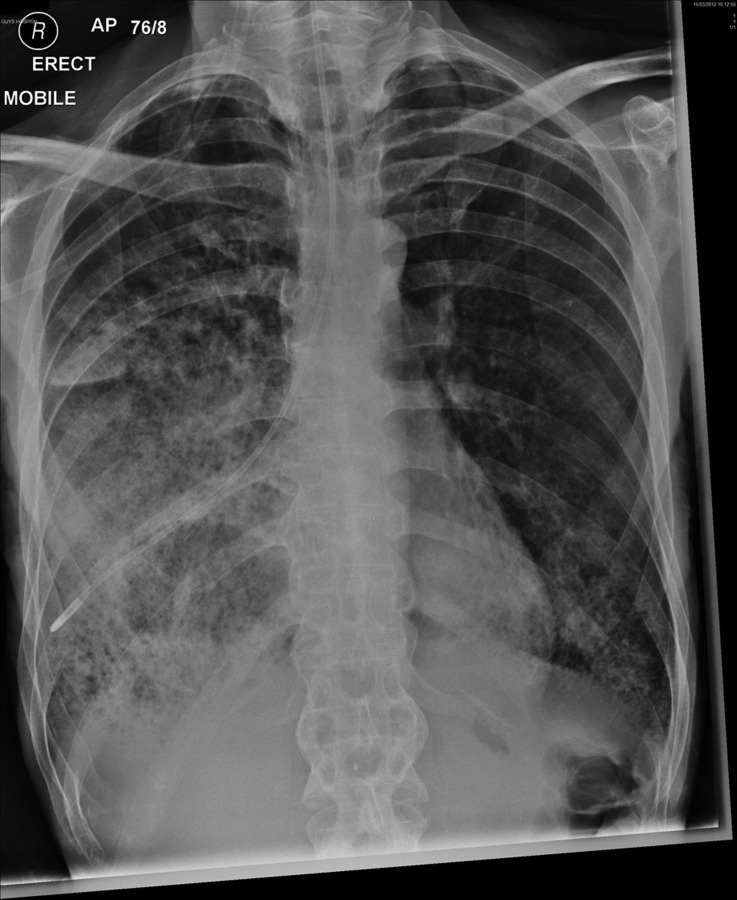
He immediately underwent clinical examination and fine nasendoscopy (FNE) on the ward which was very poorly tolerated, and an urgent mobile chest x-ray was arranged (figure 1), which revealed the NG tube to be placed within the chest.
Figure 1.
Nasogastric-tube tip is positioned within the right lung.
Treatment
He had received 540 ml of feed, medications and water flushes into lung parenchyma, and was immediately reviewed by a medical consultant and was started on intravenous antibiotics and high-flow oxygen.
Outcome and follow-up
The patient made a good recovery and was referred for palliative radiotherapy on discharge.
Discussion
NG tube placement is a routine part of clinical care for certain inpatients, including head and neck cancer patients with dysphagia, who often have distorted anatomy, impaired swallow and gag reflexes and pain related to the disease or its treatment.3 The first case of NG tube being placed into the pleural space was reported in 1978, and 3 years later, the first death related to intraparenchymal feeding through a misplaced tube was published.4 Guidelines have since been developed to aid safe verification of NG tube position, and of these methods the pH test and radiology are considered gold standard.5 According to Guys and St Thomas’ NHS Trust Guidelines, feed may be started if an aspirate of pH < 5.5 is gained.6 Certain factors are known to alter the accuracy of pH testing, including proton-pump inhibitors and H2-receptor antagonists; however, these were not present in the case presented. There was no documentation of excessive coughing, pain or breathlessness to raise suspicion of false-positive pH readings. On radiological discovery of tube misplacement, feed was immediately stopped and senior opinion sought. The decision was taken to clamp the tube but leave it in position to permit reduced inflammation and clotting of any potentially significant intraparenchymal haemorrhage. The incident was escalated appropriately and led to the alteration of trust guidelines to include the need for extra caution in head and neck patients; however, no specific change in practice was indicated.
This case highlights the fallibility of current methods for confirmation of NG tube placement, and is the second case of false-positive positioning in the Otorhinolaryngology/Maxillofacial Surgery Department at the Trust within 3 years. The pathophysiology behind acidic lung aspirates is not well described but in head and neck patients may include chronic aspiration of saliva and food particles, and/or bacterial superinfection. This may indicate the need for department-specific insertion protocols, including the consideration of reduced pH cut-offs,7 routine FNE-guided gastric placement and/or standard radiological confirmation in this patient group. Clearly, such methods raise their own issues including cost effectiveness, demand on radiology departments, exposure to potentially unnecessary radiation, and the availability of specialist ENT/maxillofacial surgeons and trainees and necessary equipment. However, the misplacement of NG tubes remains a prevalent and disastrous clinical scenario, and evidence-based practice must be consulted to ensure patient safety.
Conclusion
The false-positive confirmation of an NG feeding tube led to a critical clinical incident in a patient with head and neck cancer. Since the incidence of misplaced tubes and their attached morbidity persists, the fallibility of current placement confirmation guidelines is a serious issue. The case for reduced pH cut-offs, routine insertion of NG feeding tubes under FNE guidance or radiological confirmation may improve patient safety outcomes in head and neck cancer departments. To the best of our knowledge this is the first case of false-positive NG-tube aspiration to be published in the literature.
Learning points.
Nasogastric (NG) feeding tubes are commonly inserted to support enteral nutrition in certain patient groups.
Current national guidelines state that pH aspirates of <5.5 are the gold standard method of successful position confirmation.
In head and neck cancer patients this system is fallible.
This case demonstrates that pH aspirate can be falsely positive in certain patient groups, and a low threshold for suspicion of malpositioning should be employed.
Local trust guidelines are being reviewed at present for the potential introduction of specific NG feeding tube insertion in head and neck patients.
Footnotes
Competing interests: None.
Patient consent: Obtained.
References
- 1.NHS National Patient Safety Agency. Reducing the harm caused by misplaced nasogastric feeding tubes in adults, children and infants. National Patient Safety Alert: 10 March 2011. NPSA/2011/PSA002.
- 2.Yardley IE, Donaldson LJ. Patient safety matters: reducing the risks of nasogastric tubes. Clin Med 2010;10:228–30. [DOI] [PubMed] [Google Scholar]
- 3.Raff MH, Cho S, Dale R. A technique for positioning nasoenteral feeding tubes. J Parenter Enteral Nutr 1987;11:210–13. [DOI] [PubMed] [Google Scholar]
- 4.Torrington KG, Bowman MA. Fatal hydrothorax and empyema complicating a malpositioned nasogastric tube. Chest 1981;79:240–2. [DOI] [PubMed] [Google Scholar]
- 5.Khair J. Guidelines for testing the placing of nasogastric tubes. NursTimes 2005;101:26–7. [PubMed] [Google Scholar]
- 6.GSTT Foundation Trust Clinical Guidance. Adult naso-gastric feeding tube insertion and management. Version 4.2. Available at: www.gstt. [Google Scholar]
- 7.Gilbertson HR, Rogers EJ, Okoumunne OC. Determination of a practical pH cutoff level for reliable confirmation of nasogastric tube placement. JPEN J Parenter Enteral Nutr 2011;35:540–4. [DOI] [PubMed] [Google Scholar]


