Abstract
Cystic lesions of transient nature viz. Epstein pearls, Bohn's nodules and dental lamina cysts are frequently found in the oral cavities of newborn infants. These cysts arise from the developing dental tissues or from their remnants. These cystic lesions are not commonly seen by the dental surgeons due to their self-limiting nature and ignorance of the parents to seek the professional opinion. However, when contacted by anxious parents seeking treatment, dental surgeons should be able to explain and reassure the parents about the transient nature of these lesions and need for no treatment but regular follow-up. The present case report was written with the purpose to increase the awareness in dental surgeons about the peculiar clinical presentation and self-limiting nature of these cystic lesions, so that unnecessary surgical intervention can be avoided in such young infants.
Background
Parents of newborn infants sometimes apprehensively visit a dental surgeon or paediatrician, complaining of the presence of abnormal structures in the mouth of their infants. Many infants may have oral cysts of a transient nature, which disappear within a few days to months. Three such cysts namely Epstein pearls, Bohn's nodules and dental lamina cysts are widely reported in the literature. Epstein pearls were first described by Alois Epstein in 1880. Bohn's nodules were described by Heinrich Bohn as ‘mucous gland cysts’. Dental lamina cyst, also known as a gingival cyst of newborns, are raised nodules on alveolar ridges of infants, derived from the rests of the dental lamina which consist of keratin producing epithelial lining.1 These cysts appear as small, isolated or multiple whitish papules. These cysts can be classified into palatal (located in midline raphe) and alveolar cysts (present on the crests of alveolar ridges).2 The reported prevalence of palatal cysts in newborns is about 65%,3 while for alveolar cysts, it ranges from 25%4 to 53%.5
Despite the high prevalence, these cysts are rarely seen by the general or a paediatric dentist due to their transient nature. These cysts rupture and disappear within 2 weeks to 5 months without any treatment.2 6 7 Their transient nature is thought to be due to fusion of the cyst wall with oral epithelium and subsequent discharge of the cystic content.8 The present case report describes the characteristic clinical presentation and management of frequently found but less reported oral cysts in a newborn infant.
Case presentation
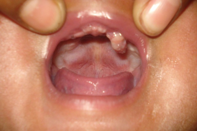
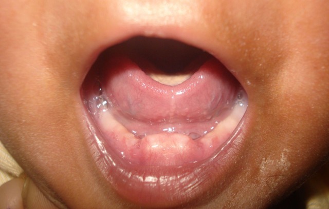
A 14-day-old male newborn infant presented to the Department of Paediatric and Preventive Dentistry, Chhatrapati Sahuji Maharaj Medical University, Lucknow with the chief complaint of swelling present on gums of both upper and lower jaws. The parents sought an opinion from a general dental surgeon, who referred them to the paediatric dentist. According to the mother, the swelling was present since birth, but without any feeding difficulty. The child was full-term born with unremarkable medical/dental history. No noticeable findings were recorded during extraoral examination. On intraoral examination, a nodular papule was present in the deciduous upper left molar region (figure 1). The papules were also present in the deciduous lower right and left central incisor regions (figure 2). The size of these papules varied from 4 to 7 mm. The infant's mother gave the history of similar appearing swelling, which was present in the deciduous upper right molar region and regressed 7 days before the infant was seen by us. A clinical examination of that area revealed the presence of small cleft on the gum pad, suggesting the previous presence of papule. No other abnormality could be found on clinical examination of labial, buccal and lingual mucosa, tongue, palate and floor of the mouth. The diagnosis of dental lamina cyst was made based on clinical examination and the characteristic appearance of the lesion.
Figure 1.
Nodular papule present in the deciduous upper left molar region.
Figure 2.
Nodular papules present in the deciduous lower central incisor region.
Differential diagnosis
In the differential diagnosis of dental lamina cysts, Epstein pearls, Bohn's nodules, congenital epulis of newborn and natal/neonatal teeth must be considered. Epstein pearls are small cystic, keratin-filled nodules, often seen on the roof of the palate and are caused by entrapped epithelium during the development of the palate. Bohn's nodules are also keratin-filled cysts, found at the junction of hard and soft palate and along buccal and lingual parts of the alveolar ridges away from the midline, and are remnants of salivary glands. All these cysts have a characteristic clinical presentation and histological findings but can be diagnosed on the basis of clinical appearance alone.
Congenital epulis of newborn is a smooth-surfaced protuberant rare benign tumour mass, present most commonly along the alveolar ridge, normal-to-reddish in colour, with a variable size of few millimetres to few centimetres in diameter and may cause feeding/respiratory problems.9–11 Treatment of the smaller lesions includes careful wait and watch/surgical intervention, although spontaneous regression is rare. Larger lesions should be surgically excised under local or general anaesthesia at the earliest as they may risk the infant by causing feeding/respiratory difficulties. Congenital epulis needs surgical management in the majority of cases, whereas dental lamina cyst is self-limiting.
Dental lamina cyst, in rare occasions, can be confused with natal/neonatal teeth which are mostly found in the mandibular anterior region.12 Natal/neonatal teeth mostly exhibit mobility as their roots are short or absent and at times, needs to be extracted as of having risk of being ingested during feeding.13
Thus, the oral cavity of newborn infants should be very carefully examined in instances of the presence of any oral lesions to rule out the self-limiting lesions requiring monitoring only such as dental lamina cysts, Epstein pearls and Bohn's nodules, with the lesions that require more aggressive treatment like natal/neonatal teeth and congenital epulis of newborn.
Treatment
The parents were explained regarding the frequent findings of these cystic lesions in newborns, were reassured about the transient nature of the lesion, and instructed to maintain the oral hygiene. No treatment of any kind was done, except for parental counselling and reassurance. It is important to note that management of all oral inclusion cysts (dental lamina cysts, Epstein pearls and Bohn's nodules) remains the same, as all these have a self-limiting nature and require no treatment.
Outcome and follow-up
The parents were advised for periodic follow-up. One-month follow-up revealed the complete regression of the cystic lesions, with no defects seen on the alveolar ridges. The infant was healthy and the parents were satisfied with the outcome.
Discussion
Fromm, in his comprehensive review of 1,367 newborn infants, found 79% of infants with oral inclusion cysts which were distinct both histologically and clinically.14 The oral cysts presented in our case are dental lamina cysts. These are the true cysts with a thin epithelial lining, and show a lumen usually filled with desquamated keratin, sometimes containing inflammatory cells.1 These cysts are more commonly found in the maxilla than in mandible. These cysts clinically appear as small, white or yellow papules along the midline, or at the junction of hard and soft palate. These cysts are found as clusters, usually in a group of 2–6 but may also occur as solitary cyst.15 The size of these cysts varies from 1 to 3 mm, however, in our case the size was larger (4–7 mm) than usually found. There is a general agreement that these cysts arise from the dental lamina. The epithelial remnants of dental lamina have the capacity to proliferate, keratinise and form small cysts. Moskow and Bloom16 noted proliferative tendency in the dental lamina, having multiple areas of microcyst formation and keratin production in the human fetus as tooth development progressed. After birth, the epithelial inclusions are usually atrophied and get resorbed. Some of the gingival cysts probably open onto the surface leaving clefts; others may be involved by the developing teeth. Some degenerate and disappear and the keratin and debris are digested by giant cells.
Although, frequent finding of these cysts suggests them to be normal structures in newborn infants, parents may be apprehensive of their presence and may seek the professional opinion. Knowledge of these frequently found lesions in infants is therefore crucial for all healthcare providers, including dental surgeons and paediatricians, who may be contacted by the parents. These cystic lesions may be easily detected by their characteristic clinical appearance in the oral cavity of the infants, making it unnecessary to have histopathological confirmation.
Since these lesions are of a transient nature, no active treatment is required for their management. Healthcare providers must reassure the parents about innocuous nature and self-limiting characteristics of these cysts, to allay the anxiety of parents towards these lesions.
Learning points.
Intraoral cystic lesions with characteristic clinical appearances are frequently found in infants.
These lesions are self-limiting, disappear within a few days to months and thus require no treatment.
Parental anxiety should be allayed by reassurance and regular follow-up should be advised.
Footnotes
Competing interests: None.
Patient consent: Obtained.
References
- 1.Shafer WG. Cysts and tumors of odontogenic origin. In: Hine MK, Levy BM, Tomrich CE, eds. Textbook of oral pathology. 4th edn India: W.B. Saunders Co, Prism (Reprint), 1993:268–9. [Google Scholar]
- 2.Paula JD, Dezan CC, Frossard WT, et al. Oral and facial inclusion cysts in newborns. J Clin Pediatr Dent 2006;31:127–9. [DOI] [PubMed] [Google Scholar]
- 3.Cataldo E, Berkman MD. Cysts of the oral mucosa in newborns. Am J Dis Child 1968;116:44–8. [DOI] [PubMed] [Google Scholar]
- 4.Friend GW, Harris EF, Mincer HH, et al. Oral anomalies in neonate, by race and gender, in urban setting. Pediatr Dent 1990;12:157–61. [PubMed] [Google Scholar]
- 5.Jorgenson RJ, Shapiro SD, Salinas CF, et al. Intraoral findings and anomalies in neonates. Pediatrics 1982;69:577–82. [PubMed] [Google Scholar]
- 6.Flinck A, Paludan A, Matsson L, et al. Oral findings in group of newborn Swedish children. Int J Clin Pediatr Dent 1994;4:67–73. [DOI] [PubMed] [Google Scholar]
- 7.Regezi JA. Cyst of jaws and neck. In: Regezi JA, Sciubba JJ, Jorden RC, eds. Oral pathology: clinical pathological correlation. 4th edn St. Louis, Missouri: W.B. Saunders Co., 1999:246. [Google Scholar]
- 8.Burke GW, Feagans WM, Jr, Elzay RP, et al. Some aspects of the origin and fate of midpalatal cysts in human fetuses. J Dent Res 1966;45:159–64. [DOI] [PubMed] [Google Scholar]
- 9.Yilmaz F, Uzuniar A, Arsian A, et al. Congenital granular cell epulis: report of 2 cases. Saudi Dent J 1999;11:24–6. [Google Scholar]
- 10.Chinidia ML, Awange DO. Congenital epulis of the newborn: a report of two cases. Br Dent J 1994;176:426–8. [DOI] [PubMed] [Google Scholar]
- 11.Merrett SJ, Crawford PJ. Congenital epulis of the newborn: a case report. Int J Paediatr Dent 2003;13:127–9. [DOI] [PubMed] [Google Scholar]
- 12.Kumar A, Grewal H, Verma M. Dental lamina cyst of newborn: a case report. J Indian Soc Pedod Prevent Dent 2008;26:175–6. [DOI] [PubMed] [Google Scholar]
- 13.Liu MH, Huang WH. Oral abnormalities in Taiwanese newborns. J Dent Child 2004;71:118–20. [PubMed] [Google Scholar]
- 14.Fromm A. Epstein pearls, Bohn's nodules and inclusion cyst of the oral cavity. J Dent Child 1967;34:275–87. [PubMed] [Google Scholar]
- 15.Neville BW, Damm DD, Allen CM, et al. Oral and maxillofacial pathology. 3rd edn St. Louis, Missouri: Saunders, 2009. [Google Scholar]
- 16.Moskow BS, Bloom A. Embryogenesis of the gingival cyst. J Clin Periodontol 1983;10:119–30. [DOI] [PubMed] [Google Scholar]


