Abstract
The case of a 59-year-old Caucasian man who presented with a 6-week history of intermittent blurring of vision and diplopia is reported. Fundoscopy revealed asymmetrical, bilateral optic disc swelling with peripapillary haemorrhages. An initial CT scan and D-dimer level were reported as normal. A subsequent MRI revealed a recanalising superior sagittal sinus thrombosis. Cerebral venous sinus thrombosis is a rare and potentially fatal condition. The author suggests that it should be part of the differential diagnosis of bilateral optic disc swelling and that a normal unenhanced CT scan and D-dimer would not rule out this potentially devastating condition.
Background
Cerebral venous sinus thrombosis (CVST) is a potentially fatal but rare condition (incidence: 3–4 cases/million/year).1 It most commonly affects the superior sagittal sinus.2
A case is described which demonstrates the importance of a wide differential diagnosis of optic disc swelling and cautions against false-negative investigations commonly used to evaluate similar presentations.
Case presentation
A 59-year-old Caucasian man presented with a 6-week history of intermittent blurring of vision and diplopia associated with a left-sided, subsiding headache.
The medical history revealed essential hypertension on triple therapy. Presenting blood pressure measured 118/70 mm Hg. His Glasgow Coma Scale was 15/15 with no sign of meningism. His visual acuities were 6/9 OU. There was no relative afferent pupillary defect. Ishihara plate reading: 13/13 OD and 12/13 OS. Facial sensation was intact. He did not have diplopia at the time of examination and his ocular motility was intact. His intraocular pressures were normal. Slit-lamp biomicroscopy of the anterior segments was normal. Fundus examination revealed bilateral optic disc swelling with peripapillary haemorrhages (figure 1).
Figure 1.

Fundus photographs showing bilateral optic disc swelling with peripapillary haemorrhages.
Investigations
Humphrey visual field testing was essentially normal.
Urgent neuroimaging was arranged. MRI was unavailable at the time, so an unenhanced CT of the head was obtained. This did not show any evidence of intracranial pathology. There was no mass-effect or midline shift and no ventricular dilatation.
Blood tests showed a normal coagulation screen, full blood count and inflammatory markers. The cardiolipin antibody screen was negative. The D-dimer measured 212 ng/ml with a normal reference value of <255.
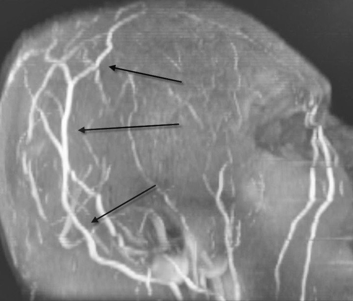
An MRV scan (magnetic resonance venography) was arranged as an outpatient with the false reassurances of minimal symptoms, a normal CT of the head and negative D-dimer. This revealed superior sagittal sinus thrombosis with early recanalisation (figures 2 and 3).
Figure 2.
Axial T2 MRI scan of the brain demonstrating hyperintensity within the superior sagittal sinus (SSS), suggesting thrombosis. Note the small, hypointense focus at the anterior aspect of the SSS reflecting partial recanalisation.
Figure 3.
Three-dimensional magnetic resonance venography showing prominent flow within cortical veins due to shunting to alternative venous drainage pathways.
Differential diagnosis
Idiopathic intracranial hypertension.
Intracranial mass with hydrocephalus.
Malignant hypertension.
Treatment
Anticoagulation with warfarin was initiated.
Outcome and follow-up
The patient's symptoms resolved spontaneously soon after our initial assessment.
The patient will continue to be monitored by neurology and ophthalmology.
Discussion
CT may show the classic ‘delta sign’ but there is evidence that it may be completely normal despite CVST.2
A negative D-dimer has traditionally been seen as a reliable way to rule out certain thrombotic conditions such as deep vein thrombosis and pulmonary embolism.3 There is little evidence demonstrating its sensitivity in cerebral venous thrombosis. Our patient had a normal D-dimer.
A prospective study of 33 patients showed that recanalisation occurs only within the first 4 months following cerebral venous thrombosis, irrespective of oral anticoagulation.4 This correlates with the clinical history of our patient in whom the symptoms resolved within 8 weeks and recanalisation was evident on the MRV prior to the initiation of warfarin.
CVST should be considered as part of the differential diagnosis for any patient presenting with bilateral optic disc swelling and contrast-enhanced CT or MRI may be essential for the diagnosis.
Learning points.
CT scan may be normal despite serious intracranial pathology. A contrast-enhanced CT or MRI may be essential in the evaluation of cerebral venous sinus thrombosis.
The D-dimer was normal despite superior sagittal sinus thrombosis. A negative result cannot rule out the condition.
Superior sagittal sinus thrombosis is a rare but important differential diagnosis of bilateral optic disc swelling.
Acknowledgments
I would like to thank Nitin Anand (Consultant Ophthalmologist, Calderdale and Huddersfield NHS trust) and Gavin Durra (Diagnostic Radiologist, Rosebank Clinic, Johannesburg) for their invaluable assistance.
Footnotes
Competing interests: None.
Patient consent: Obtained.
References
- 1.Gupta RK, Jamjoom AA, Devkota UP. Superior sagittal sinus thrombosis presenting as a continuous headache: a case report and review of the literature. Cases J 2009;2:9361. [DOI] [PMC free article] [PubMed] [Google Scholar]
- 2.Anand N, Chan C, Wang NE. Cerebral venous thrombosis: a case report. J Emerg Med 2009;36:132–7. [DOI] [PubMed] [Google Scholar]
- 3.Penaloza A, Roy PM, Kline Jet al. Performance of age-adjusted D-dimer cut-off to rule out pulmonary embolism. J Thromb Haemostasis: JTH 2012;10:1291–6. Epub 2012/05/10. [DOI] [PubMed] [Google Scholar]
- 4.Baumgartner RW, Studer A, Arnold M, et al. Recanalisation of cerebral venous thrombosis. J Neurol, Neurosurg, Psychiatry 2003;74:459–61. [DOI] [PMC free article] [PubMed] [Google Scholar]


