Abstract
In the present study, we isolated preadipocytes from the adipose tissue of Peking duck and subsequently cultured them in vitro. Cell counting kit-8 assay was employed to establish the growth curve of duck primary preadipocytes. Meanwhile, after the cells reaching full confluency, they were induced to differentiate into mature adipocytes by the addition of a cocktail containing dexamethasone, insulin, 3-isobutyl-1-methylxanthine, and oleic acid for 8 days. Successful differentiation was demonstrated by the development of lipid droplets and the expression of key marker genes including peroxisome proliferator-activated receptor-γ (PPARγ), CCAAT/enhancer binding protein-α (CEBP/α) and adipocyte fatty acid-binding protein (FABP4). Our results showed that duck primary preadipocytes began to adhere 12 h after seeding as short spindle shapes or litter triangles, which grew quickly 3 days post attachment and maintained stable after day 7. After 8 days the preadipocytes were induced to differentiate into mature adipocytes, which were stained red by oil red O. Additionally, it showed that during preadipocyte differentiation PPARγ mRNA was highly expressed at day 3, while CEBP/α and FABP4 mRNA peaked at day 5 and 8, respectively. These results indicate that we have successfully isolated and cultured Peking duck preadipocytes and successfully induced them to differentiate into mature adipocytes. This work could lay a foundation for further research into waterfowl adipogenesis.
Keywords: Differentiation, Isolate and culture, Peking duck, Preadipocytes
Introduction
Adipose tissue is a complex organ that regulates and coordinates energy homeostasis (Kershaw and Flier 2004). The growth of adipose tissue is primarily due to the increase in adipocyte cell number (hyperplasia) and the enlargement of adipocytes (hypertrophy). It is commonly known that adipocytes are derived from pluripotent mesenchymal stem cells (MSCs) in a process called adipogenesis. MSCs have the ability to develop into several cell types, such as adipocytes, chondrocytes, osteoblasts, and myocytes (Covas et al. 2008; Lin et al. 2010). In the past, researchers have successfully isolated and cultured preadipocytes from many species in vitro, and from this our knowledge of preadipocyte differentiation has been enhanced (Shi et al. 2011; Ren et al. 2012; Mueller 2014).
The biological process of preadipocyte differentiation has been extensively studied by using a variety of cell lines (such as 3T3L1, 3T3-F442A, Ob17) and primary preadipocytes cultured in vitro (Arsenijevic et al. 2012; Mueller 2014). By treating the cells with a hormonal cocktail that includes dexamethasone (DEX), insulin, and 3-isobutyl-1-methylxanthine (IBMX), preadipocytes of most species can differentiate into mature adipocytes. During this time specific adipogenic genes are expressed and triacylglycerol (TG) accumulates and is stored in the cells. However, in poultry due to the low capacity of adipose tissue to perform de novo lipogenesis (DNL) (Ding et al. 2012), the addition of oleic acid is needed to supplement the hormonal cocktail to induce preadipocyte differentiation in vitro (Matsubara et al. 2008; Wang et al. 2011; He et al. 2012). The role of oleic acid in promoting adipogenesis is unclear but research conducted by many investigators has provided compelling evidence that oleic acid can significantly activate adipocyte-related genes. Moreover, it has been shown that oleic acid by itself can promote adipogenesis (Shi et al. 2010, 2011; Wang et al. 2011; He et al. 2012).
Activation of the transcriptional processes promoting preadipocyte differentiation requires many key genes such as peroxisome proliferator-activated receptor-γ (PPARγ), CCAAT/enhancer binding protein-α (CEBP/α), and adipocyte fatty acid-binding protein (FABP4). PPARγ is a member of the nuclear-receptor superfamily and is required for adipogenesis in both adipocytes (Karbiener et al. 2009) and fibroblasts (Tontonoz et al. 1994), and no factor has been discovered that promotes adipogenesis in the absence of PPARγ (Farmer 2006; Rosen and MacDougald 2006). CEBP/α induces many adipogenic genes directly, such as PPARγ, and in vivo studies demonstrate an important role for this transcription factor in the development and maintenance of adipose tissue (Zhang et al. 2004). In addition, ectopic expression of CEBP/α alone can surprisingly induce adipogenesis in fibroblasts (Freytag et al. 1994). FABP4 is a marker for lipid accumulation in adipose tissue. In duck, knockdown of FABP4 was proven to inhibit expression of genes involved in preadipocyte differentiation (He et al. 2012). In chicken, knockdown of FABP4 leads to decreased lipid accumulation and upregulated expression of PPARγ in adipocytes (Shi et al. 2011). Surprisingly, high expression of FABP4 induces high lipolytic rate and decreases the abdominal fat mass in chicken (Shi et al. 2010). Up to now, investigations on adipogenesis were studied using chicken preadipocytes (Matsubara et al. 2005, 2008) and chicken embryonic fibroblasts (Liu et al. 2010). However, little is known about the molecular mechanism of adipogenesis in waterfowl.
Peking duck (Anas platyrhynchos) is a world famous waterfowl breed that is able to accumulate large quantities of lipid in the subcutaneous adipose tissue (Attia et al. 2012; Ding et al. 2012). It has been well established that the development of duck adipose tissue is due to a combination of adipocyte hyperplasia and hypertrophy from hatching until 4 weeks of age, after which adipose expansion is accomplished exclusively by adipocyte hypertrophy (Kou et al. 2012). However, the molecular mechanism involving the growth and differentiation of duck preadipocytes remains unknown. Therefore, the present study was designed to isolate the preadipocytes from duck adipose tissue and maintain them in culture. From this work we can gain information about the growth characteristics and the differentiation process of duck preadipocytes. Currently, preadipocytes from 1 week old Peking duck were isolated, cultured, and induced to differentiate, and expression patterns of three marker genes (PPARγ, CEBP/α, FABP4) were determined during duck adipogenesis. This work will provide a foundation for further research involving the mechanisms of duck adipogenesis. In addition, it can be used to explore novel methods that control fat deposition in poultry.
Materials and methods
Isolation and culture of duck preadipocytes
Duck preadipocytes were isolated from 1 week old Peking duck after hatching (random selected from female or male ducks) according to the method of Matsubara et al. (2005) with some modifications. Briefly, Ducks were rapidly decapitated and subcutaneous adipose tissue was sterilely dissected from the leg. Then the adipose tissue was minced into fine sections using scissors and incubated in digestion buffer [PBS (−), 0.1 % collagenase type Ι (Gibco, Grand Island, NY, USA), 4 % BSA (Gibco)] at 37 °C in a shaking water bath for 40–60 min. The growth medium [Dulbecco’s modified Eagle’s Medium/nutrient mixture Ham’s F12 (V/V, 1:1; Gibco), 10 % fetal calf serum (FBS; Gibco), 100 μg/mL streptomycin and 100 U/mL penicillin (Gibco)] was used to end the digestion. The resulting mixture was filtered through 20-μm nylon screens to remove undigested tissue and large cell aggregates. These filtered cells were then centrifuged at 300×g for 10 min at room temperature to separate floating adipocytes from the stromal-vascular cells. The preadipocytes were seeded at a density of 1 × 10⁴ cells/cm2 and cultured in a humidified atmosphere of 5 % CO2 and 95 % air at 37 °C. Cells were passaged when confluent at day 3–4.
Cell proliferation assay and preadipocytes morphological observations
Duck preadipocytes were seeded on 96-well plates and cell proliferation was determined on every other day using a CCK-8 (Beijing Zomanbio biotechnology, China) as previously described (Zhao et al. 2013). 10 μL of CCK-8 reagent was added to each well and incubated at 37 °C for 2–4 h until the medium became yellow. Absorbance at 450 nm was measured using a microplate reader. The CCK assays were performed every other day (from day 1 to day 11) to establish the growth curve of cells. Before measuring cell activities, the characteristics of preadipocytes were observed using a microscope (Olympus, Tokyo, Japan).
Induction of duck preadipocyte differentiation
Duck preadipocytes were cultured in a manner similar to chicken preadipocytes with little modifications (Matsubara et al. 2005). The fully confluent preadipocytes were incubated for another 2 days before differentiation (day 0). Then cells were differentiated for 8 days into mature adipocytes by introducing the combination of 1 μM DEX (Sigma, St. Louis, MO, USA), 10 μg/mL insulin (Sigma) and 0.5 mM IBMX (Sigma) supplemented with 300 μM oleic acid (Sigma) in Dulbecco’s modified Eagle’s Medium/Ham’s F12 medium (V/V, 1:1) for the first 3 days. From day 4 to day 6 growth medium was supplemented with insulin only. From day 6 on only growth medium was used. Also during this time, the morphologic characteristics of the adipocytes were observed under the microscope.
Oil red O staining
Changes in the cell morphology and visual accumulation of cytoplasmic lipid droplets were detected and photographed using a phase contrast microscope. Oil red O (Sigma) staining was used to characterize lipid accumulation of the mature adipocytes according to Matsubara et al. (2008). Briefly, the adipocytes were fixed with 4 % formalin for 1 h after being washed with PBS for 3 times. After fixation, the cells were washed with distilled water, and stained with oil red O (0.5 % oil red O dissolved in isopropyl alcohol) for 30 min at room temperature. Then cells were washed again and culture dishes containing oil red O stain were photographed under the microscope.
QPCR of marker genes for preadipocyte differentiation
Total RNAs were extracted from the cultured cells at different differentiation stages using Trizol reagent (Invitrogen, Carlsbad, CA, USA). First strand cDNA was synthesized from 10 μg of total RNA using a cDNA synthesis kit following the manufacturer’s instructions (TaKaRa, Shiga, Japan).
The mRNA expression levels of several genes were detected using a SYBR PrimerScript™ RT-PCR kit (TaKaRa) and the CFX96™ Real-Time System (Bio-Rad, Hercules, CA, USA). The PCR was carried out in a 25 μL reaction volume, including 2.0 μL cDNA, 12.5 μL of SYBR Premix EX Taq, 8.5 μL of sterile water, and 1.0 μL of each gene-specific primer. The calibrator-normalized relative quantification method using the 2−△△CT method was employed (Livak and Schmittgen 2001). To normalize the target genes in similar cDNA samples, β-actin and ribosomal 18S rRNA were selected as the reference genes. All reactions were completed in triplicate and the data represented are the means of three independent experiments. The specific primers for genes are listed in Table 1.
Table 1.
Primer sequences for QPCR
| Gene name | Forward primer (5′–3′) | Reverse primer (5′–3′) | Product size (bp) | GenBank ID |
|---|---|---|---|---|
| C/EBPα | GTGCTTCATGGAGCAAGCCAA | TGTCGATGGAGTGCTCGTTCT | 191 | KF471128 |
| PPARγ | CCTCCTTCCCCACCCTATT | CTTGTCCCCACACACACGA | 108 | EF546801 |
| FABP4 | CCCAATGTAACCATCAGC | ACTTCTGCACCTGCTTCAG | 178 | HQ640428 |
| 18S RNA* | TTGGTGGAGCGATTTGTC | ATCTCGGGTGGCTGAACG | 129 | AF173614 |
| β-Actin* | CAACGAGCGGTTCAGGTGT | TGGAGTTGAAGGTGGTCTCG | 92 | EF667345 |
* Reference gene for data normalization
Statistical analysis
Results are presented as the mean ± standard deviation (SD). The data were subjected to ANOVA testing, and the means were assessed for significance by Tukey’s test using SPSS (version 17). A p value less than 0.05 (p < 0.05) was considered significant in all statistical analysis.
Results
Dynamic observation and growth curve of duck preadipocytes
Morphologic characteristics of in vitro cultured duck preadipocytes at different stages are shown in Fig. 1. At day 1, cells were observed as bright spots with a round shape in the visual fields, and they were suspended in the medium [Fig. 1a(d1)]. After 24 h incubation, most of the preadipocytes began to adhere to the bottom of the flasks with a notable spindle shape. At day 3, more spindle shaped cells adhered at the bottom of the flasks and they grew very fast [Fig. 1b(d3)]. During the growth phase, preadipocytes morphologically resembled fibroblasts. Until day 4–5, cells spread all over the bottom of the flasks and they reached full confluency [Fig. 1c(d5)]. It should be noted that the morphologic characteristics of passaged cells were similar with primary cells cultured in vitro, which grew much faster than the spindle shaped primary cells [Fig. 1d(d2)].
Fig. 1.

Morphologic characteristics of duck preadipocytes at different stages cultured in vitro (under microscope, ×100). At day 1, cells could be observed as bright spots with a round shape in the visual fields. At day 3, cells had a fusiform shape with a single nucleus and grew very fast. At day 5, cells confluenced together and interlaced as a reticulation shape. Passaged cells shared morphology similar with primary preadipocytes
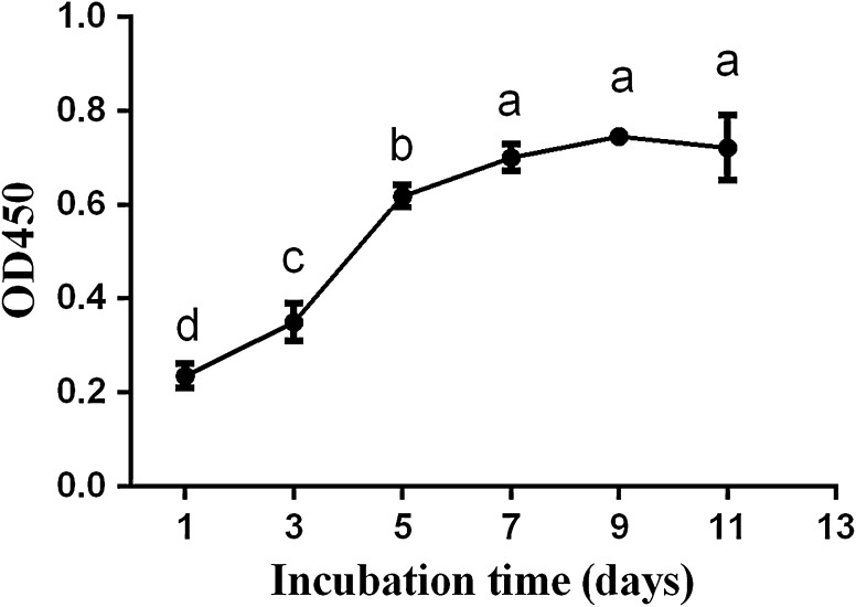
As shown in Fig. 2, growth curve of cells reflected that duck preadipocytes underwent a latency period before day 3. After that, cells entered into the logarithmic phase until day 7, during which activities of the cells increased fast and significantly (p < 0.05). After the cells were cultured for 7 days they entered into the plateau phase, during which the activities remained stable. Finally, the cell activities began to decrease after day 9.
Fig. 2.
Growth curve of duck primary preadipocytes cultured in vitro. Different letters on every other day represent significant differences (p < 0.05)
Dynamic observation and characterization of duck preadipocyte differentiation
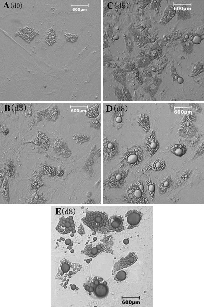
Changes in cell morphology and cytoplasmic lipid droplets were observed and photographed under the microscope during cell differentiation. Two days after cell adhesion, a few small lipid droplets would be seen in the medium [Fig. 3a(d0)]. Three days after the addition of the inducer, most cells got an oval morphology with more lipid droplets filling in the center of the cells [Fig. 3b(d3)]. Then cells were cultured with medium containing insulin only for 2 days, more lipid droplets accumulated in the cells that became bigger than before [Fig. 3c(d5)]. When cells differentiated to day 8, these little lipid droplets became huge lipid droplets filling in the cell, of which the nucleus was located at one side instead of the center [Fig. 3d(d8)]. At that time the cells could be stained red by oil red O stain [Fig. 3e(d8)]. These mature adipocytes would grow bigger if they were cultured longer, however, at this stage cells might fall off from the dishes and float in the medium easily.
Fig. 3.
Morphologic characteristics of duck preadipocytes during differentiation in vitro under microscope (×100). a Morphology of the differentiating cells at day 0. b Morphology of the differentiating cells at day 3. c Morphology of differentiating cells at day 5. d Morphology of the differentiating cells at day 8. e Oil red O staining of differentiating cells at day 8
Expression profiles of three maker genes during duck preadipocyte differentiation
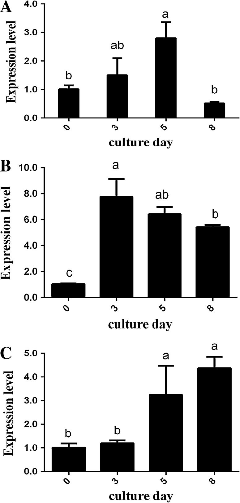
The expression profiles of the three maker genes during duck preadipocyte differentiation are shown in Fig. 4. Obviously, all of these marker genes, including C/EBPα, PPARγ, and FABP4, were expressed in the cells during preadipocyte differentiation, with different expression patterns. Both C/EBPα and PPARγ showed an increased-decrease expression pattern during adipogenesis. In particular, expression of C/EBPα increased to the top point at day 5, which was significantly higher than that of other culture days (p < 0.05), while PPARγ reached the highest level at day 3, after which the expression decreased to lower levels (p < 0.05). In contrary, expression of FABP4 gradually increased up to day 8, peaking at the end of the whole differentiation stage (p < 0.05).
Fig. 4.
The relative mRNA expression of three maker genes for duck preadipocytes differentiation. a Expression pattern of C/EBPα mRNA; b expression pattern of PPARγ mRNA; and c expression pattern of FABP4 mRNA during the differentiation of duck preadipocytes. The mRNA expression level at day 0 was assigned as control. The different lowercase letters at the top of each bar indicate significant differences between the expression of the gene at different culture days (p < 0.05)
Discussion
To date, there is very little information about duck adipogenesis, which may be attributed to the fact that until now no culture system was available. Recently, He et al. began to research functions of genes related to adipogenesis in duck primary preadipocytes (He et al. 2012). However, the specific procedures of isolation and differentiation of duck preadipocytes have not yet been reported. In the present study, we successfully isolated and obtained preadipocytes by means of collagenase digestion on the adipose tissue of 7 days old healthy Peking duck. In the literature, there are some differences in the method of isolating primary preadipocytes and culturing among different species, such as how long the adipose tissue is exposed to collagenase, centrifugal conditions, seeding density, and so on. In humans, preadipocytes were cultured after collagenase digestion for 45 min followed by centrifugation at 1,000 rpm for 5 min (Collins et al. 2011). In chicken, preadipocytes were obtained by digesting for 60 min and centrifugating at 800×g for 10 min (Liu et al. 2009), while in goose centrifugal condition was 300×g for 10 min instead (Wang et al. 2011). In the current research, Peking duck preadipocytes, unlike Sheldrake ducks (He et al. 2012), could be isolated by collagenase digestion for 50 min and centrifugating at 300×g for 10 min with promising results.
Similar with situations in other species, duck preadipocytes also went through a series of developmental stages with changes in cell morphology (Cristancho and Lazar 2011). During the early growth phase, duck preadipocytes were similar to fibroblasts. However at 100 % confluency, treatment with a hormonal cocktail to induce differentiation leads to drastic changes in cell morphology. The preadipocytes converted to a spherical shape, accumulated lipid droplets, and progressively became mature white adipocytes. Situations were the same with the passaged cells, which shared similar morphologies with primary preadipocytes. In the current research, we found that some preadipocytes would change their shapes after being passaged 4 times and had diminished ability to adhere to the culture dishes. Therefore, we recommend that research using preadipocytes be conducted before the fourth passage.
In the past decades, a variety of differentiation protocols have been developed for preadipocytes among different species. In most cases, primary preadipocytes or cell lines can differentiate into mature adipocytes in the presence of a hormonal cocktail consisting of insulin, IBMX, and DEX (Arsenijevic et al. 2012). In particular, it is reported that human preadipocyte differentiation need insulin, DEX, IBMX, thyroid hormone, and troglitazone (Collins et al. 2011), while in goat and pig, insulin, DEX, IBMX, and troglitazone are regular inducers for preadipocyte differentiation (Liu et al. 2012; Ren et al. 2012). It should be noted that this cocktail is modified in poultry because the addition of oleic acid is necessary to induce adipogenesis (Matsubara et al. 2008; Wang et al. 2011). We conducted a series of experiments to optimize the induction method of duck preadipocytes and here we report that although the cells might accumulate lipid droplets and differentiate into mature adipocytes using a hormonal cocktail lacking oleic acid (data not shown), a higher differentiating efficiency would be obtained when oleic acid was present in the medium. In the presence of the hormonal cocktail supplemented with oleic acid, more than 90 % of the preadipocytes differentiated into mature adipocytes as shown by oil red O staining [Fig. 3e(d8)]. Our results support previous finding in chicken (Liu et al. 2009) and goose (Wang et al. 2011), and suggest that exogenous oleic acid plays a critical role in promoting preadipocyte differentiation in poultry.
Earlier research has identified some marker genes that are essential for preadipocyte differentiation, including FABP4, lipoprotein lipase (LPL), PPARγ, C/EBPα etc. Matsubara et al. found that FABP4, PPARγ, and C/EBPα are highly expressed in chicken preadipocytes during adipogenesis at 9, 24, and 9 h post differentiation, respectively. This demonstrates that in poultry PPARγ may be a key regulator in the early stages of adipogenesis (Matsubara et al. 2005). In our study, the developmental expression profiles of PPARγ, C/EBPα, and FABP4 were also detected in duck preadipocytes at different stages. During adipogenesis, PPARγ was highly expressed at day 3, when the preadipocytes began to accumulate lipid, demonstrating that it is highly likely that PPARγ plays a critical role in regulating the early stages of duck adipogenesis (Mueller 2014). Additionally, it is reported that C/EBPα followed PPARγ as a master regulator of preadipocyte differentiation, which was positively regulated by PPARγ during adipogenesis (Tamori et al. 2002; Siersbæk et al. 2010). In our study, expression level of C/EBPα peaked at day 5, which was later than that of PPARγ. These findings indicate that duck C/EBPα might function in the same manner as in other species, in which C/EBPα could be promoted by PPARγ, and they may cooperatively stimulate preadipocyte differentiation (Matsubara et al. 2008). Moreover, research has demonstrated that FABP4 might play an important role in preadipocyte differentiation (Shi et al. 2010; He et al. 2012) and was recognized as a middle to late marker of preadipocyte differentiation (MacDougald and Lane 1995). Currently, we found that FABP4 was highly expressed on day 8, thus we speculate that FABP4 may affect duck preadipocyte differentiation and can be used as a late marker. Taken together, the results above suggest that during duck preadipocyte differentiation PPARγ, C/EBPα, and FABP4 affect adipogenesis at different phases.
Conclusions
We report for the first time a procedure for isolating and differentiating Peking duck preadipocytes. After collection of preadipocytes from adipose tissue, it takes 3–4 days for the cells to reach full confluency and 8 additional days for them to differentiate into mature adipocytes. To induce differentiation, a cocktail consisting of DEX, insulin, IBMX, and oleic acid is added to the medium. In addition, PPARγ, C/EBPα, and FABP4 exhibit different expression patterns during duck preadipocyte differentiation. Therefore, these genes can be used as markers to identify the different stages of preadipocyte differentiation.
Acknowledgments
This work was supported by the earmarked fund for China Agriculture Research Service (No. CARS-43-6).
Abbreviations
- CCK-8
Cell counting kit-8
- CEBP/α
CCAAT/enhancer binding protein-α
- DEX
Dexamethasone
- DNL
De novo lipogenesis
- FABP4
Adipocyte fatty acid-binding protein
- FBS
Fetal bovine serum
- IBMX
3-Isobutyl-1-methylxanthine
- LPL
Lipoprotein lipase
- MSCs
Mesenchymal stem cells
- PPARγ
Peroxisome proliferator-activated receptor-γ
- SD
Standard deviation
- TG
Triacylglycerol
References
- Arsenijevic T, Gregoire F, Delforge V, Delporte C, Perret J. Murine 3T3-L1 adipocyte cell differentiation model: validated reference genes for qPCR gene expression analysis. PLoS One. 2012;7:e37517. doi: 10.1371/journal.pone.0037517. [DOI] [PMC free article] [PubMed] [Google Scholar]
- Attia Y, Qota E, Zeweil H, Bovera F, Abd Al-Hamid A, Sahledom M. Effect of different dietary concentrations of inorganic and organic copper on growth performance and lipid metabolism of White Pekin male ducks. Br Poult Sci. 2012;53:77–88. doi: 10.1080/00071668.2011.650151. [DOI] [PubMed] [Google Scholar]
- Collins JM, Neville MJ, Pinnick KE, Hodson L, Ruyter B, van Dijk TH, Reijngoud DJ, Fielding MD, Frayn KN. De novo lipogenesis in the differentiating human adipocyte can provide all fatty acids necessary for maturation. J Lipid Res. 2011;52:1683–1692. doi: 10.1194/jlr.M012195. [DOI] [PMC free article] [PubMed] [Google Scholar]
- Covas DT, Panepucci RA, Fontes AM, Silva WA, Jr, Orellana MD, Freitas MC, Neder L, Santos AR, Peres LC, Jamur MC. Multipotent mesenchymal stromal cells obtained from diverse human tissues share functional properties and gene-expression profile with CD146+ perivascular cells and fibroblasts. Exp Hematol. 2008;36:642–654. doi: 10.1016/j.exphem.2007.12.015. [DOI] [PubMed] [Google Scholar]
- Cristancho AG, Lazar MA. Forming functional fat: a growing understanding of adipocyte differentiation. Nat Rev Mol Cell Biol. 2011;12:722–734. doi: 10.1038/nrm3198. [DOI] [PMC free article] [PubMed] [Google Scholar]
- Ding F, Pan ZX, Kou J, Li L, Xia L, Hu QS, Liu HH, Wang JW. De novo lipogenesis in the liver and adipose tissues of ducks during early growth stages after hatching. Comp Biochem Physiol B Biochem Mol Biol. 2012;163:154–160. doi: 10.1016/j.cbpb.2012.05.014. [DOI] [PubMed] [Google Scholar]
- Farmer SR. Transcriptional control of adipocyte formation. Cell Metab. 2006;4:263–273. doi: 10.1016/j.cmet.2006.07.001. [DOI] [PMC free article] [PubMed] [Google Scholar]
- Freytag SO, Paielli DL, Gilbert JD. Ectopic expression of the CCAAT/enhancer-binding protein alpha promotes the adipogenic program in a variety of mouse fibroblastic cells. Gene Dev. 1994;8:1654–1663. doi: 10.1101/gad.8.14.1654. [DOI] [PubMed] [Google Scholar]
- He J, Tian Y, Li JJ, Shen JD, Tao ZR, Fu Y, Niu D, Lu LZ. Expression pattern of adipocyte fatty acid-binding protein gene in different tissues and its regulation of genes related to adipocyte differentiation in duck. Poult Sci. 2012;91:2270–2274. doi: 10.3382/ps.2012-02149. [DOI] [PubMed] [Google Scholar]
- Karbiener M, Fischer C, Nowitsch S, Opriessnig P, Papak C, Ailhaud G, Dani C, Amri EZ, Scheideler M. microRNA miR-27b impairs human adipocyte differentiation and targets PPARgamma. Biochem Biophys Res Commun. 2009;390:247–251. doi: 10.1016/j.bbrc.2009.09.098. [DOI] [PubMed] [Google Scholar]
- Kershaw EE, Flier JS. Adipose tissue as an endocrine organ. J Clin Endocr Metab. 2004;89:2548–2556. doi: 10.1210/jc.2004-0395. [DOI] [PubMed] [Google Scholar]
- Kou J, Wang WX, Liu HH, Pan ZX, He T, Hu JW, Li L, Wang JW. Comparison and characteristics of the formation of different adipose tissues in ducks during early growth. Poult Sci. 2012;91:2588–2597. doi: 10.3382/ps.2012-02273. [DOI] [PubMed] [Google Scholar]
- Lin CS, Xin ZC, Deng CH, Ning H, Lin G, Lue TF. Defining adipose tissue-derived stem cells in tissue and in culture. Histol Histopathol. 2010;25:807–815. doi: 10.14670/HH-25.807. [DOI] [PubMed] [Google Scholar]
- Liu S, Wang L, Wang N, Wang Y, Shi H, Li H. Oleate induces transdifferentiation of chicken fibroblasts into adipocyte-like cells. Comp Biochem Phys A Mol Integr Physiol. 2009;154:135–141. doi: 10.1016/j.cbpa.2009.05.011. [DOI] [PubMed] [Google Scholar]
- Liu S, Wang Y, Wang L, Wang N, Li Y, Li H. Transdifferentiation of fibroblasts into adipocyte-like cells by chicken adipogenic transcription factors. Comp Biochem Phys A Mol Integr Physiol. 2010;156:502–508. doi: 10.1016/j.cbpa.2010.04.003. [DOI] [PubMed] [Google Scholar]
- Liu HF, Gui MX, Dong H, Wang X, Li XW. Differential expression of AdipoR1, IGFBP3, PPARgamma and correlative genes during porcine preadipocyte differentiation. In Vitro Cell Dev Biol Anim. 2012;48:54–60. doi: 10.1007/s11626-011-9468-6. [DOI] [PubMed] [Google Scholar]
- Livak KJ, Schmittgen TD. Analysis of relative gene expression data using real-time quantitative PCR and the 2(−Delta Delta C(T)) Method. Methods. 2001;25:402–408. doi: 10.1006/meth.2001.1262. [DOI] [PubMed] [Google Scholar]
- MacDougald OA, Lane MD. Transcriptional regulation of gene expression during adipocyte differentiation. Annu Rev Biochem. 1995;64:345–373. doi: 10.1146/annurev.bi.64.070195.002021. [DOI] [PubMed] [Google Scholar]
- Matsubara Y, Sato K, Ishii H, Akiba Y. Changes in mRNA expression of regulatory factors involved in adipocyte differentiation during fatty acid induced adipogenesis in chicken. Comp Biochem Physiol A Mol Integr Physiol. 2005;141:108–115. doi: 10.1016/j.cbpb.2005.04.013. [DOI] [PubMed] [Google Scholar]
- Matsubara Y, Endo T, Kano K. Fatty acids but not dexamethasone are essential inducers for chick adipocyte differentiation in vitro. Comp Biochem Phys A Mol Integr Physiol. 2008;151:511–518. doi: 10.1016/j.cbpa.2008.07.002. [DOI] [PubMed] [Google Scholar]
- Mueller E (2014) Understanding the variegation of fat: novel regulators of adipocyte differentiation and fat tissue biology. Biochim Biophys Acta 1842:352–357 [DOI] [PubMed]
- Ren Y, Wu H, Zhou X, Wen J, Jin M, Cang M, Guo X, Wang Q, Liu D, Ma Y. Isolation, expansion, and differentiation of goat adipose-derived stem cells. Res Vet Sci. 2012;93:404–411. doi: 10.1016/j.rvsc.2011.08.014. [DOI] [PubMed] [Google Scholar]
- Rosen ED, MacDougald OA. Adipocyte differentiation from the inside out. Nat Rev Mol Cell Biol. 2006;7:885–896. doi: 10.1038/nrm2066. [DOI] [PubMed] [Google Scholar]
- Shi H, Wang Q, Wang Y, Leng L, Zhang Q, Shang Z, Li H. Adipocyte fatty acid-binding protein: an important gene related to lipid metabolism in chicken adipocytes. Comp Biochem Physiol B: Biochem Mol Biol. 2010;157:357–363. doi: 10.1016/j.cbpb.2010.08.005. [DOI] [PubMed] [Google Scholar]
- Shi H, Zhang Q, Wang Y, Yang P, Wang Q, Li H. Chicken adipocyte fatty acid-binding protein knockdown affects expression of peroxisome proliferator-activated receptor γ gene during oleate-induced adipocyte differentiation. Poult Sci. 2011;90:1037–1044. doi: 10.3382/ps.2010-01161. [DOI] [PubMed] [Google Scholar]
- Siersbæk R, Nielsen R, Mandrup S. PPARγ in adipocyte differentiation and metabolism—novel insights from genome-wide studies. FEBS Lett. 2010;584:3242–3249. doi: 10.1016/j.febslet.2010.06.010. [DOI] [PubMed] [Google Scholar]
- Tamori Y, Masugi J, Nishino N, Kasuga M. Role of peroxisome proliferator-activated receptor-gamma in maintenance of the characteristics of mature 3T3-L1 adipocytes. Diabetes. 2002;51:2045–2055. doi: 10.2337/diabetes.51.7.2045. [DOI] [PubMed] [Google Scholar]
- Tontonoz P, Hu E, Spiegelman BM. Stimulation of adipogenesis in fibroblasts by PPAR gamma 2, a lipid-activated transcription factor. Cell. 1994;79:1147–1156. doi: 10.1016/0092-8674(94)90006-X. [DOI] [PubMed] [Google Scholar]
- Wang F, Lu L, Yuan H, Tian Y, Li J, Shen J, Tao Z, Fu Y. Molecular cloning, expression, and regulation of goose leptin receptor gene in adipocytes. Mol Cell Biochem. 2011;353:267–274. doi: 10.1007/s11010-011-0795-4. [DOI] [PubMed] [Google Scholar]
- Zhang JW, Klemm DJ, Vinson C, Lane MD. Role of CREB in transcriptional regulation of CCAAT/enhancer-binding protein beta gene during adipogenesis. J Biol Chem. 2004;279:4471–4478. doi: 10.1074/jbc.M311327200. [DOI] [PubMed] [Google Scholar]
- Zhao CF, Liu Y, Que HP, Yang SG, Liu ZQ, Weng XC, Hui HD, Liu SJ. SCIRR39 promotes differentiation of oligodendrocyte precursor cells and regulates expression of myelin-associated inhibitory factors. J Mol Neurosci. 2013;50:533–541. doi: 10.1007/s12031-013-9983-x. [DOI] [PubMed] [Google Scholar]


