

Brown adipose tissue (BAT) is present in human adults and when present visualised with [18F]-FDG PET. It contains large amounts of mitochondriae, which gives a higher water content compared to white adipose tissue and a high FDG avidity when activated [1].
Five adults underwent PET/MR. BAT was cold-activated using a water-perfused vest, with injection of 100Mbq FDG after 60 minutes [2]. Hereafter cervical regions were scanned on an Siemens Biograph mMR 3T. PET was performed as single bed 10 minutes acquisition.
MRI included DIXON sequence for attenuation correction and axial T2 weighted sequences with and without water suppression, the latter for calculation of water percentage (W%).
ROIs were drawn on T2 images omitting vessels and muscle and propagated to PET images and to calculated water percentage maps (Figure 1). Subcutaneous fat regions were used as reference (SC).
Figure 1.
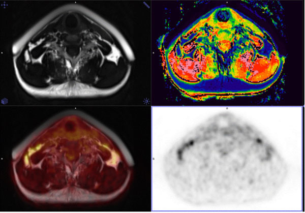
T2, water % , T2/PET and PET images.
FDG-PET identified activated BAT in 3 of 5 subjects. On each scan eight ROIs were drawn, including one reference region (SC). SUVmean for BAT, nBAT and SC were 1.60 (SD 0.26), 0.38 ( 0.12) and 0.64 (0.12) respectively (Figure 2). Calculated water percentage MRI maps for these regions were 19.6% (SD2.9%), 16.5% (4.2%) and 16.8% (1.6%) respectively (Figure 3). No significant difference in W% was found between BAT and nBAT.
Figure 2.
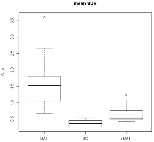
Box plot SUV value BAT, SC, nBAT
Figure 3.
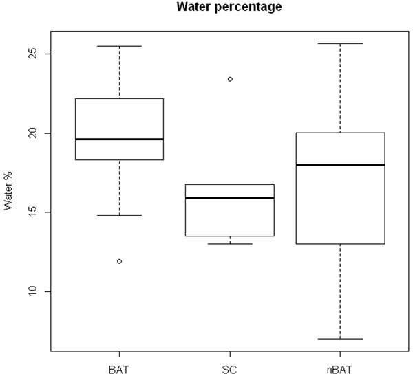
Box plot W% BAT, SC, nBAT
No significant correlation was found between water percentage in fat tissue with or without BAT, maybe because the two modalities measures different physiological properties. Accordingly, BAT can be present but not activated and therefore measurements with both modalities seems necessary for fully characterization of BAT.
References
- 1.Cypess AM, Lehman S, et al. Identification and importance of brown adipose tissue in adult humans. N Engl J Med. 2009;360(15):1509–17. doi: 10.1056/NEJMoa0810780. [DOI] [PMC free article] [PubMed] [Google Scholar]
- 2.van Rooijen BD, van der Lans AA, et al. Imaging cold-activated brown adipose tissue using dynamic T2*-weighted magnetic resonance imaging and 2-deoxy-2-[18F]fluoro-D-glucose positron emission tomography. Invest Radiol. 2013;48(10):708–14. doi: 10.1097/RLI.0b013e31829363b8. [DOI] [PubMed] [Google Scholar]
- 3.Chen YC, Cypess AM, et al. Measurement of human brown adipose tissue volume and activity using anatomic MR imaging and functional MR imaging. J Nucl Med. 2013;54(9):1584–7. doi: 10.2967/jnumed.112.117275. [DOI] [PMC free article] [PubMed] [Google Scholar]