Summary
Numerous thiol-reactive compounds cause oxidative stress where cells counteract by activation of survival strategies regulated by thiol-based sensors. In Streptomyces coelicolor, a model actinomycete, a sigma/antisigma pair SigR/RsrA controls the response to thiol-oxidative stress. To unravel its full physiological functions, chromatin immuno-precipitation combined with sequence and transcript analyses were employed to identify 108 SigR target genes in S. coelicolor and to predict orthologous regulons across actinomycetes. In addition to reported genes for thiol homeostasis, protein degradation and ribosome modulation, 64 additional operons were identified suggesting new functions of this global regulator. We demonstrate that SigR maintains the level and activity of the housekeeping sigma factor HrdB during thiol-oxidative stress, a novel strategy for stress responses. We also found that SigR defends cells against UV and thiol-reactive damages, in which repair UvrA takes a part. Using a refined SigR-binding sequence model, SigR orthologues and their targets were predicted in 42 actinomycetes. This revealed a conserved core set of SigR targets to function for thiol homeostasis, protein quality control, possible modulation of transcription and translation, flavin-mediated redox reactions, and Fe-S delivery. The composition of the SigR regulon reveals a robust conserved physiological mechanism to deal with thiol-oxidative stress from bacteria to human.
Introduction
Cells are exposed to various forms of oxidative stressors such as reactive oxygen species (ROS), natural or xenobiotic redox-active compounds, or some antibiotics, which elicit the formation of ROS inside the cell. The response mechanism involves various enzymes to remove such oxidants, systems to repair and recycle damaged cell components, and to maintain optimal cell physiology (Imlay, 2008; Zuber, 2009). One form of oxidative damage that frequently occurs in proteins and small molecules is the oxidation of cysteine thiols by ROS (Kiley and Storz, 2004; Jacob et al., 2006). Cysteine thiols can also be modified by reactive nitrogen species (RNS) and thiol-reactive electrophiles (Hess et al., 2005; Rudolph and Freeman, 2009)
The presence of ROS, RNS and other thiol-reactive compounds can be sensed directly through thiol-based sensor-regulators, which modulate the expression of genes encoding functions that constitute the biological stress response (Antelmann and Helmann, 2011). The best studied examples of thiol-based sensor-regulators that respond to ROS in bacteria include H2O2-sensing OxyR in Escherichia coli, organic peroxide-sensing OhrR in Bacillus subtilis, and the anti-sigma factor RsrA that senses thiol oxidation and modulates regulator SigR activity in Streptomyces coelicolor (Paget and Buttner, 2003; D’Autreaux and Toledano, 2007; Antelmann and Helmann, 2011). In eukaryotes, thiol-based redox switches have been well exemplified in Yap1 of Saccharomyces cerevisiae and the mammalian Nrf2/Keap1 system (D’Autreaux and Toledano, 2007; Kensler et al., 2007; Brandes et al., 2009). Representative target genes of these thiol-based regulators are those that encode thiol homeostasis system such as thioredoxin (Trx), glutaredoxin (Grx) and small molecular thiol systems (regulated by OxyR, SigR/RsrA, Yap1, Nrf2/Keap1), catalases and peroxidases (by OxyR and Yap1), organic hydroperoxidase (by OhrR), detoxification of electrophiles (by SigR and Nrf2), and some proteolytic system (by OxyR, SigR, Yap1, Nrf2) (Kensler et al., 2007; den Hengst and Buttner, 2008; Imlay, 2008; Antelmann and Helmann, 2011).
A zinc-containing anti-sigma (ZAS) factor RsrA that binds an ECF (group 4) sigma factor SigR in S. coelicolor responds to diamide-induced thiol oxidation by forming disulphide bonds, releasing SigR to transcribe its target genes (Kang et al., 1999; Li et al., 2003b; Bae et al., 2004). In addition to thiol-oxidants, the presence of non-oxidative thiol-reactive compounds also induces expression of the SigR regulon, suggesting that RsrA may respond to these compounds either directly through thiolation of reactive cysteines or indirectly through changes in reduced thiol pools such as mycothiol, the functional equivalent in actinomycetes of glutathione (Newton et al., 2008; Park and Roe, 2008). To date, around 44 SigR target genes have been identified based on promoter sequence similarity, S1 mapping and microarray expression profiling (Paget et al., 1998; 2001; Park and Roe, 2008; Kallifidas et al., 2010). The products of known SigR target genes include thiol-redox proteins such as thioredoxin systems (TrxBA, TrxC), the first enzyme in mycothiol synthesis (MshA), and a glutaredoxin-like protein called mycoredoxin (Mrx). They also include proteolytic components of protein quality control (PepN, SsrA, ClpP1P2, ClpX, ClpC, Lon), cysteine production (CysM), methionine reduction (MsrA, MsrB), guanine synthesis (GuaB), ribosome-associated function (RpmE, RelA), and electrophile detoxification (Mca) (Paget et al., 2001; Park and Roe, 2008; Kallifidas et al., 2010). Therefore, perturbation of intracellular thiols, as sensed through RsrA, induces expression of gene products that contributes to protein quality control, detoxification of thiol-conjugative xenobiotics, and modulation of transcription and translation.
Because SigR/RsrA activity controls the cellular response to a variety of stressors, we expect the SigR regulon to be large and encompass additional stress response functions in S. coelicolor. Therefore, we performed chromatin immuno-precipitation on a chip (ChIP-chip) assays and discovered > 100 direct SigR target promoters transcribing > 160 genes. The comprehensive characterization of the S. coelicolor SigR regulon helped us understand in more details the global effects of and cellular response to thiol-oxidative stress. Furthermore, to identify functions that are most critical to survive thiol-oxidative stress response, we used the SigR-binding consensus sequence to predict orthologous thiol-oxidative stress regulons in other actinomycetes. The analysis revealed a core conserved regulon as well as lineage-specific adaptations.
Results and discussion
Identification of SigR target genes by chromatin immuno-precipitation on a chip (ChIP-chip) and sequence analysis
To identify SigR target sites in the S. coelicolor genome we performed a ChIP-chip experiment using anti-SigR serum with wild-type cell cultures treated with diamide to activate SigR activity. We also performed the same ChIP-chip experiment with a ΔsigR mutant to subtract non-specific chromatin enrichment because S. coelicolor possesses many additional sigma factors that may cross-react with the anti-SigR serum. After subtracting non-specific signal, we identified 122 regions significantly enriched by the immuno-precipitation of SigR (P-value ≤ 0.05). Next, to identify more precisely putative SigR promoters, we searched within the enriched genomic regions for putative SigR promoters by detecting sequences that differed from the previously proposed SigR promoter (GGAAT-N18-GTT; (Paget et al., 2001) by at most 2 nucleotides with either of 2 possible spacer lengths (18 and 19 nucleotides) on either DNA strand. Only 5 of the 122 enriched regions did not contain at least one sequence matching our criteria, increasing the confidence that the regions enriched by the ChIP-chip assay are indeed SigR binding sites. Overall, 176 putative SigR promoter sequences were detected, and 108 of them are oriented towards annotated genes within 500 nt from the start codon.
In Table 1, we summarized genes predicted to be transcribed from these 108 promoters. Detailed information about the position of SigR-bound regions (enriched ChIP peaks), position and sequence of the predicted SigR promoters is presented in Table S1. Considering operon structures (http://scocyc.streptomyces.org.uk), more than 163 genes are predicted to be under the direct control of SigR. Among these, 44 promoters have been previously reported to produce SigR-dependent transcripts by S1 mapping experiments (Paget et al., 2001; Park and Roe, 2008; Kallifidas et al., 2010) or microarray studies (Kallifidas et al., 2010). Re-examination of the transcriptional microarray data (Kallifidas et al., 2010), using slightly different criteria (see Experimental procedures), predicts SigR-dependent transcripts from a total of 62 promoters within 57 of the regions bound by SigR in vivo. We marked these promoters in Table S1. Therefore, our ChIP-chip analysis revealed between 46 and 64 new SigR target promoters, undetectable from transcriptome profiling alone. The lack of detection in microarray analysis alone could be due partly to low level of gene expression and/or additional transcription from one or more SigR-independent promoter.
Table 1.
The SigR regulon genes.
| Ga | Numberb | Name | Description | Confirmc |
|---|---|---|---|---|
| 3 | SCO0569 | rpmJ | 50S ribosomal protein L36 | |
| 3 | SCO0570* | rpmG3 | 50S ribosomal protein L33 (C-type) | (S1) |
| 8 | SCO0882* | Hypothetical protein, possible dithiol-disulphide isomerase | (S1) | |
| 8 | SCO0884* | Probable oxidoreductase, probable pyridine nucleotide-disulphide oxidoreductases | ||
| 1 | SCO0885* | trxC | Thioredoxin | (S1, A) |
| 8 | SCO0917* | Possible oxygenase | ||
| 7 | SCO0973* | Probable integral membrane protein | ||
| 1 | SCO1084* | trxA4 | Putative thioredoxin | S1 |
| 12 | SCO1085* | Probable phospholipid/glycerol acyltransferase, divergent from trxA4 (1084) | ||
| 8 | SCO1142* | Possible oxidoreductase, probable FAD-dependent pyridine nucleotide-disulphide oxidoreductase | ||
| 5 | SCO1238* | Putative ATP-dependent Clp protease subunit | ||
| 6 | SCO1425* | Possible AsnC-family transcriptional regulatory protein | ||
| 14 | SCO1426* | Hypothetical protein | ||
| 3 | SCO1513* | relA | GTP pyrophosphokinase for ppGpp synthesis | (S1) |
| 3 | SCO1598 | rplT | 50S ribosomal protein L20 | |
| 3 | SCO1599 | rpmI | 50S ribosomal protein L35 | |
| 3 | SCO1600* | infC | Translation initiation factor IF-3 | S1 |
| 6 | SCO1618 | Hypothetical protein, similar to cobalamin binding protein BtuF | ||
| 6 | SCO1619* | Possible transcriptional regulatory protein of HTH-XRE family | ||
| 5 | SCO1643 | prcA | 20S proteasome alpha-subunit | |
| 5 | SCO1644 | prcB | 20S proteasome beta-subunit | |
| 5 | SCO1645 | Hypothetical protein | ||
| 5 | SCO1646 | pup | Prokaryotic ubiquitin-like protein | |
| 5 | SCO1647 | pafD | Proteasome accessory factor | |
| 5 | SCO1648* | mpa | Probable AAA ATPase similar to mycobacterium proteasome-associated ATPase | S1 |
| 3 | SCO1758* | engA | GTP-binding protein with multiple functions | S1 |
| 8 | SCO1869* | Conserved hypothetical, probable protein dithiol-disulphide isomerase | (S1, A) | |
| 11 | SCO1919 | Probable metal-sulphur cluster biosynthetic enzyme | (A) | |
| 11 | SCO1920* | sufU | Similar to NifU in SUF-type Fe-S assembly gene cluster (1926-1919) | (A), S1 |
| 9 | SCO1936* | tal2 | Probable transaldolase, a component of non-oxidative pentose phosphate pathway | S1 |
| 9 | SCO1937 | zwf2 | Glucose 6-phosphate 1-dehydrogenase | |
| 9 | SCO1938 | Hypothetical protein | ||
| 9 | SCO1939 | pgl | 6-phosphogluconolactonase | |
| 10 | SCO1958* | uvrA | ABC excision nuclease subunit A | (A), S1 |
| 14 | SCO1995* | Unknown | (S1, A) | |
| 11 | SCO1996 | coaE | Dephospho-CoA kinase | (A) |
| 11 | SCO1997* | Unknown | (S1, A) | |
| 7 | SCO2124* | Possible hydrophobic membrane spanning regions | ||
| 7 | SCO2154* | Integral membrane protein | ||
| 11 | SCO2161* | sufA | HesB/YadR/Yfh family protein, probable SufA | (S1), S1 |
| 11 | SCO2162* | nadA | Quinolinate synthetase A subunit for nicotinamide synthesis, divergent from sufA (2161). 4Fe-4S enzyme | |
| 11 | SCO2194* | lipA | Putative lipoyl synthase | S1 |
| 7 | SCO2254* | Possible transmembrane efflux protein of MFS(major facilitator superfamily) | S1 | |
| 7 | SCO2310* | Possible transmembrane efflux protein similar to TcmA | ||
| 6 | SCO2331* | Possible MarR family transcriptional regulator | ||
| 6 | SCO2481* | Hypothetical protein, putative transcriptional regulator | ||
| 6 | SCO2537* | Possible DNA-binding protein | (A), S1 | |
| 6 | SCO2538 | Hypothetical protein | ||
| 6 | SCO2539 | era | GTP-binding protein era | |
| 3 | SCO2595* | obgE | GTPase, Obg subfamily, ObgE | S1 |
| 5 | SCO2617 | clpX | ATP-dependent Clp protease ATP binding subunit | |
| 5 | SCO2618 | clpP2 | ATP-dependent Clp protease proteolytic subunit 2 | (A) |
| 5 | SCO2619* | clpP1 | ATP dependent Clp protease proteolytic subunit 1 | (S1, A) |
| 8 | SCO2634* | Hypothetical protein, contains DsbA-like thioredoxin domain | (S1, A) | |
| 5 | SCO2635* | Probable aminopeptidase | ||
| 14 | SCO2642* | Unknown | ||
| 5 | SCO2643* | pepN | Aminopeptidase N | (S1, A) |
| 7 | SCO2763* | ABC transporter protein, ATP-binding component | S1 | |
| 8 | SCO2816* | Conserved hypothetical protein, flavin utilizing monooxygenase superfamily | ||
| 8 | SCO2849* | Conserved hypothetical protein, flavin utilizing monooxygenase superfamily | (S1, A) | |
| 2 | SCO2910 | cysM | Cysteine synthase | (A) |
| 2 | SCO2911* | moaD | Putative MoaD-family protein with ubiquitin fold | (A) |
| 3 | SCOs02* | ssrA | Transfer messenger RNA (tmRNA) | (S1) |
| 7 | SCO3083* | Possible integral membrane protein | (S1, A) | |
| 12 | SCO3091* | cfa | Cyclopropane-fatty-acyl-phospholipid synthase, SAM-dependent methylase | (A), S1 |
| 13 | SCO3162* | Possible esterase, β-lactamase superfamily | (S1, A) | |
| 14 | SCO3187* | Hypothetical protein | (S1) | |
| 7 | SCO3206* | Probable transmembrane efflux protein | (A) | |
| 6 | SCO3207* | TetR family transcriptional regulator | ||
| 8 | SCO3295 | Possible oxidoreductase | ||
| 8 | SCO3296* | Possible oxidoreductase, similar to F420-dependent alcoholdehydrogenase | S1 | |
| 5 | SCO3373* | clpC | Probable Clp-family ATP-binding protease similar to MecB/ClpC | (S1), S1 |
| 11 | SCO3403* | folE | GTP cyclohydrolase I for tetrahydrofolate biosynthesis | (S1) |
| 1 | SCO3442* | mrxB | Glutaredoxin-like protein, probable mycoredoxin | S1 |
| 6 | SCO3449 | rsrA2-1 | Putative ZAS (zinc-containing anti-sigma) factor | |
| 6 | SCO3450* | sigR2 | ECF (group 4) sigma factor close to SigR | |
| 6 | SCO3451* | rsrA2-1 | Putative ZAS (zinc-containing anti-sigma) factor | |
| 14 | SCO3509* | Hypothetical protein | ||
| 7 | SCO3764 | Integral membrane protein | (A) | |
| 7 | SCO3765* | Possible integral membrane protein with CBS domain | ||
| 14 | SCO3766* | Hypothetical protein | ||
| 14 | SCO3767* | Hypothetical protein, similar to tellurite resistance protein TerB | ||
| 1 | SCO3889 | trxA | Thioredoxin | (A) |
| 1 | SCO3890* | trxB | Thioredoxin reductase | (S1, A) |
| 14 | SCO4039* | Unknown | (A) | |
| 14 | SCO4040* | Unknown | ||
| 8 | SCO4109* | Possible oxidoreductase, probable aldo/keto reductases | ||
| 14 | SCO4203* | Hypothetical protein | ||
| 1 | SCO4204* | mshA | Glycosyltransferase for mycothiol synthesis | (S1, A) |
| 1 | SCO4205 | Conserved hypothetical protein | ||
| 8 | SCO4297* | Possible oxidoreductase with FMN binding site, old yellow enzyme family | (A) | |
| 8 | SCO4298 | Carboxylesterase | ||
| 8 | SCO4299 | Integral membrane protein | ||
| 11 | SCO4418 | pdxH2 | Pyridoxamine 5′-phosphate oxidase oxidase | |
| 11 | SCO4419* | pdxH3 | Conserved hypothetical, probable pyridoxamine 5′-phosphate oxidase for pyridoxal phosphate synthesis | |
| 14 | SCO4420* | Hypothetical protein | ||
| 13 | SCO4561* | NLP/P60 family protein | ||
| 4 | SCO4770* | guaB | Inosine 5′ monophosphate dehydrogenase | (S1, A) |
| 4 | SCO4771 | guaB1 | Inosine 5′ monophosphate dehydrogenase | |
| 10 | SCO4797* | uvrD | Probable ATP-dependent DNA helicase | S1 |
| 8 | SCO4833 | Phosphorylmutase | ||
| 8 | SCO4834 | Hypothetical protein | ||
| 8 | SCO4835* | Conserved hypothetical protein with alkylhydroperoxidase-like AhpD/CMD domain | ||
| 2 | SCO4956* | msrA | Probable peptide methionine S-sulphoxide reductase | (S1, A), S1 |
| 14 | SCO4966* | Possible membrane protein | ||
| 1 | SCO4967* | mca | Mycothiol-S-conjugate amidase | (S1, A) |
| 1 | SCO4968 | Hypothetical protein | (A) | |
| 9 | SCO5042* | fumC | Fumarate hydratase, class II family (iron-independent) | (A) |
| 6 | SCO5065* | Possible transcriptional regulator, LuxR family | ||
| 14 | SCO5163* | Unknown | (S1) | |
| 11 | SCO5178* | moeB | Possible sulphurylase related to molybdopterin synthesis | (S1, A) |
| 1 | SCO5187* | mrxA | Glutaredoxin-like protein, probable myc917oredoxin | (S1, A) |
| 10 | SCO5188* | uvrD2 | ATP-dependent DNA helicase (UvrD/REP type), divergent from mrxA (5187) | |
| 6 | SCO5216* | sigR | ECF (group 4) sigma factor sR | (S1, A) |
| 6 | SCO5217 | rsrA | ZAS (zinc-containing antisigma) factor for sR | (A) |
| 14 | SCO5284* | Hypothetical protein | ||
| 5 | SCO5285* | lon | ATP-dependent protease | (S1) |
| 6 | SCO5357* | rho | Transcription termination factor, an RNA–DNA helicase | |
| 3 | SCO5359* | rpmE1 | Ribosomal proteins L31 | (S1, A) |
| 3 | SCO5360 | prfA | Peptide release factor A | |
| 3 | SCO5361 | hemK | Probable release factor-specific methyltransferase | |
| 8 | SCO5465* | Conserved hypothetical, probable F420-dependent NADP oxidoreductase | (S1, A) | |
| 14 | SCO5490* | Hypothetical protein | ||
| 14 | SCO5545* | hypothetical protein | (A) | |
| 6 | SCO5552* | ndgR | IclR family transcription regulator | |
| 3 | SCO5705* | Hypothetical protein, first gene in translation-related gene cluster | S1 | |
| 3 | SCO5706 | infB | Translation initiation factor IF-2 | |
| 3 | SCO5707 | Hypothetical protein | ||
| 3 | SCO5708 | rbfA | Ribosome binding factor | |
| 3 | SCO5709 | truB | Probable tRNA pseudouridine synthase | |
| 10 | SCO5754* | cinA | Competence-DNA damage-induced protein, neighbouring clgR (5755) | (S1, A) |
| 3 | SCO5796* | hflX | Conserved ribosome binding GTPase | (S1, A) |
| 6 | SCO5820* | hrdB | Major vegetative sigma factor | S1 |
| 14 | SCO5864* | Hypothetical protein | ||
| 14 | SCO5865 | Hypothetical protein | ||
| 2 | SCO6061* | msrB | Probable peptide methionine R-sulphoxide reductase | (S1, A), S1 |
| 14 | SCO6126* | Hypothetical protein | ||
| 13 | SCO6127* | Probable carboxylesterase, probable phenylcarbamate hydrolase | ||
| 11 | SCO6423* | Putative lipoate-protein ligase | (A) | |
| 8 | SCO6551* | Probable oxidoreductase, aldo/keto reductase family | (S1, A) | |
| 12 | SCO6759* | phyA/hopE | Probable phytoene (hopanoid) synthase | (S1, A), S1 |
| 12 | SCO6760 | phyB/hopD | Squalene/phytoene synthase | |
| 12 | SCO6761 | Small (52 aa) hypothetical protein of unknown function | ||
| 12 | SCO6762 | phyC/hopC | Squalene/phytoene dehydrogenase | |
| 12 | SCO6763 | phyD/hopB | Polyprenyl diphosphate synthase | |
| 12 | SCO6764 | phyE/hopA | Squalene–hopene cyclase | |
| 12 | SCO6765 | Lipoprotein | ||
| 12 | SCO6766 | Hypothetical protein | ||
| 12 | SCO6767 | ispG | 4-hydroxy-3-methyl-2-en-1-yl diphosphate synthase | |
| 12 | SCO6768 | 1-deoxy-d-xylulose-5-phospate synthase | ||
| 12 | SCO6769 | Aminotransferase | ||
| 12 | SCO6770 | DNA-binding protein | ||
| 12 | SCO6771 | Small hydrophobic protein | ||
| 6 | SCO6775* | Probable transcriptional regulator, TetR family | ||
| 14 | SCO6776* | Unknown | ||
| 6 | SCO7140* | Probable DNA-binding protein | ||
| 14 | SCO7631* | Possible secreted protein | ||
| 1 | SCO7632* | Probable amidase similar to mshB and mca | (A), S1 | |
| 8 | SCO7784* | Possible oxidoreductase, probable nitro/flavin reductase | S1 | |
| 8 | SCO7785 | Transcriptional regulator |
Genes were grouped according to predicted functions such as thiol homeostasis (1), sulphur metabolism (2), modulation of ribosome and/or translation (3), guanine nucleotide metabolism (4), protein degradation (5), transcriptional regulators or DNA binding proteins (6), transporter or integral membrane proteins (7), oxidoreductases (8), energy metabolism (9), DNA damage repair and/or recombination (10), cofactor metabolism (11), lipid metabolism (12), others (13), or function unpredictable (14).
Genes with SigR-binding promoters were marked with asterisks (*).
The genes whose SigR-dependent expression was previously confirmed by S1 or microarray (A) were marked with S1 and A in parenthesis (Paget et al., 2001; Park and Roe, 2008; Kallifidas et al., 2010). Those confirmed in this study were noted with S1.
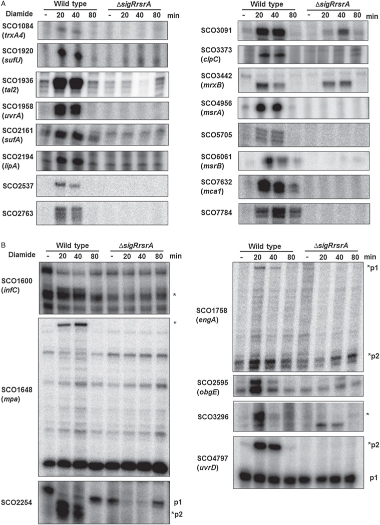
When 108 SigR target operons were classified on the basis of known and predicted functions, many of them fall in the functional groups related with maintenance of thiol redox homeostasis and sulphur-containing amino acids (11 operons; groups 1 and 2 in Table 1), protein folding and degradation (7 operons; group 5), and oxidoreductases (15 operons; group 8) (Table 1), fortifying previous proposals that the SigR regulon functions to achieve redox homeostasis and protein quality control under conditions of thiol-oxidative stress (Paget et al., 2001; Park and Roe, 2008; Kallifidas et al., 2010). Within these groups, we noticed newly added genes encoding a putative mycoredoxin (SCO3442), and a eukaryotic-type proteasome system that degrades proteins ligated with prokaryotic ubiquitin-like protein Pup (Mpa, PafD, Pup, PrcAB) first reported in M. tuberculosis (Pearce et al., 2008; Burns and Darwin, 2010). This gene cluster encodes a putative AAA ATPase (Mpa), an accessory factor (PafD), prokaryotic ubiquitin-like protein (Pup), 20S proteasome alpha and beta subunits (PrcAB) similar to those encoded by Mycobacterium tuberculosis (Pearce et al., 2008; Darwin, 2009). S1 mapping analysis demonstrates that mpa (SCO1648) has a SigR-dependent promoter, which is diamide-induced, in addition to ones that are independent of SigR (Fig. 1). Since the thiol-oxidant diamide can cause protein misfolding and aggregation (Kallifidas et al., 2010), induction of proteolytic systems by diamide-triggered SigR matches biological necessity.
Fig. 1.
S1 nuclease mapping of selected SigR-dependent promoter regions. RNA samples were prepared from exponentially growing wild-type and ΔsigRrsrA cells treated with 0.5 mM diamide for 0, 20, 40 and 80 min.
A. Transcript patterns of genes whose primary transcripts are from SigR-dependent promoters.
B. Results from genes which produce transcripts from other promoters independent of SigR. The bands with marked asterisks in panel B are from predicted SigR-binding promoters.
We also found a large number of SigR target genes that may function in modulating ribosome-associated processes (9 operons; group 3). Newly identified genes in this group encode probable translation initiation factors (IF-2, IF-3), 50S ribosomal proteins (L20, L35), ribosome-associated GTP-binding proteins (EngA, ObgE, Era), a ribosome binding factor RbfA, and a tRNA pseudouridine synthase (TruB). Observation of many ribosome-associated genes in the SigR regulon leads us to postulate that thiol-oxidative stress may somehow hinder translation processes, against which cells induce a SigR-mediated response. Induction of tmRNA (ssrA) by SigR under thiol-oxidative stress suggests an increase in truncated mRNA in the A-site of ribosome in the cell. It has been shown in E. coli that ribosome stalling induces A-site bound RelE or RelE-like RNA interferases to cut mRNA (Neubauer et al., 2009; Christensen-Dalsgaard et al., 2010), whose continued translation requires tmRNA that adds the SsrA tag to the truncated protein for degradation. In this respect, a recent discovery of the role of tmRNA in relieving polyribosomal stalling in dnaK and other stress-related mRNA to facilitate their translation in streptomycetes highlights a potential new link between ribosomal modulation and stress response (Barends et al., 2010). Increased demand for guanine nucleotides due to elevation in activity of RelA and GTP-binding proteins during stress could be met by increased GMP synthesis by guaB and guaB1 gene products. Increased demand for guanine nucleotide can also be explained by the fact that guanine is most vulnerable to oxidation among four DNA bases due to its lower oxidation-reduction potential (Kawanishi et al., 2001).
Many transcription factors (15 operons; group 6) are predicted to be SigR targets, implying that SigR is a global regulator that can change the transcription profile through cascades of affected transcriptional regulators. Among newly identified SigR targets are those encoding the major housekeeping sigma factor HrdB (SCO5820) and a SigR-like ECF sigma factor SigR2 (SCO3450) and its putative partner RsrA2-2 (SCO3451). The gene for an IclR-like transcription factor (SCO5552) called ‘NdgR’ that regulates amino-acid-dependent growth and antibiotic production (Yang et al., 2009) also contains a SigR-binding promoter sequence.
Synthesis of a number of redox-sensitive cofactors (such as Fe-S, CoA, NAD, lipoate, folic acid, pyridoxal phosphate and molybdopterin) appears to be regulated by SigR (9 operons; group 11). In addition to the previously identified sufA, coaE, folE and moeB genes [encoding a putative Fe-S assembly factor, coenzyme A kinase, GTP cyclohydrolase I for folate biosynthesis, and sulphurylase possibly involved in molybdopterin synthesis (Paget et al., 2001) respectively], we found that sufU, nadA, lipA and pdxH2/3 [encoding putative NifU-type Fe-S assembly factor, quinolinate synthetase A subunit for nicotinamide synthesis, lipoic acid synthase, and phospho-pyridoxamine synthetase for pyridoxal phosphate (vitamin B6)] were all direct targets of SigR. It is interesting to note that some of these proteins are either putative Fe-S assembly components (SufA, SufU) or Fe-S containing enzymes (NadA, LipA). In addition, the cofactors they synthesize contain sulphur atoms (Fe-S, folate, CoA, lipoic acid), or involve sulphur-mediated reactions (LipA, MoeB). Exposed Fe-S cofactors are labile to oxidation, so we propose that increased SigR activity contributes to replenishing Fe-S cofactors, Fe-S-containing proteins, and/or other oxidation-labile S-containing factors upon oxidative stress.
S1 nuclease mapping confirms predicted SigR promoters
To provide additional experimental evidence for some of the newly identified SigR targets, we examined 25 selected transcripts by S1 nuclease mapping at different times after exposing S. coelicolor cells to the thiol oxidant diamide. We observed SigR-specific transcripts from 24 promoters whose 5′ ends match with the predicted location of the promoters (Table S1). This supports the hypothesis that nearly all the SigR regulon genes presented in Table 1 produce SigR-dependent transcripts. We detected new SigR-dependent transcripts from 15 promoters whose expression has not been previously reported (SCO1084, 1600, 1648, 1758, 1936, 2194, 2254, 2595, 2763, 3296, 3442, 4797, 5705, 5820, 7784), confirming that ChIP-chip analysis detects SigR target genes with high sensitivity. Figure 1 shows S1 mapping analysis of transcripts from these promoters. Some genes produce transcripts initiated primarily from SigR-dependent promoters (Fig. 1A; 1084, 1920, 1936, 1958, 2161, 2194, 2537, 2763, 3091, 3373, 4956, 5705, 6061, 7632, 7784). Others utilize additional SigR-independent promoters (Fig. 1B; 1600, 1758, 2254, 2595, 3296, 4797).
Examination of genes with multiple promoters reveals diverse patterns of SigR-dependent modulation. For example, SCO1758 (engA) encoding a putative GTPase produces p1 transcript as predicted (250 nt upstream from the start codon; Table S1) and also the downstream unpredicted p2 transcripts, whose promoter sequence (GGAT-N16-GTT; ggatcacccggtaaaggggtgtt) resembles the SigR consensus but with a shorter spacing of 16 nucleotides. This implies a flexibility of spacing between −35 and −10 elements of SigR-dependent promoters. In some genes such as SCO2254 the dominant transcripts produced under non-stressed condition disappear transiently following stress independent of SigR, during which period the SigR-dependent transcripts are induced.
In promoters such as p2 of SCO1758 (engA; Fig. 1B) and SCO3442 (mrxB; Fig. 1A) (encoding a paralogue of mycoredoxin and a glutaredoxin-like protein) the SigR-induced transcripts still appear in the ΔsigR-rsrA mutant but with delayed response. A similar phenomenon was also observed for SCO3091 (cfa) encoding a putative cyclopropane-fatty acyl phospholipid synthase (Fig. 1A). The delayed induction kinetics in ΔsigR-rsrA mutant suggests that these SigR-dependent promoters may also be recognized by SigR paralogues that recognize similar promoter sequences as SigR. Similar induction pattern has been reported for transcripts encoding Lon protease, which are induced rapidly by SigR but with slower kinetics in the absence of SigR (Kallifidas et al., 2010). SCO3450 and SCO3451, which are SigR regulated genes encoding homologues of SigR (SigR2) and RsrA (RsrA2-1), respectively, show a similar pattern of induction (data not shown). A likely candidate for a sigma factor that shares similar promoter recognition sequence with SigR is SCO5147 (SigR1), which is a close homologue of SigE from M. tuberculosis (MtbSigE) as exemplified in the phylogenic comparison of ZAS-linked ECF sigma factors from S. coelicolor and M. tuberculosis (Fig. S2). The promoter for sigR1 has GGAAC-N18-GTT sequence that is not bound by SigR in our ChIP analysis. It is induced by diamide with slower kinetics (data not shown), and we propose that it is recognized by SigR1 itself. These observations indicate that a subset of SigR regulon genes can be recognized by at least one closely related SigR paralogue in addition to or in the absence of SigR.
SigR maintains the level and activity of the housekeeping sigma factor HrdB during thiol-oxidative stress
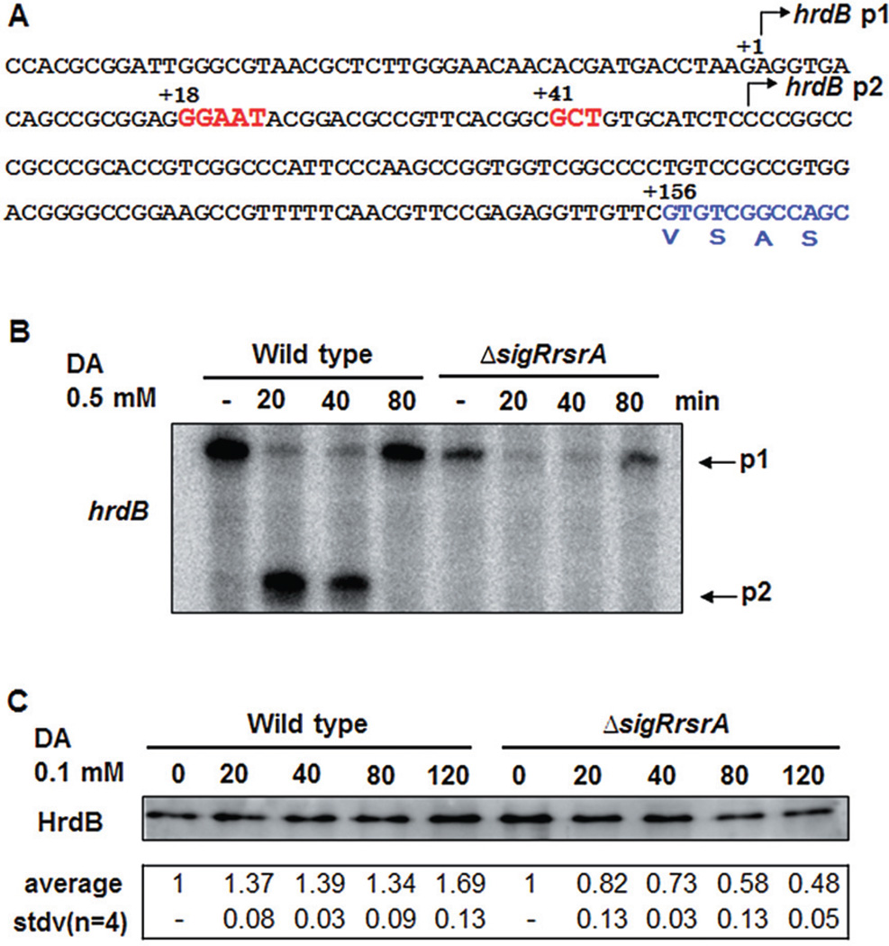
The SigR regulon includes several known or predicted transcription factors (SCO1425, 1619, 2331, 2481, 2537, 3207, 3450, 5065, 5552, 6775, 7140), which may significantly increase the number of genes regulated in response to thiol oxidative stress. In addition, we found that the major housekeeping sigma factor HrdB (SCO5820) possesses a previously unrecognized SigR-dependent promoter (GGAAT-N18-GCT) about 50 nt downstream of the known SigR-independent promoter (p1) (Buttner et al., 1990) (Fig. 2A). S1 mapping analysis of hrdB transcripts revealed that p1 is indeed the primary promoter in the absence of stress. However, diamide stress rapidly decreased the amount of p1 transcripts and increased the SigR-dependent transcripts from the predicted down-stream p2 promoter (Fig. 2B). The decrease in p1 transcripts upon oxidative stress occurred independently of SigR. This indicates that SigR functions to elevate hrdB transcription under oxidative stress conditions. Existence of a second promoter to compensate for loss of transcription from the primary promoter is also observed in SCO2254 encoding a putative efflux membrane protein (Fig. 1B), suggesting that this phenomenon may not be rare among SigR-regulated genes. Whereas some primary promoters such as p1 of SCO5820 (hrdB) and p1 of SCO2254 are affected dramatically by diamide treatment, the primary promoter p1 of SCO4797 (uvrD) is not affected at all. Therefore, the inhibitory effect of oxidative stress on transcription from a major p1 or housekeeping promoter may be a specific response at a subset of genes.
Fig. 2.
SigR-dependent transcription activation of the hrdB gene encoding the major sigma factor in S. coelicolor.
A. The position of the SigR-dependent promoter p2 upstream of the hrdB coding region predicted from ChIP-chip and sequence pattern. The previously reported promoter p1 resides further upstream.
B. S1 nuclease mapping of hrdB transcripts from diamide-treated cells revealing two alternative transcripts.
C. Western blot analysis of HrdB protein. Crude extracts from cells treated with diamide (0.1 mM) for up to 120 min were analysed to quantify the amount of HrdB protein as described in Experimental procedures. For each lane, an aliquot of 1 µg protein extract plus 1 µg BSA in lysis buffer was analysed. Results from four independent experiments were quantified to estimate changes in the level of HrdB protein upon oxidative stress, presented with the average value ± standard deviations (SD).
To test whether SigR controls the amount of HrdB protein during oxidative stress, as predicted from transcript analysis, we monitored the amount of HrdB protein by analytical immunoblot analysis. To ensure quantitative detection of HrdB, we diluted cell extract with cell lysis buffer containing non-specific BSA protein as a protein buffer that could minimize uncontrolled loss of proteins in cell extract during liquid handling and electrophoresis. Results in Fig. 2C demonstrate that HrdB protein is maintained during oxidative stress condition at a level slightly higher than that under non-stressed condition in the wild type. On the contrary, the level of HrdB decreased significantly in ΔsigRrsrA mutant upon oxidative stress condition. Even though it is not clear why the level of HrdB protein increases in the wild type during the stress, it is obvious that SigR does contribute to maintain the level of HrdB protein, as predicted from transcript analysis. We further monitored the activity of HrdB by examining transcripts from HrdB target promoters (rrnDp1, p3 and p4) in the ribosomal RNA operon rrnD, in the wild type and mutant. All four promoters of the rrnD operon share the consensus sequence for HrdB recognition, especially in the −10 region, to varying extent (Baylis and Bibb, 1988; Hahn and Roe, 2007). In vitro transcription from rrnD promoters with reconstituted holoenzyme containing HrdB (EσHrdB) has demonstrated that all four promoters can be transcribed by EσHrdB, even though transcription from p3 and p4 promoters requires additional cellular factors (Hahn and Roe, 2007). We found that the amounts of the abundant p3 and p4 transcripts were maintained throughout the stress period and even increased up to 50% and 30%, respectively, during 40–80 min diamide treatment. On the other hand, they both decreased continuously in the ΔsigRrsrA mutant to about 40% and 60% level, respectively, during the 2 h of stress (Fig. S3). Transcripts from the p1 promoter decreased to 20% level during the stress in the mutant, whereas the level was maintained in the wild type (Fig. S3). These results clearly demonstrate that SigR is necessary to maintain the level and activity of the major housekeeping sigma factor during thiol-oxidative stresses.
The UvrA protein is necessary to cope with UV and thiol-oxidative stresses
Among the newly discovered SigR targets are SCO1958, SCO4797 and SCO5188, which encode putative UvrA, UvrD and a paralogue of UvrD (UvrD2), respectively, predicted to function in nucleotide excision repair. The UvrABC endonuclease complex with UvrD (DNA helicase II) has been shown to repair UV-induced DNA damages, the most prominent of which are pyrimidine dimers (Sinha and Hader, 2002). However, whether the UvrABC system is needed under thiol-oxidative stress condition and whether thiol-oxidative stress causes DNA damage have not been known. Consensus SigR promoters (GGCAT-N18-GTT for uvrA, GGCAT-N18-GTT for uvrD, GGAAA-N19-GTC for uvrD2) are located 103, 31 and 7 nt upstream from the start codons of respective genes (Table S1). The uvrA gene is transcribed at a very low level under unstressed conditions. However, upon diamide stress, it is drastically induced from a SigR-dependent promoter (Fig. 1A). On the contrary, the uvrD gene is transcribed at a relatively high level from a SigR-independent promoter p1 during unstressed condition. Upon induction, the upstream SigR-dependent promoter p2 is induced (Fig. 1B). The uvrD2 transcripts were not detectable by S1 analysis, probably due to low amount.
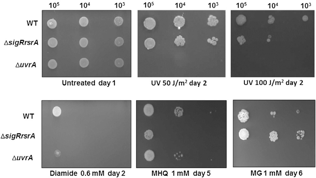
In order to examine whether UvrA indeed confers UV resistance, and whether SigR and UvrA have related functions in conferring resistance to UV and thiol-reactive compounds in S. coelicolor, we made a ΔuvrA strain and analysed its sensitivity to UV and thiol-reactive compounds along with ΔsigR-rsrA mutant. Figure 3 demonstrates that ΔuvrA has increased sensitivity to UV irradiation, and ΔsigR-rsrA is also sensitive to UV, though not as dramatically as ΔuvrA. Therefore, SigR is necessary to achieve optimal UV resistance possibly through inducing UvrA system. The SigR regulon genes were not induced by UV irradiation (data not shown), suggesting that UV itself is not capable of activating SigR function. SigR can be activated by thiol-reactive oxidants/electrophiles such as diamide and methyl hydroquinone (MHQ), as predicted from the induction of thiol-specific oxidative stress response by these compounds in B. subtilis (Antelmann et al., 2008; Nguyen et al., 2009). Since some electrophiles can react not only with thiols in proteins and small molecules but also with DNA, we examined whether UvrA is needed to cope with chemical stresses caused by thiol-reactive electrophiles such as diamide, methyl hydroquinone (MHQ) and methyl glyoxal (MG). It has been reported that MG induces not only thiol-specific oxidative stress response but also SOS response, suggesting some DNA damage in B. subtilis. (Nguyen et al., 2009). MG is known to cause DNA damage by reacting with DNA bases to form N2-(1-carboxylethyl)-2′-deoxyguanosine (CEdG) (Synold et al., 2008). Whether this kind of damage is repaired by the Uvr system has not been reported. Whether thiol-reactive diamide and MHQ could elicit DNA damage has not been reported either. The result in Fig. 3 shows that UvrA is needed to defend cells against these electrophiles. As expected, ΔsigR-rsrA mutant became very sensitive to thiol-reactive electrophiles. Dramatic resistance of ΔsigR-rsrA toward MG is unexpected. Inability to induce many thiol-homeostatic components may somehow confer better survival relative to the wild type in the presence of MG in plate culture. When the sensitivity of ΔsigR-rsrA toward MG was monitored in liquid culture during early exponential phase (OD ~ 0.3), the mutant was more sensitive to MG than the wild type, suggesting that the resistance phenotype on plate culture could depend on treatment method (data not shown). Overall, the finding that SigR induces DNA repair proteins expands our understanding of the function of SigR to accommodate response towards UV- and some electrophile-incurred damages.
Fig. 3.
Sensitivity of ΔuvrA and ΔsigRrsrA mutants to UV and thiol-reactive oxidants and electrophiles. Serially diluted spores of wild-type (WT), ΔsigRrsrA, ΔuvrA strains were spotted on NA plates with or without added chemicals such as diamide (0.6 mM), methyl hydroquinone (MHQ, 1 mM) and methyl glyoxal (MG, 1 mM). For UV irradiation, the spotted plates were exposed to UV light from UV cross-linker for 30 seconds. Plates were incubated at 30°C for 1 (untreated), 2 (UV, diamide), 5 (MHQ) or 6 days (MG).
The phylogenetic analysis of SigR orthologues and their predicted respective regulons reveals a core conserved thiol oxidative stress regulon in Actinomycetes
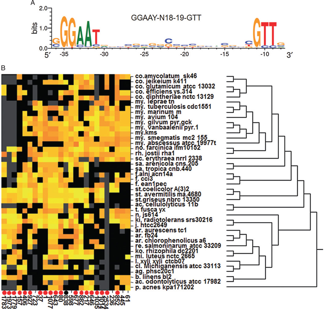
The characterization of this larger SigR regulon in S. coelicolor furthers our understanding of the organism’s response to thiol oxidative stress. However, it has been previously observed that the composition of a given regulon in a given organism may result from specific adaptations to its ecological niche (Dufour et al., 2008; 2010a; Perez and Groisman, 2009). Therefore, the SigR regulon in S. coelicolor may comprise many functions that are specific only to this organism, and not shared by other organisms. Therefore, comparison of predicted SigR regulons in other organisms may indicate functions that are commonly required to cope with thiol oxidative stress in general apart from those specifically required for this organism. To test this hypothesis and predict which genes found in the S. coelicolor SigR regulon are most likely to be directly relevant to thiol oxidative stress, we performed a phylogenetic analysis of SigR-RsrA orthologues and their regulons across a group of Actinomycetes using a computational method previously described (Dufour et al., 2008; 2010a). Since this phylogenetic analysis relies on the accurate computational detection of putative SigR promoter sequences, we constructed a refined SigR promoter model by aligning the sequences of predicted promoters corresponding to 57 enriched regions in the ChIP-chip assay that matches with genes transcribed in a SigR-dependent manner (Table S1). Figure 4A demonstrates a new SigR consensus logo, with a representative sequence of GGAAT-N18-19-GTT.
Fig. 4.
The top 25 orthologous sets of genes predicted to be SigR targets across 42 selected Actinomycete species from 22 genera.
A. Sequence logo of the SigR promoter motif in S. coelicolor. The logo was derived from the alignment of 58 SigR-dependent promoters detected by ChIP-chip. The heights of the letters represent their conservation at the particular position of the multiple sequence alignment. The −35 and −10 regions are separated by a non-conserved spacer region of 18 or 19 base pair. The logo was produced using WebLogo (http://weblogo.berkeley.edu/).
B. The heatmap indicates whether the promoter region of the corresponding gene contains a putative SigR promoter for each species (row) and each set of orthologous genes (column). Orange and yellow indicate moderate and strong match to the SigR promoter model respectively. Black indicates that the corresponding species possess a gene belonging to the corresponding orthologous set but that no match to the SigR promoter model, while grey indicates that the species lacks a gene representing the orthologous set. Species are organized according to their phylogenetic relationship as indicated by the dendrogram on the right. Sets of orthologous genes are identified by arbitrary numbers (Table 2). Sets of orthologous genes containing confirmed SigR target genes in S. coelicolor are indicated by circles at the bottom. Red circles indicate genes detected by SigR ChIP-chip and experimentally verified for SigR-dependent expression. The black circle indicates a gene that is associated with a SigR ChIP-chip peak without experimental verification.
We first detected and aligned the protein sequences of all SigR orthologues found in 42 Actinomycetes to determine the conservation of their DNA binding domains (only Mycobacterium leprae TN did not possess SigR and RsrA orthologues). The multiple sequence alignment showed that the protein domains determining promoter sequence specificity (regions σ2.4 and σ4.2) of SigR orthologues are well conserved across all species (Fig. S1). Therefore, we assumed that all SigR orthologues recognize very similar if not identical promoter sequences. Thus, we identified putative SigR targets across all the selected genomes by searching in each genome for sequences matching S. coelicolor SigR promoter model (Fig. 4A) in the upstream of annotated genes. With the promoter score cut-off chosen for this analysis, we captured 72 of the 108 SigR promoters defined by our ChIP-chip analysis in S. coelicolor. Finally, putative SigR target genes across the 42 genomes were grouped by orthology and clustered according to the occurrence of predicted SigR-dependent promoters.
The emerging phylogenetic patterns predicted that the top 25 sets of orthologous genes appear to be regulated by SigR orthologues across most of the 42 Actinomycetes, thus, forming a core conserved regulon for the response to thiol oxidative stress (Fig. 4B, Table 2). The predictions resulting from this comparative genomics analysis are well supported by the experimental data presented in this report because 20 of the S. coelicolor genes corresponding to the top 25 conserved SigR targets are associated with SigR ChIP-chip peaks (Table 2). Excluding the regulator pair SigR-RsrA, the predicted functions of these genes fall in 6 categories: (i) thiol redox homeostasis with trxB (#80), trxC (#217), mrxA (#927), msrA (#157) and 2 Dsb-like proteins (#1014, #1783); (ii) protein quality control with dnaK (#61), grpE (#445), clgR (#646), clpC (#1) and htpX (#723); (iii) flavin-mediated oxidoreductases (#838, #1779, #1973); (iv) translational modulation with prfA (#236) and rpmE1 (#37); (v) transcriptional modulation with rbpA (#598) and Mtb-sigB (#2534); (vi) iron-sulphur cluster assembly with sufA (#677). Additionally, hypothetical proteins of unpredictable functions (#852, #1035, #1997, #5796) were included. Table 2 summarizes the description of each set of orthologous genes, by presenting its representative gene and the genera in which it occurs among 22 genera examined. The analysis predicts that the thiol redox homeostasis and protein quality control are the two most outstanding functions in the core SigR regulon across actinomycetes, followed by transcriptional and translational modulations. Mtb-sigB, a group 2 sigma factor, controls general stress response and is induced by a variety of stressors and by multiple sigma factors such as SigH and SigE in Mycobacterium tuberculosis (Sachdeva et al., 2010). Therefore, in many actinomycetes, it is likely that SigR orthologues control the expression of another general stress factor of non-ECF family, suggesting that the stress signal initiates a transcriptional cascade, which includes multiple DNA binding proteins. The function of RbpA is not well understood except that it associates with RNA polymerase and confers resistance to rifampicin in actinomycetes (Newell et al., 2006; Dey et al., 2010). PrfA terminates translation as a peptide release factor and recycles ribosome whereas RpmE1 is the ribosomal protein L31. How the induction of genes for PrfA and RpmE1 is beneficial under thiol-oxidative conditions is not clear. The rpmE1 gene encoding an isoform of L31 with cysteine residues for zinc-binding is induced by SigR, whereas the cysteine-negative paralogue rpmE2 is induced by Zur under zinc-depleted condition in S. coelicolor (Owen et al., 2007; Shin et al., 2007). Whether thiol oxidations in ribosomal components or translational complex causes increased demand for PrfA and RpmE1 and whether zinc-containing RpmE1 is sensitive to oxidative stress deserve future studies.
Table 2.
Top 25 genes that are highly conserved as SigR regulon members among actinomycetes.
| Orthologues No. |
Gene No.a | Gene name | Function | Genus (frequency)b |
|---|---|---|---|---|
| 61 | SCO3671 | dnaK | Hsp70 family chaperone, no direct SigR target in Sco, First gene in dnaK-grpE-dnaJ-hspR cluster (3671-3668), a SigE target in Mtb | Co, My, No, Rh, Sc, F, T, N, Ki, J, Mi, B, Ac, P (14/22) |
| 445 | SCO3670 | grpE | Co-chaperone | Co, My, No, Rh, Sc, F, T, N, Ki, J,, B, Ac (12/22) |
| 852 | SCO3083* | Possible integral membrane protein | Co, My, No, Rh, Sc, F, St, T, N, Ki, B, P (12/22) | |
| 236 | SCO5360* | prfA | Peptide chain release factor 1 (RF-1), rpmE1-prfA operon | My, Rh, Sc, F, St, T, N, Ki (8/22) |
| 2534 | MT2783 | Mtb-sigB | Sigma factor SigB in M. tuberculosis regulated by SigE | Co, My, No, Rh, Sc (5/22) |
| 1014 | SCO2634* | Hypothetical protein, contains DsbA-like thioredoxin domain | Co, My, No, Rh, Sc, F, St, T, N, Ar, Re (11/22) | |
| 1035 | SCO3187* | Hypothetical protein | My, No, Rh, F, St, J, Ar, Re (8/22) | |
| 646 | SCO5755* | clgR | Activator of clpP and lon protease synthetic genes. Neighbouring a SigR-target gene (5754; cinA) | Co, My, No, Rh, Sc, St, N, Ki, Ar, Ko, Mi, Cl, Ag, Ac, P (15/22) |
| 217 | SCO0885* | trxC | Thioredoxin | Co, My, No, Rh, Sa, St, Ac, N, Ki, Ko, Mi, Ag, Ac, P (14/22) |
| 882 | SCO1997* | Unknown | Co, My, No, Rh, Sa, F, St, J, Ar, Re, Ko, Mi, P (13/22) | |
| 677 | SCO2161* | sufA | HesB domain, probable Iron-sulphur cluster assembly protein | Co, My, No, Rh, Sc, Sa, St, T, N, Ki, J, Ar, Re, Ko, Mi, Cl, B (17/22) |
| 598 | SCO1421 | rpbA | RNA polymerase binding protein | Co, My, No, Rh, Sc, Sa, F, St, Ac, T, N, Ki, J, Ar, Re, Ko, Mi, B (18/22) |
| 838 | SCO2816# | Conserved hypothetical protein, Flavin utilizing monooxygenase superfamily | Co, My, No, Rh, Sc, F, St, Ki, J, Ar, Ko, Mi, Cl, B (14/22) | |
| 80 | SCO3890* | trxB | Thioredoxin reductase | Co, My, No, Rh, Sc, Sa, F, St, Ac, T, Ki, J, Ar, Re, Ko, Mi, L, Cl, B, Ac, P (21/22) |
| 643 | SCO5216* | sigR | Sigma factor R | Co, My, No, Rh, Sc, Sa, F, St, Ac, T, N, Ki, J, Ar, Re, Ko, Mi, Cl, B, Ac, P (21/22) |
| 1077 | SCO5217* | rsrA | Anti-SigR factor | Co, My, No, Rh, Sc, Sa, F, St, Ac, T, N, Ki, J, Ar, Re, Ko, Mi, Cl, Bl, P (20/22) |
| 1 | SCO3373* | clpC | Clp-family ATP-binding protease similar to MecB/ClpC | Co, My, No, Rh, Sc, Sa, F, St, Ac, T, N, Ki, J, Mi, L, Cl, B, Ac (18/22) |
| 37 | SCO5359* | rpmE1 | Ribosomal proteins L31 | My, Rh, Sc, Sa, F, St, T, N, Ki, J (10/22) |
| 723 | SCO4609 | htpX | Possible peptidase, M. tuberculosis HtpX homologue, no SigR target, a SigE target in Mtb | My, No, Rh, Sc, F, St, N, Ki, J, Ar (10/22) |
| 927 | SCO5187* | mrxA | Mycoredoxin, glutaredoxin-like protein | My, No, Rh, Sa, F, St, Ac, N, Ki, Ar, Re (11/22) |
| 406 | SCO5796* | Unknown | Co, My, Sa, F, St, T, Ki, L, Cl, Ag (10/22) | |
| 157 | SCO4956* | msrA | Methionine S-sulphoxide reductase | Co, No, F, St, N, Ki, J, Ar, Ko, Mi, Cl, Ag, B, P (14/22) |
| 1779 | SCO3296* | Possible oxidoreductase similar to F420-dependent alcoholdehydrogenase | No, Rh, Sc, St, T, N, Ki, J, Ar, Ko, Mi, L, B (13/22) | |
| 1973 | SCO3295* | Possible oxidoreductase containing flavin | No, St, T, N, Ki, J, Ar, Re, Mi, L, B (11/22) | |
| 1783 | SCO1869* | Conserved hypothetical protein, probable DsbA-like dithiol-disulphide isomerase (FrnE subfamily) | Co, Rh, Sa, F, St, T, N, Ki, J, Ar, Re, Cl, Ag, B (14/22) |
The representative gene from S. coelicolor (SCO) or M. tuberculosis (MT) in each orthologue cluster was presented. The SCO genes that were determined to be SigR-dependent by ChIP-chip and supported by additional S1 and microarray data are marked with asterisks (*). The genes detected by ChIP-chip but not by additional expression data are marked with ‘#’.
The genera that possess the corresponding SigR target gene in each orthologous cluster are presented with abbreviations; Co (Corynebacterium), My (Mycobacterium), No (Norcadia), Rh (Rhodococcus), Sc (Saccharopolyspora), Sa (Salinispora), F (Frankia), St (Streptomyces), T (Thermobifida), N (Nocardioides), Ki (Kineococcus), J (Janibacter), Ar (Arthrobacter), Re (Renibacterium), Ko (Kocuria), Mi (Micrococcus), L (Leifsonia), Cl (Clavibacter), Ag (Agreia), B (Brevibacterium), Ac (Actinomyces), and P (Propionibacterium). The frequency in parenthesis represents the number of genera that posses the corresponding SigR target gene among 22 genera, whose 42 genomes were examined in this study.
When we extended the analysis to include less conserved genes up to 250 gene sets, we found that some gene sets are still conserved significantly across actinomycetes, whereas others are more restricted to certain genera. Figure 5 displays the clustered pattern of these gene sets to exhibit a relatively common group (E) and genus-specific groups (A, B, C, D, F, G), distributed to a specific genus such as Streptomyces (group A&E), Salinispora (group B&E), Frankia (group C&E), Corynebacterium (group D), Mycobacterium (group E&F) and Arthrobacter (group G). The so-called extended regulon of S. coelicolor (group A&E) contains 79 genes, and among genes with predicted functions the main functions represented are: electron transport (9 genes), biosynthesis of cofactors (9 genes), transcriptional regulators (6 genes), and protein homeostasis (5 genes). However, only 29 of these 79 putative targets are associated with a ChIP-chip peak from our analysis, suggesting that the computational detection of SigR targets that are not broadly conserved across the phylogeny may be impaired by a high rate of false-positive in this analysis (Fig. 5, Table S2).
Fig. 5.
The top 250 orthologous sets of genes predicted to be SigR targets across 42 selected Actinomycetes. The heatmap colour coding is identical to Fig. 4. Species are organized in the same order as in Fig. 4. Red marks indicate genes that are associated with a SigR ChIP-chip peak and differentially expressed under diamide stress, while black marks indicate genes that are only associated with a SigR ChIP-chip peak. The broader characterization of the conservation of SigR targets predicts in addition to the core regulon the existence of extended SigR regulons that are specific to lineages of related species. Lineage specific SigR regulons are indicated by letters on top, and the gene composition of each group is summarized in Table S2.
SigR and its paralogues with shared promoter selectivity – Overlap of SigR regulon with other regulons
Among the top 25 conserved SigR regulon members, dnaK (#61) and grpE(#445) are not directly dependent on SigR in S. coelicolor. The consensus SigR logo is not found upstream of the dnaK-grpE-dnaJ-hspR operon in S. coelicolor as well as in all the other Streptomyces genomes examined in this study. In M. tuberculosis, transcription of the dnaK operon is activated in response to heat shock and oxidative stress by SigE, a paralogue of SigH (SigR orthologue) (Fig. S2), with similar recognition sequence (Song et al., 2008). Mtb-sigB gene (#2534) is also recognized by SigE in M. tuberculosis. Since Mtb-SigH induces the expression of the sigE gene (Raman et al., 2001; Manganelli et al., 2002), there exist two tiers of overlap between regulons of SigH and SigE in M. tuberculosis, arising from similar recognition sequence and from hierarchically sequential induction. Likewise, it is conceivable that the dnaK operon in S. coelicolor is regulated by a SigR paralogue that resembles Mtb-SigE. If this were true, SigR1 (SCO5147) is the most likely candidate for activating the dnaK operon in S. coelicolor (Fig. S2). SCO5147 could play a similar role in other species since it is found among the top 50 conserved sets of SigR-regulon members in actinomycetes (Table S2, group E). In M. tuberculosis, the SigE-binding consensus (GGAACY-N17-18-GTT) overlaps significantly with SigH (SigR orthologue)-binding consensus (GGAAYA-N17-18-GTT) (Sachdeva et al., 2010). It has been reported that a single position in the −35 element primarily distinguish the recognition specificity of SigE from that of SigH (Song et al., 2008). Considering this, it is likely that the list of bioinformatically predicted SigR regulon genes includes not only those directly recognized by SigR orthologues, but also some that are recognized by SigE-type paralogues. In addition, since sigE transcription requires SigH in M. tuberculosis, the predicted SigR/SigH regulon members may include indirectly regulated ones due to regulator cascade. The extent of overlap between SigR orthologues and their close paralogues in sequence specificity as well as hierarchical regulation may differ in different organisms. Experimental verification may require not only the comparison of transcripts between the wild-type and regulator mutants, but also regulator binding studies in vivo and in vitro.
Involvement of ClgR in the core SigR regulon is intriguing (Table 2). The clgR gene (SCO5755) is co-transcribed with SCO5754, a SigR target gene which encodes a homologue of CinA known to be induced by DNA damage and competence in Streptococcus pneumoniae (Martin et al., 1995). In S. coelicolor ClgR positively regulates genes for proteases (clpP1/P2, lon, clpC) that are direct targets of SigR (Bellier and Mazodier, 2004). In C. glutamicum, ClgR controls transcription of genes involved in proteolysis and DNA repair such as ptrB encoding endopeptidase, hflX encoding a GTP-binding protein, recR for recombinational repair of UV-induced DNA damage (Engels et al., 2004). Inclusion of known ClgR targets in the SigR regulon of S. coelicolor demonstrates an interesting regulatory circuit that involves double positive regulators. The contribution of SigR appears critical to allow promoter recognition as well as provision of ClgR in transcribing ClgR target genes.
Functional overlap between prokaryotic SigR regulon and eukaryotic Nrf2 regulon suggests a broad conservation of the thiol oxidative stress response
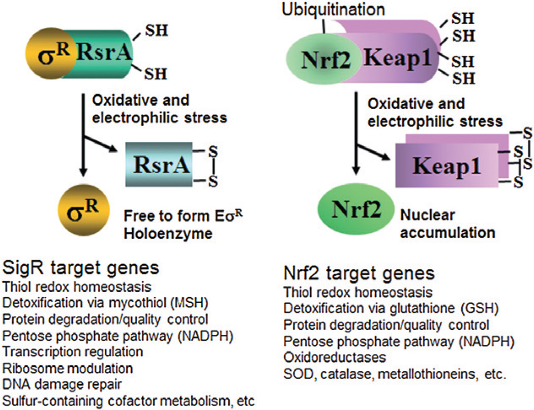
Examination of SigR regulon genes in S. coelicolor reveals a striking functional similarity with an analogous system in mammals as summarized in Fig. 6. The Nrf2-Keap1 system in higher eukaryotes constitutes a defence mechanism against stresses imposed by electrophiles and oxidants. Nrf2 is associated with its inhibitor Keap1, which facilitates ubiquitination and proteosomal degradation of Nrf2. Attacked by electrophiles or oxidants, Nrf2-Keap1 interaction is disrupted, and the released Nrf2 induces its regulon genes through binding to antioxidant response elements (ARE) (Kensler et al., 2007). Nrf2 protein itself does not have any sequence homology with SigR. However, the mechanism by which it is activated involves thiol-disulphide isomerization of its inhibitor Keap1 as occurs in the activity modulation of SigR by RsrA (Dinkova-Kostova et al., 2002). Similarly to RsrA, Keap1 has many cysteine residues (27 cysteines) and responds to oxidative and electrophilic stressors. Functions of the downstream target genes also overlap significantly between the SigR/RsrA and Nrf2-Keap1 systems. When activated, Nrf2 induces genes for various thiol homeostatic proteins, such as glutathione peroxidase, γ-glutamyl-cystein synthetase, thioredoxin reductase and peroxiredoxin 1, which are functionally related with thiol redox proteins, such as thioredoxins and their reductase, peroxiredoxin, mycothiol synthetic enzymes, and mycoredoxins regulated by SigR (Thimmulappa et al., 2002; Rangasamy et al., 2004). Just as Nrf2 increases synthesis of proteasome subunits and γ-glutamyl peptidase, SigR activates expression of lon, pepN, clp proteases and mpa (Kwak et al., 2003). The amino acid sequence of pepN is homologous to γ-glutamyl peptidase. Both Nrf2 and SigR regulate pentose phosphate pathway enzymes such as glucose 6-phosphate dehydrogenase and transaldolase for generating reducing potential NADPH. The SCO2763 gene product, a putative ATP-binding cassette (ABC) transporter, is a close homologue of multidrug resistance-associated protein 1 (Mrp1/Abcc1) whose expression depends on Nrf2 and is responsible for cellular extrusion of GSH-conjugates (Hayashi et al., 2003). Whether SCO2763 has a function in exporting mycothiol (MSH)-S-conjugates of electrophiles deserves experimental study.
Fig. 6.
Induction and function of SigR regulon in comparison with mammalian anti-oxidative Nrf2-Keap1 system. SigR regulon is induced in response to thiol-reactive oxidants and electrophilic compounds, through inactivation of antisigma RsrA by forming intramolecular disulphide bond. Nrf2 regulon is induced by similar stressors, through inactivation of inhibitory factor Keap1 that facilitates degradation of Nrf2, by forming multiple intra- and inter-molecular disulphide bonds, and S-alkylation. Functions of SigR target genes overlap with those of Nrf2 targets to a large extent, which include thiol redox homeostasis, small thiol-mediated detoxification, protein quality control, and pentose phosphate pathway for generating NADPH. The scheme for Nrf2 activation mechanism is simplified to highlight analogy with SigR-RsrA (Kensler et al., 2007; Antelmann and Helmann, 2011).
Conclusions
Genome-wide identification of direct SigR binding sites under thiol-oxidative stress conditions, combined with transcript and sequence analyses, revealed many new genes of SigR regulon in S. coelicolor. The wide spectrum of target genes that include cascades of regulators indicates that SigR is a global regulator. As expected, SigR induces many genes related with homeostasis of thiols in proteins and small molecules (mycothiols), and proper protein quality control, which are most prominently affected by thiol-reactive/oxidative stressors. Additionally, SigR induces many genes for ribosome-associated components, transcriptional regulators including sigma factors, numerous oxidoreductases (many of which are predicted to contain flavin), DNA damage repair, cofactor metabolism that include several Fe-S containing enzymes/proteins, and lipid synthesis. The full list of SigR target genes enabled prediction and experimental demonstration of new physiological functions of the SigR regulon under thiol-oxidative stress condition. We found that SigR directly contributes to maintain the level and activity of the major housekeeping sigma factor HrdB through increasing its transcription during oxidative stress. SigR also confers resistance to UV and thiol-reactive compounds, likely by inducing enzymes of DNA damage repair. Generation of a refined SigR promoter model enabled comparative genomics analysis of the SigR regulon, which revealed the existence of a core SigR regulon conserved across 42 selected Actinomycetes. The most prominent functions of the conserved SigR-orthologue regulons are related with thiol redox homeostasis, including the maintenance of sulphur-containing amino acids, and protein quality control involving chaperones and proteolytic systems. In addition, genes for translational modulation, transcriptional regulatory cascades, oxidoreductases and Fe-S assembly functions are also included. The bioinformatic analysis also predicts the existence of variable extended regulons specific for each genus. These results complement the experimental characterization of the SigR regulon in S. coelicolor to identify genes and functions that are most likely to be critical for the biological response to thiol oxidative stress. Comparison of SigR-regulated gene functions with those of thiol-oxidative stress response system (Nrf2-Keap1) in mammals reveals many shared gene functions, suggesting a robust physiological mechanism to deal with thiol-reactive stresses across distantly related life forms.
Experimental procedures
Strains and media
Spores of S. coelicolor A3(2) strain M145 were inoculated in YEME liquid medium containing 5 mM MgCl2•6H2O and 10% sucrose, and were grown at 30°C (Kieser et al., 2000). NA plates (0.8% nutrient broth, 2% agar powder) were used for spotting test. E. coli was grown in LB broth (1% tryptone, 0.5% yeast extract and 1% NaCl). E. coli ET12567, a non-methylating strain containing pUZ8002 for donor functions and BW25113 harbouring pIJ790 for linear DNA recombination were used for disruption as recommended (Gust et al., 2002).
Chromatin immunoprecipitation on a chip (ChIP-chip) analysis
The probes used to fabricate the custom-made 385 K NimbleGen microarray (Roche NimbleGen, Madison WI) for the ChIP-chip experiment were designed using the ‘chipD’ software (Dufour et al., 2010b). The parameters were set to obtain complete tilling of the S. coelicolor A3(2) genome with overlapping isothermal probes that ranged from 35 to 75 bases with an average spacing of 24 bases. In addition, probes alternate between the forward and reverse orientation to capture both DNA strands. The probe sequences and the ChIP-chip data are available at the NCBI’s Gene Expression Omnibus database with the accession number GPL14825 (http://www.ncbi.nlm.nih.gov/geo/query/acc.cgi?acc=GPL14825).
Streptomyces coelicolor cells (wild-type M145 and ΔsigRrsrA mutant strains) were grown to exponential phase (OD ~ 0.3) and were treated with 0.5 mM diamide for 20 min to prepare samples for the ChIP-chip experiment. The samples were prepared as previously described (Dufour et al., 2008). Briefly, the cell cultures were fixed with 1% formaldehyde for 4 min at 30°C followed by quenching with 0.125 M glycine for 30 min in ice, washed with phosphate-buffered saline, and frozen. Cells were suspended in 100 mMTris (pH 8.0), 300 mM NaCl, 2% Triton X-100, and 1 mM phenylmethylsulphonyl fluoride, and sonicated to lyse cells and fragment the genomic DNA. Micrococcal nuclease (100 U ml−1) and RNase A (1 µg ml−1) were added to the preparation to further fragment the genomic DNA and degrade RNA. Cell debris was removed by centrifugation and an aliquot was removed to analyse DNA fragmentation by agarose gel eletrophoresis (desired size of ~200–1000 bp with enrichment for ~500 bp molecules). Anti-SigR rabbit polyclonal serum (7.5 µl) was used to immunoprecipitate cross-linked DNA fragments for each sample prepared from about 50 ml culture. A mock immunoprecipitation was performed on the ΔsigRrsrA cells using the same serum to detect non-specific interactions. The enriched DNA was amplified via ligation-mediated PCR amplification using oligonucleotide linkers (oJW102: GCGGTGACCCGGGAGATCTGAATTC; oJW103: GAATTCAGATC). The DNA enriched using anti-SigR serum was labelled with Cy5 dye while DNA from the mock precipitation was labelled with Cy3. Labelled DNA from three independent immuno-precipitation experiments were pooled together in equal proportion to obtain the 4 µg of DNA that was hybridized on a microarray. Sample hybridization and microarray scanning were performed by Roche NimbleGen (Madison, WI) according to their standard protocol.
For each array, the Cy3 and Cy5 signals were normalized using the vsn method (Huber et al., 2002) in the R statistical software environment before calculating the log2 ratio of the experimental signal (Cy5) over the control signal (Cy3). A plot of the intensity ratios versus intensity averages revealed no dye bias. Therefore, no further correction was applied to the data. To remove the signal generated by non-specific DNA immuno-precipitation, the signal from the ΔsigRrsrA cells was subtracted from the signal obtained from the wild-type cells. Only one array was hybridized for each S. coelicolor strain. Regions of the genome enriched for occupancy by SigR were detected using the TAMALPAIS method (Bieda et al., 2006) at P-value ≤ 0.05 using a threshold set at the 99th percentile of the log2 ratio. The relative peak intensities were calculated by averaging the 10 highest consecutive probe signals for each selected peak.
Re-examination of expression profiling experiments under diamide stress
The microarray data of Kallifidas et al. (2010) were re-examined. To increase the detection sensitivity of SigR-dependent transcripts, the change in transcript abundances in the wild-type strain (M600) was divided by the change in transcript abundances in the ΔsigR mutant strain (J2139) measured 20 min after the addition of diamide. Transcripts that were at least 1.5-fold more abundant in the wild-type strain were selected to identify putative members of the SigR regulon. Taking the ratio of the changes in abundance may help identify SigR-dependent transcripts that did not show net increase in abundances in the wild-type strain because higher activity of the SigR-dependent promoter may be accompanied by lower activity of an alternative promoter.
S1 nuclease mapping
Cells were treated with 0.1 ~ 0.5 mM diamide at the exponential growth phase (OD ~ 0.3) for 20–120 min. After harvest, they were disrupted by sonication in Kirby mix. RNA isolation and S1 nuclease mapping was performed according to a standard procedure (Kieser et al., 2000). Promoter regions (from −500 to +60 nt relative to each start codon of SigR regulon gene) were amplified by PCR and used as probes. Probes were labelled with [γ-32P]-ATP and hybridized with 5–50 µg RNA at 50°C. After S1 nuclease treatment, protected DNAs were separated on 5% polyacrylamide gel containing 7 M urea. Signals were detected and quantified by BAS-2500 (Fuji film).
Western blot analysis
Cells were grown and treated with diamide as described for sample preparation for S1 mapping. Harvested cells were re-suspended in lysis buffer [20 mM Tris-HCl (pH 7.9), 10% (v/v) glycerol, 5 mM EDTA, 0.1 mM DTT, 10 mM MgCl2, 1 mM PMSF, 0.15 M NaCl]. The suspension was sonicated with ultrasonicator (Sonics and Materials), and cleared by centrifugation. Protein concentration in crude cell extract was determined by Bradford reagent solution (Bio-Rad) using BSA as a standard. For each sample, ~ 10 µl cell extract containing 50 µg protein was further diluted to the final concentration of 0.125 µg µl−1 with lysis buffer (up to 400 µl) that additionally contain 50 µg BSA to serve as a protein buffer. Aliquots of 8 µl containing 1 µg crude protein extract and the same amount of BSA were resolved on 10% SDS-PAGE. Immuno-detection by polyclonal mouse antibody against HrdB protein and the anti-mouse IgG secondary antibody at 1:5000 dilution ratio, followed by ECL detection system (Amersham Life Science), was done as described previously (Kim et al., 2009).
Sensitivity test
The disruption mutant of SCO1958 (uvrA) was obtained by using PCR-targeting mutagenesis protocol (Gust et al., 2002) as described previously (Kim et al., 2009). The wild-type and mutant spores (105, 104, 103) were spotted on NA media. For UV sensitivity assay, the spotted spores were exposed to UV in the CL-1000 UV cross-linker at 50 or 100 J m−2 before incubating. For chemical sensitivity, spores were spotted on NA media containing 0.6 mM diamide, 1 mM methylhydroquinone or 1 mM methylglyoxal, and incubated from 2 to 6 days at 30°C.
Determination of orthologous genes and phylogenetic tree reconstruction
All the genome sequences were obtained from the Integrated Microbial genome database on September 2008 (http://img.jgi.doe.gov) (Markowitz et al., 2006). Groups of orthologous genes were determined using the OrthoMCL 1.4 software (http://www.orthomcl.org/) using the default parameters (Li et al., 2003a). To determine the species phylogenetic tree, we first selected about 20 proteins from groups of orthologues that have only one member in each genome and aligned the amino acid sequences with the MUSCLE 3.7 software (http://www.drive5.com/muscle/) (Edgar, 2004). The alignments were then concatenated and poorly aligned positions were removed using the GBlocks 0.91b software (http://molevol.cmima.csic.es/castresana/Gblocks.html) (Talavera and Castresana, 2007). Finally, the phylogenetic tree was computed from the multiple alignment using the Fasttree 1.0.0 software (http://www.microbesonline.org/fasttree/) set to default parameters (Price et al., 2009).
Comparative genomics reconstruction of the SigR regulon
The phylogenetic analyses used to predict the conservation of SigR target genes across related species was conducted as previously described (Dufour et al., 2010a). The SigR promoter sequence model used to predict target genes across all the genomes sequences was constructed by aligning 58 SigR promoter sequences from S. coelicolor using the HMMER 1.8.5 software (http://hmmer.janelia.org/). To detect putative SigR-dependent promoters we scored the 300 base pair sequence upstream of every annotated gene in each genome. The distribution of scores of each genome was normalized to a normal distribution to eliminate the influence of varying base composition in the background sequences among species. In addition, because bacterial genomes are organized in polycistronic operons, the promoter scores were propagated to the remaining genes of the operon according to the probability that consecutive genes are co-transcribed. Operon predictions were obtained from the VIMSS database (http://www.microbesonline.org/operons/) (Price et al., 2005). Finally, genes with a z-score of ≥ 2.0 were selected as putative SigR targets.
Acknowledgements
This work was supported by grants for NRL of Molecular Microbiology to J.H. Roe (NRF-2009–0079278) and for the Intelligent Synthetic Biology Center of Global Frontier Project from MEST (2011–0031960). Ji-Sun Yoo received doctoral fellowship from the second-stage BK21 Program for Life Sciences at SNU.
Footnotes
Supporting information
Additional supporting information may be found in the online version of this article.
Please note: Wiley-Blackwell are not responsible for the content or functionality of any supporting materials supplied by the authors. Any queries (other than missing material) should be directed to the corresponding author for the article.
References
- Antelmann H, Helmann JD. Thiol-based redox switches and gene regulation. Antioxid Redox Signal. 2011;14:1049–1063. doi: 10.1089/ars.2010.3400. [DOI] [PMC free article] [PubMed] [Google Scholar]
- Antelmann H, Hecker M, Zuber P. Proteomic signatures uncover thiol-specific electrophile resistance mechanisms in Bacillus subtilis. Expert Rev Proteomics. 2008;5:77–90. doi: 10.1586/14789450.5.1.77. [DOI] [PubMed] [Google Scholar]
- Bae JB, Park JH, Hahn MY, Kim MS, Roe JH. Redox-dependent changes in RsrA, an anti-sigma factor in Streptomyces coelicolor: zinc release and disulfide bond formation. J Mol Biol. 2004;335:425–435. doi: 10.1016/j.jmb.2003.10.065. [DOI] [PubMed] [Google Scholar]
- Barends S, Zehl M, Bialek S, de Waal E, Traag BA, Willemse J, et al. Transfer-messenger RNA controls the translation of cell-cycle and stress proteins in Streptomyces. EMBO Rep. 2010;11:119–125. doi: 10.1038/embor.2009.255. [DOI] [PMC free article] [PubMed] [Google Scholar]
- Baylis HA, Bibb MJ. Transcriptional analysis of the 16S rRNA gene of the rrnD gene set of Streptomyces coelicolor A3(2) Mol Microbiol. 1988;2:569–579. doi: 10.1111/j.1365-2958.1988.tb00065.x. [DOI] [PubMed] [Google Scholar]
- Bellier A, Mazodier P. ClgR, a novel regulator of clp and lon expression in Streptomyces. J Bacteriol. 2004;186:3238–3248. doi: 10.1128/JB.186.10.3238-3248.2004. [DOI] [PMC free article] [PubMed] [Google Scholar]
- Bieda M, Xu X, Singer MA, Green R, Farnham PJ. Unbiased location analysis of E2F1-binding sites suggests a widespread role for E2F1 in the human genome. Genome Res. 2006;16:595–605. doi: 10.1101/gr.4887606. [DOI] [PMC free article] [PubMed] [Google Scholar]
- Brandes N, Schmitt S, Jakob U. Thiol-based redox switches in eukaryotic proteins. Antioxid Redox Signal. 2009;11:997–1014. doi: 10.1089/ars.2008.2285. [DOI] [PMC free article] [PubMed] [Google Scholar]
- Burns KE, Darwin KH. Pupylation versus ubiquitylation: tagging for proteasome-dependent degradation. Cell Microbiol. 2010;12:424–431. doi: 10.1111/j.1462-5822.2010.01447.x. [DOI] [PMC free article] [PubMed] [Google Scholar]
- Buttner MJ, Chater KF, Bibb MJ. Cloning, disruption, and transcriptional analysis of three RNA polymerase sigma factor genes of Streptomyces coelicolor A3(2) J Bacteriol. 1990;172:3367–3378. doi: 10.1128/jb.172.6.3367-3378.1990. [DOI] [PMC free article] [PubMed] [Google Scholar]
- Christensen-Dalsgaard M, Jorgensen MG, Gerdes K. Three new RelE-homologous mRNA interferases of Escherichia coli differentially induced by environmental stresses. Mol Microbiol. 2010;75:333–348. doi: 10.1111/j.1365-2958.2009.06969.x. [DOI] [PMC free article] [PubMed] [Google Scholar]
- D’Autreaux B, Toledano MB. ROS as signalling molecules: mechanisms that generate specificity in ROS homeostasis. Nat Rev Mol Cell Biol. 2007;8:813–824. doi: 10.1038/nrm2256. [DOI] [PubMed] [Google Scholar]
- Darwin KH. Prokaryotic ubiquitin-like protein (Pup), proteasomes and pathogenesis. Nat Rev Microbiol. 2009;7:485–491. doi: 10.1038/nrmicro2148. [DOI] [PMC free article] [PubMed] [Google Scholar]
- Dey A, Verma AK, Chatterji D. Role of an RNA polymerase interacting protein, MsRbpA, from Mycobacterium smegmatis in phenotypic tolerance to rifampicin. Microbiology. 2010;156:873–883. doi: 10.1099/mic.0.033670-0. [DOI] [PubMed] [Google Scholar]
- Dinkova-Kostova AT, Holtzclaw WD, Cole RN, Itoh K, Wakabayashi N, Katoh Y, et al. Direct evidence that sulfhydryl groups of Keap1 are the sensors regulating induction of phase 2 enzymes that protect against carcinogens and oxidants. Proc Natl Acad Sci USA. 2002;99:11908–11913. doi: 10.1073/pnas.172398899. [DOI] [PMC free article] [PubMed] [Google Scholar]
- Dufour YS, Landick R, Donohue TJ. Organization and evolution of the biological response to singlet oxygen stress. J Mol Biol. 2008;383:713–730. doi: 10.1016/j.jmb.2008.08.017. [DOI] [PMC free article] [PubMed] [Google Scholar]
- Dufour YS, Kiley PJ, Donohue TJ. Reconstruction of the core and extended regulons of global transcription factors. PLoS Genet. 2010a;6:e1001027. doi: 10.1371/journal.pgen.1001027. [DOI] [PMC free article] [PubMed] [Google Scholar]
- Dufour YS, Wesenberg GE, Tritt AJ, Glasner JD, Perna NT, Mitchell JC, Donohue TJ. chipD: a web tool to design oligonucleotide probes for high-density tiling arrays. Nucleic Acids Res. 2010b;38:W321–W325. doi: 10.1093/nar/gkq517. [DOI] [PMC free article] [PubMed] [Google Scholar]
- Edgar RC. MUSCLE: multiple sequence alignment with high accuracy and high throughput. Nucleic Acids Res. 2004;32:1792–1797. doi: 10.1093/nar/gkh340. [DOI] [PMC free article] [PubMed] [Google Scholar]
- Engels S, Schweitzer JE, Ludwig C, Bott M, Schaffer S. clpC and clpP1P2 gene expression in Corynebacterium glutamicum is controlled by a regulatory network involving the transcriptional regulators ClgR and HspR as well as the ECF sigma factor sigmaH. Mol Microbiol. 2004;52:285–302. doi: 10.1111/j.1365-2958.2003.03979.x. [DOI] [PubMed] [Google Scholar]
- Gust B, Kieser T, Chater K. Redirect Technology: PCR-targeting System in Streptomyces coelicolor. Norwich: John Innes Centre; 2002. [Google Scholar]
- Hahn MY, Roe JH. Partial purification of factors for differential transcription of the rrnD promoters for ribosomal RNA synthesis in Streptomyces coelicolor. J Microbiol. 2007;45:534–540. [PubMed] [Google Scholar]
- Hayashi A, Suzuki H, Itoh K, Yamamoto M, Sugiyama Y. Transcription factor Nrf2 is required for the constitutive and inducible expression of multidrug resistance-associated protein 1 in mouse embryo fibroblasts. Biochem Biophys Res Commun. 2003;310:824–829. doi: 10.1016/j.bbrc.2003.09.086. [DOI] [PubMed] [Google Scholar]
- den Hengst CD, Buttner MJ. Redox control in actinobacteria. Biochim Biophys Acta. 2008;1780:1201–1216. doi: 10.1016/j.bbagen.2008.01.008. [DOI] [PubMed] [Google Scholar]
- Hess DT, Matsumoto A, Kim SO, Marshall HE, Stamler JS. Protein S-nitrosylation: purview and parameters. Nat Rev Mol Cell Biol. 2005;6:150–166. doi: 10.1038/nrm1569. [DOI] [PubMed] [Google Scholar]
- Huber W, von Heydebreck A, Sultmann H, Poustka A, Vingron M. Variance stabilization applied to microarray data calibration and to the quantification of differential expression. Bioinformatics. 2002;18(Suppl. 1):S96–S104. doi: 10.1093/bioinformatics/18.suppl_1.s96. [DOI] [PubMed] [Google Scholar]
- Imlay JA. Cellular defenses against superoxide and hydrogen peroxide. Annu Rev Biochem. 2008;77:755–776. doi: 10.1146/annurev.biochem.77.061606.161055. [DOI] [PMC free article] [PubMed] [Google Scholar]
- Jacob C, Knight I, Winyard PG. Aspects of the biological redox chemistry of cysteine: from simple redox responses to sophisticated signalling pathways. Biol Chem. 2006;387:1385–1397. doi: 10.1515/BC.2006.174. [DOI] [PubMed] [Google Scholar]
- Kallifidas D, Thomas D, Doughty P, Paget MS. The sigmaR regulon of Streptomyces coelicolor A32 reveals a key role in protein quality control during disulphide stress. Microbiology. 2010;156:1661–1672. doi: 10.1099/mic.0.037804-0. [DOI] [PubMed] [Google Scholar]
- Kang JG, Paget MS, Seok YJ, Hahn MY, Bae JB, Hahn JS, et al. RsrA, an anti-sigma factor regulated by redox change. EMBO J. 1999;18:4292–4298. doi: 10.1093/emboj/18.15.4292. [DOI] [PMC free article] [PubMed] [Google Scholar]
- Kawanishi S, Hiraku Y, Oikawa S. Mechanism of guanine-specific DNA damage by oxidative stress and its role in carcinogenesis and aging. Mutat Res. 2001;488:65–76. doi: 10.1016/s1383-5742(00)00059-4. [DOI] [PubMed] [Google Scholar]
- Kensler TW, Wakabayashi N, Biswal S. Cell survival responses to environmental stresses via the Keap1-Nrf2-ARE pathway. Annu Rev Pharmacol Toxicol. 2007;47:89–116. doi: 10.1146/annurev.pharmtox.46.120604.141046. [DOI] [PubMed] [Google Scholar]
- Kieser T, Bibb M, Buttner M, Chater K, Hopwood D. Practical Streptomyces Genetics. Norwich: The John Innes Foundation; 2000. [Google Scholar]
- Kiley PJ, Storz G. Exploiting thiol modifications. PLoS Biol. 2004;2:e400. doi: 10.1371/journal.pbio.0020400. [DOI] [PMC free article] [PubMed] [Google Scholar]
- Kim MS, Hahn MY, Cho Y, Cho SN, Roe JH. Positive and negative feedback regulatory loops of thiol-oxidative stress response mediated by an unstable isoform of sigmaR in actinomycetes. Mol Microbiol. 2009;73:815–825. doi: 10.1111/j.1365-2958.2009.06824.x. [DOI] [PubMed] [Google Scholar]
- Kwak MK, Wakabayashi N, Greenlaw JL, Yamamoto M, Kensler TW. Antioxidants enhance mammalian proteasome expression through the Keap1-Nrf2 signaling pathway. Mol Cell Biol. 2003;23:8786–8794. doi: 10.1128/MCB.23.23.8786-8794.2003. [DOI] [PMC free article] [PubMed] [Google Scholar]
- Li L, Stoeckert CJ, Roos DS. OrthoMCL: identification of ortholog groups for eukaryotic genomes. Genome Res. 2003a;13:2178–2189. doi: 10.1101/gr.1224503. [DOI] [PMC free article] [PubMed] [Google Scholar]
- Li W, Bottrill AR, Bibb MJ, Buttner MJ, Paget MS, Kleanthous C. The role of zinc in the disulphide stress-regulated anti-sigma factor RsrA from Streptomyces coelicolor. J Mol Biol. 2003b;333:461–472. doi: 10.1016/j.jmb.2003.08.038. [DOI] [PubMed] [Google Scholar]
- Manganelli R, Voskuil MI, Schoolnik GK, Dubnau E, Gomez M, Smith I. Role of the extracytoplasmic-function sigma factor sigma(H) in Mycobacterium tuberculosis global gene expression. Mol Microbiol. 2002;45:365–374. doi: 10.1046/j.1365-2958.2002.03005.x. [DOI] [PubMed] [Google Scholar]
- Markowitz VM, Korzeniewski F, Palaniappan K, Szeto E, Werner G, Padki A, et al. The integrated microbial genomes (IMG) system. Nucleic Acids Res. 2006;34:D344–D348. doi: 10.1093/nar/gkj024. [DOI] [PMC free article] [PubMed] [Google Scholar]
- Martin B, Garcia P, Castanie MP, Claverys JP. The recA gene of Streptococcus pneumoniae is part of a competence-induced operon and controls lysogenic induction. Mol Microbiol. 1995;15:367–379. doi: 10.1111/j.1365-2958.1995.tb02250.x. [DOI] [PubMed] [Google Scholar]
- Neubauer C, Gao YG, Andersen KR, Dunham CM, Kelley AC, Hentschel J, et al. The structural basis for mRNA recognition and cleavage by the ribosome-dependent endonuclease RelE. Cell. 2009;139:1084–1095. doi: 10.1016/j.cell.2009.11.015. [DOI] [PMC free article] [PubMed] [Google Scholar]
- Newell KV, Thomas DP, Brekasis D, Paget MS. The RNA polymerase-binding protein RbpA confers basal levels of rifampicin resistance on Streptomyces coelicolor. Mol Microbiol. 2006;60:687–696. doi: 10.1111/j.1365-2958.2006.05116.x. [DOI] [PubMed] [Google Scholar]
- Newton GL, Buchmeier N, Fahey RC. Biosynthesis and functions of mycothiol, the unique protective thiol of Actinobacteria. Microbiol Mol Biol Rev. 2008;72:471–494. doi: 10.1128/MMBR.00008-08. [DOI] [PMC free article] [PubMed] [Google Scholar]
- Nguyen TT, Eiamphungporn W, Mader U, Liebeke M, Lalk M, Hecker M, et al. Genome-wide responses to carbonyl electrophiles in Bacillus subtilis: control of the thiol-dependent formaldehyde dehydrogenase AdhA and cysteine proteinase YraA by the MerR-family regulator YraB (AdhR) Mol Microbiol. 2009;71:876–894. doi: 10.1111/j.1365-2958.2008.06568.x. [DOI] [PubMed] [Google Scholar]
- Owen GA, Pascoe B, Kallifidas D, Paget MS. Zinc-responsive regulation of alternative ribosomal protein genes in Streptomyces coelicolor involves zur and sigmaR. J Bacteriol. 2007;189:4078–4086. doi: 10.1128/JB.01901-06. [DOI] [PMC free article] [PubMed] [Google Scholar]
- Paget MS, Buttner MJ. Thiol-based regulatory switches. Annu Rev Genet. 2003;37:91–121. doi: 10.1146/annurev.genet.37.110801.142538. [DOI] [PubMed] [Google Scholar]
- Paget MS, Kang JG, Roe JH, Buttner MJ. sigmaR, an RNA polymerase sigma factor that modulates expression of the thioredoxin system in response to oxidative stress in Streptomyces coelicolor A3(2) EMBO J. 1998;17:5776–5782. doi: 10.1093/emboj/17.19.5776. [DOI] [PMC free article] [PubMed] [Google Scholar]
- Paget MS, Molle V, Cohen G, Aharonowitz Y, Buttner MJ. Defining the disulphide stress response in Streptomyces coelicolor A3(2): identification of the sigmaR regulon. Mol Microbiol. 2001;42:1007–1020. doi: 10.1046/j.1365-2958.2001.02675.x. [DOI] [PubMed] [Google Scholar]
- Park JH, Roe JH. Mycothiol regulates and is regulated by a thiol-specific antisigma factor RsrA and sigma(R) in Streptomyces coelicolor. Mol Microbiol. 2008;68:861–870. doi: 10.1111/j.1365-2958.2008.06191.x. [DOI] [PubMed] [Google Scholar]
- Pearce MJ, Mintseris J, Ferreyra J, Gygi S, Darwin KH. Ubiquitin-like protein involved in the proteasome pathway of Mycobacterium tuberculosis. Science. 2008;322:1104–1107. doi: 10.1126/science.1163885. [DOI] [PMC free article] [PubMed] [Google Scholar]
- Perez JC, Groisman EA. Evolution of transcriptional regulatory circuits in bacteria. Cell. 2009;138:233–244. doi: 10.1016/j.cell.2009.07.002. [DOI] [PMC free article] [PubMed] [Google Scholar]
- Price MN, Huang KH, Alm EJ, Arkin A. A novel method for accurate operon predictions in all sequenced prokaryotes. Nucleic Acids Res. 2005;33:880–892. doi: 10.1093/nar/gki232. [DOI] [PMC free article] [PubMed] [Google Scholar]
- Price MN, Dehal PS, Arkin A. FastTree: computing large minimum evolution trees with profiles instead of a distance matrix. Mol Biol Evol. 2009;26:1641–1650. doi: 10.1093/molbev/msp077. [DOI] [PMC free article] [PubMed] [Google Scholar]
- Raman S, Song T, Puyang X, Bardarov S, Jacobs WR, Husson RN. The alternative sigma factor SigH regulates major components of oxidative and heat stress responses in Mycobacterium tuberculosis. J Bacteriol. 2001;183:6119–6125. doi: 10.1128/JB.183.20.6119-6125.2001. [DOI] [PMC free article] [PubMed] [Google Scholar]
- Rangasamy T, Cho CY, Thimmulappa RK, Zhen L, Srisuma SS, Kensler TW, et al. Genetic ablation of Nrf2 enhances susceptibility to cigarette smoke-induced emphysema in mice. J Clin Invest. 2004;114:1248–1259. doi: 10.1172/JCI21146. [DOI] [PMC free article] [PubMed] [Google Scholar]
- Rudolph TK, Freeman BA. Transduction of redox signaling by electrophile-protein reactions. Sci Signal. 2009;2:re7. doi: 10.1126/scisignal.290re7. [DOI] [PMC free article] [PubMed] [Google Scholar]
- Sachdeva P, Misra R, Tyagi AK, Singh Y. The sigma factors of Mycobacterium tuberculosis: regulation of the regulators. FEBS J. 2010;277:605–626. doi: 10.1111/j.1742-4658.2009.07479.x. [DOI] [PubMed] [Google Scholar]
- Shin JH, Oh SY, Kim SJ, Roe JH. The zinc-responsive regulator Zur controls a zinc uptake system and some ribosomal proteins in Streptomyces coelicolor A3(2) J Bacteriol. 2007;189:4070–4077. doi: 10.1128/JB.01851-06. [DOI] [PMC free article] [PubMed] [Google Scholar]
- Sinha RP, Hader D. UV-induced DNA damage and repair: a review. Photochem Photobiol Sci. 2002;1:225–236. doi: 10.1039/b201230h. [DOI] [PubMed] [Google Scholar]
- Song T, Song SE, Raman S, Anaya M, Husson RN. Critical role of a single position in the −35 element for promoter recognition by Mycobacterium tuberculosis SigE and SigH. J Bacteriol. 2008;190:2227–2230. doi: 10.1128/JB.01642-07. [DOI] [PMC free article] [PubMed] [Google Scholar]
- Synold T, Xi B, Wuenschell GE, Tamae D, Figarola JL, Rahbar S, Termini J. Advanced glycation end products of DNA: quantification of N2-(1-Carboxyethyl)-2′-deoxyguanosine in biological samples by liquid chromatography electrospray ionization tandem mass spectrometry. Chem Res Toxicol. 2008;21:2148–2155. doi: 10.1021/tx800224y. [DOI] [PMC free article] [PubMed] [Google Scholar]
- Talavera G, Castresana J. Improvement of phylogenies after removing divergent and ambiguously aligned blocks from protein sequence alignments. Syst Biol. 2007;56:564–577. doi: 10.1080/10635150701472164. [DOI] [PubMed] [Google Scholar]
- Thimmulappa RK, Mai KH, Srisuma S, Kensler TW, Yamamoto M, Biswal S. Identification of Nrf2-regulated genes induced by the chemopreventive agent sulforaphane by oligonucleotide microarray. Cancer Res. 2002;62:5196–5203. [PubMed] [Google Scholar]
- Yang YH, Song E, Kim EJ, Lee K, Kim WS, Park SS, et al. NdgR, an IclR-like regulator involved in amino-acid-dependent growth, quorum sensing, and antibiotic production in Streptomyces coelicolor. Appl Microbiol Biotechnol. 2009;82:501–511. doi: 10.1007/s00253-008-1802-x. [DOI] [PubMed] [Google Scholar]
- Zuber P. Management of oxidative stress in Bacillus. Annu Rev Microbiol. 2009;63:575–597. doi: 10.1146/annurev.micro.091208.073241. [DOI] [PubMed] [Google Scholar]


