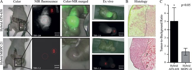
Figure 4. In vivo images and TBRs of mice bearing human orthotopic tumors.

A) UPPER ROW: Representative images of a mouse injected with 75 μg/0.5 nmol of hybrid ATN-658. The tumor on the cecum is exposed using a laparotomy, and measured after 72 h. Fluorescent signals in the colon surrounding the tumor are due to ingested agents as the signals is disappeared when the colon is emptied as seen on the ex vivo images. White arrow indicate the tumor. LOWER ROW: Mouse injected with 75 μg/0.5 nmol hybrid MOPC-21 which showed no specific NIR fluorescent signals. White arrow indicate the tumor. Red dotted circles = Regions of interests (with B as Background). B) Merged images (NIR fluorescent microscopy and histology) show co-localization of the uPAR specific multimodal agent, especially at the tumor border (border surrounded with dashed line). Apart from minor fluorescent signals in necrotic areas (black arrow), no tumor-specific signals are seen with the control agent. (magnification 40x) C) Graph shows the mean TBR and SD (n=3 for each group) of mice with orthotopic colorectal tumors after 72 h (P < 0.05).
