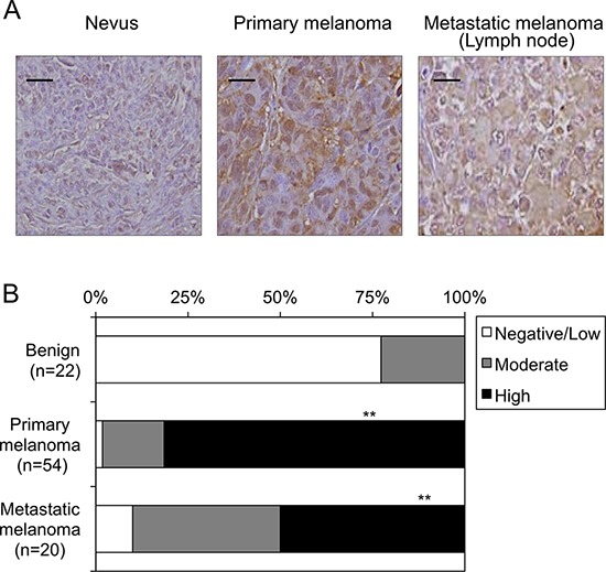
Figure 3. Expression levels of DTX3L in nevi and melanoma tissues in humans.

Representative photographs A. and results of statistical analysis B. of Dtx3l protein expression levels in nevi, primary melanomas and metastatic melanomas for lymph nodes in humans by immunohistochemical analysis are presented. White, gray and black columns show negative/low, moderate and high expression levels, respectively, of DTX3L protein expression levels in nevus, primary melanoma and metastatic melanoma tissues in humans B. Densitometric evaluation for the immunohistochemical results was performed using the software program WinROOF (MITANI Corporation) as previously reported (25). Number of DTX3L negatively/lowly, moderately and highly expressed cells was divided by number of total cells in five fields with 200-fold magnification in each tissue. Significantly different (**, p < 0.01) from nevi by Fisher's exact test. Scale bar, 25 μM.
