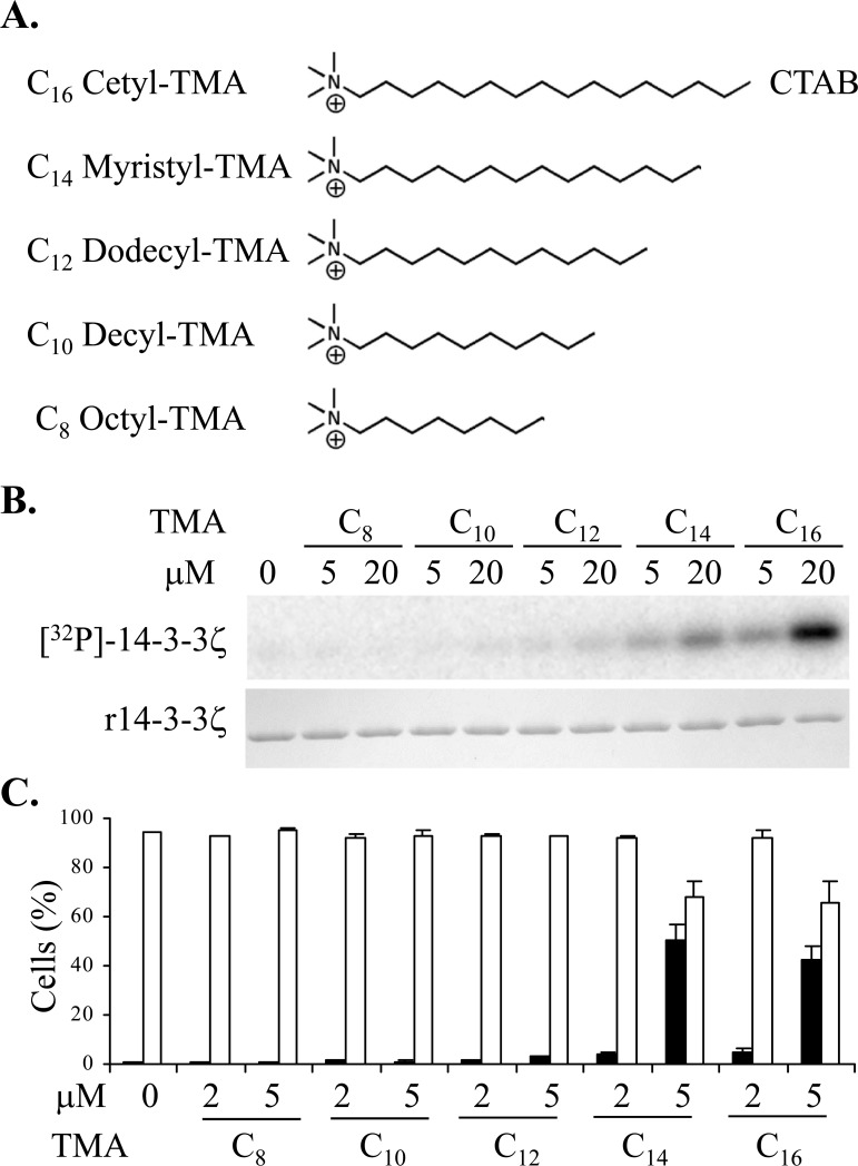
Figure 1.
A. Structures of the trimethylammonium (TMA) compounds assessed for 14-3-3 modulating activity. B. Phosphorylation of 14-3-3 by PKA in vitro in presence or absence of TMA compounds at the concentrations shown. The upper panel is [32P]-phospho-labeled 14-3-3ζ and the lower panel is Coomassie stained 14-3-3 protein. C. Effect of TMA compounds on Jurkat cell after 20 h treatment at the concentrations shown. Cell viability is shown in open bars and TMRE negative staining cells are shown in black bars. The error bars show the range of duplicate determinations: and the results are representative of multiple experiments.

