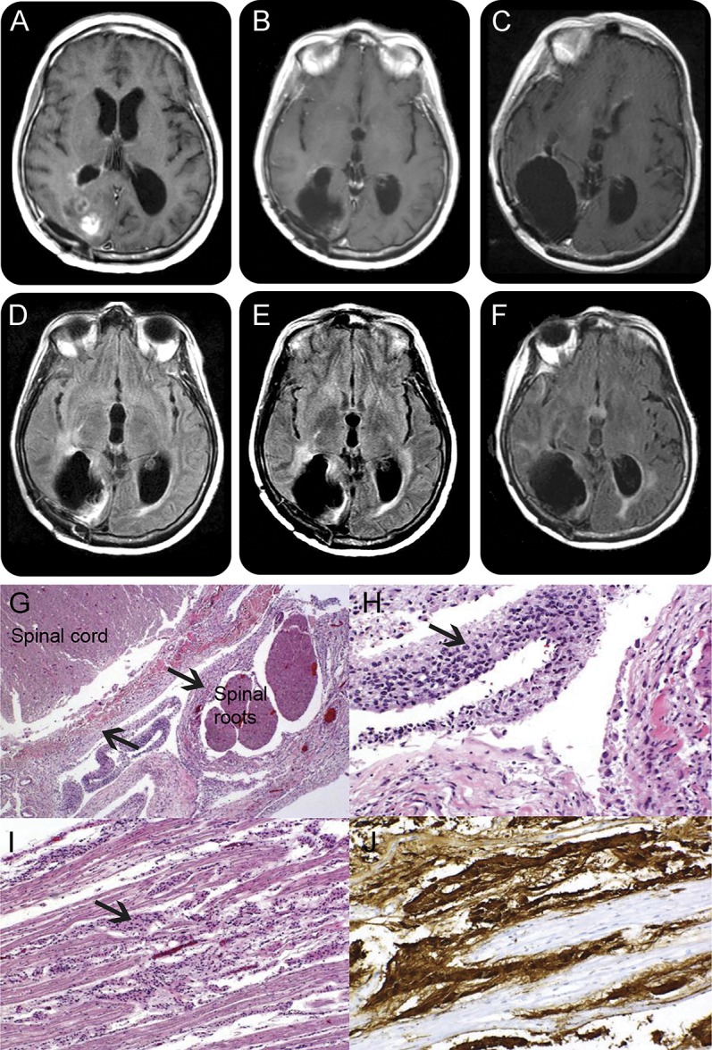
Figure. MRI and autopsy findings.

Brain MRI T1 postgadolinium images 7 months after the diagnosis show mild ventricular dilation and hyperintense signal corresponding to residual tumor (A). In spite of progressive clinical worsening, subsequent scans done 11 (B) and 12 months (C) after diagnosis show no significant progression of enhancing tumor. Corresponding fluid-attenuated inversion recovery sequences at 7 months (D), 11 months (E), and 12 months (F) after diagnosis confirm absence of progression of nonenhancing tumors, as occasionally seen in patients on bevacizumab. This dissociation between neurologic deterioration and absence of neurologic worsening poses a diagnostic dilemma. Autopsy shows extensive leptomeningeal spread of the tumor, with hematoxylin & eosin low power (G) and high power (H) demonstrating the coating of spinal cord and nerve roots by glioma (arrows). (I) Tumor cells coating the cauda equina; glial fibrillary acid protein immunohistochemistry further demonstrates the glial nature of the tumor, shown in brown (J). MRI of the spine (not shown) had normal results, reflecting bevacizumab masking effects.
