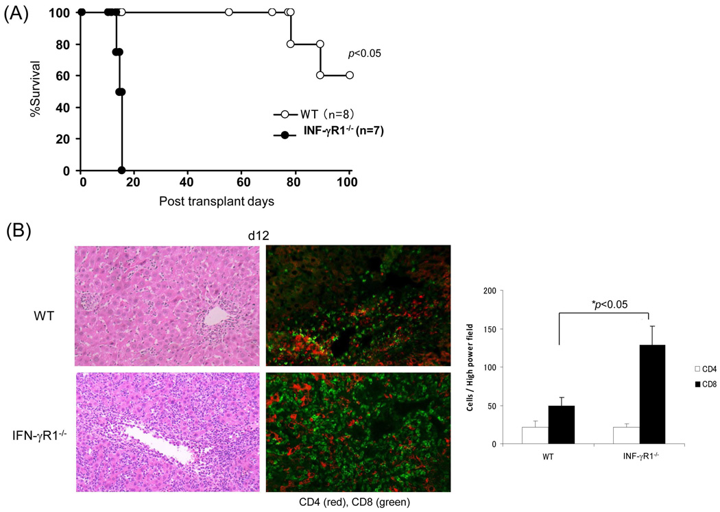
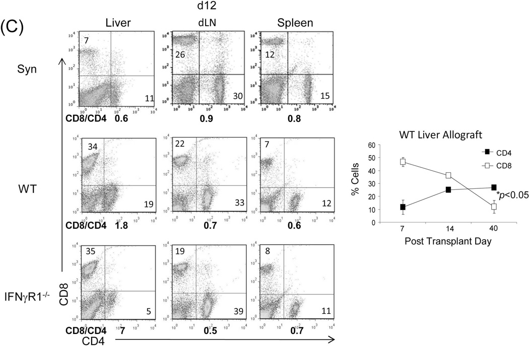
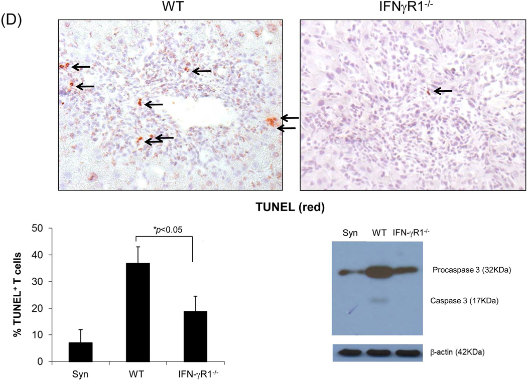
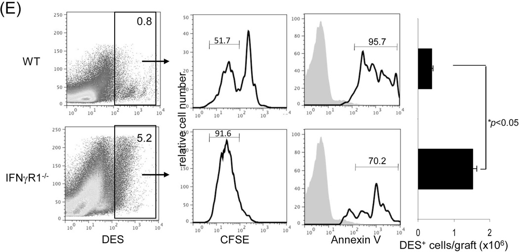
Figure 1. Liver transplant tolerance is mediated by IFN-γ signaling dependent elimination of effector T cells.
Liver grafts from WT or IFN-γR1−/− mice (both B6, H2b) were transplanted into C3H (H2k) recipients. (A) Survival of the liver allografts was compared between WT (N=8) and IFN-γR1−/− (n=7) groups. (B) Histology of liver allo-transplants on POD12. The graft sections were stained with H&E (left panels) and immunohistochemistry for CD4 (red) & CD8 (green) (middle panels), and the CD4+ and CD8+ cells were counted and expressed as mean cell number/high power field ± SD (n=3) (right panel). (C) Flow analyses of liver graft infiltrating T cells. Left panels: lymphocytes isolated from the WT or IFN-γR1−/− liver allografts on POD 12 were stained for CD4 and CD8. Syngeneic B6 liver grafts served as control (Syn). The number is the percentage of T cell subset in total lymphocytes. CD8/CD4 ratio was calculated and displayed at the bottom. Right panel: Percentage of CD4 and CD8 cells in graft infiltrating lymphocytes isolated from the recipients bearing WT liver allograft for 7, 14 or 40 days, and expressed as mean ± SD (n=3). (D) Apoptotic activity in the liver graft. Upper panels: the sections of liver grafts were stained for TUNEL (red). Lower panels: T cells isolated from the liver graft were stained for TUNEL and analyzed by flow cytometry (left). Protein isolated from these T cells was analyzed for expression of caspase 3 by Western blot (right). (E) Impact of IFN-γ signaling on specific CD8 T cell response. On POD 3, recipients were intravenously given 1.5 × 10⁷CFSE-labeled T cells from DES TCR transgenic mice (H2k). Three days later, lymphocytes were isolated from liver grafts and stained with anti-DES and -annexin V mAbs, and analyzed by flow cytometry gated on DES+ cells and expressed as histograms. The number is the percentage of dividing cells and annexin V+ cells, respectively. The shaded area is isotype control. The absolute number of DES+ cells were calculated based on flow data and expressed as mean/graft ± SD (n=3).


