Abstract
Based on the super light trapping property of butterfly Trogonoptera brookiana wings, the SiO2 replica of this bionic functional surface was successfully synthesized using a simple and highly effective synthesis method combining a sol–gel process and subsequent selective etching. Firstly, the reflectivity of butterfly wing scales was carefully examined. It was found that the whole reflectance spectroscopy of the butterfly wings showed a lower level (less than 10 %) in the visible spectrum. Thus, it was confirmed that the butterfly wings possessed a super light trapping effect. Afterwards, the morphologies and detailed architectures of the butterfly wing scales were carefully investigated using the ultra-depth three-dimensional (3D) microscope and field emission scanning electronic microscopy (FESEM). It was composed by the parallel ridges and quasi-honeycomb-like structure between them. Based on the biological properties and function above, an exact SiO2 negative replica was fabricated through a synthesis method combining a sol–gel process and subsequent selective etching. At last, the comparative analysis of morphology feature size and the reflectance spectroscopy between the SiO2 negative replica and the flat plate was conducted. It could be concluded that the SiO2 negative replica inherited not only the original super light trapping architectures, but also the super light trapping characteristics of bio-template. This work may open up an avenue for the design and fabrication of super light trapping materials and encourage people to look for more super light trapping architectures in nature.
Keywords: Bio-template fabrication, Butterfly wing scales, Light trapping materials
Background
Nature offers a variety of surfaces with brilliant properties and is a source of inspiration for numerous applications and techniques. A number of researchers have paid close attention to the fantastic surfaces triggered by nature, such as the anisotropy of the rice leaves [1, 2], the self-cleaning of the lotus leaves [3–5], the antireflection of moth eyes [6–8], the fog collection system in cactus [9, 10], the reversible adhesive of the gecko feet [11, 12], the superhydrophobicity of the water strider legs [13, 14], and the iridescence of the butterfly wings [15–17]. Mimicking or studying the basic principles of the sophisticated tactics from nature is of significant importance for the design of artificial analogs.
During the last few decades, much effort has been directed toward the brightest and most vivid structure-based colors in nature that arise from the interaction of light with surfaces on the micro- and nanoscale. The architectures on the surface of butterfly wings not only formed the gorgeous appearance, but also made the butterfly wings with appealing properties, such as observable optical response to temperature [18], highly selective vapor response [19, 20], and light trapping effect [21, 22]. Although many scientists have done a lot of research on the architectures of butterfly wings and their multi-functional features, little attention has been paid to the black color in spite of its ubiquity. Many scientists have ascribed the blackness of butterfly wings to melanin solely, until recent research results indicated that the nanostructures of scales also play a key role in the exhibition of blackness [23–25]. In addition, seeking for light trapping surfaces has been a great challenge for researchers and now the research on light trapping materials has attracted more and more people’s attention because of the increasing importance of light trapping materials. Considering the above, we chose butterfly Trogonoptera brookiana black wings as a natural model and revealed its properties for super light trapping.
On the other hand, although the color mechanism and structural characterizations have been well investigated for a long time [26], the studies on bionic preparation of the subtle nanostructures on butterfly wing scales have been greatly restricted. In fact, the exact combination of the three-dimensional (3D) structure and cuticle complex refractive index [n* = (1.55 ± 0.05) + i (0.06 ± 0.01)] is beyond the capabilities of existing nanofabrication techniques [27]. In spite of this, the potential valuable application prospect still inspired scientists to devote themselves to mimicking the distinctive surface nanostructure of butterfly wings. The colorful butterfly wing surface has been fabricated using soft lithography technique [28], low-temperature atomic layer deposition [29], colloidal self-assembly, sputtering and atomic layer deposition, even combining all these layer deposition techniques together [30]. Not only sorts of surface processing technology but also a variety of materials were employed to fabricate replicas of the multi-layered scales on the surface of butterfly wing, such as polyelectrolyte multilayer films [31], carbon nanotube [32], polypyrrole [33], oxidizing material include TiO2 [34], Bi2WO6 [35], Fe3O4 [36], SnO2 [19], and so on. Even so, exact replica of such biological structures by an artificial synthesis route are difficult; what is more, the existing studies about butterflies mainly focus on the structural colors, photonic crystal structures, and the replica of photonic structures in butterfly wings. Few researchers showed solicitude for the bionic fabricating of the super light trapping architectures in butterfly wings. So, it is urgently necessary to develop a high-efficiency and low-cost technique to realize the outstanding light trapping architectures.
In this work, we selected the butterfly Trogonoptera brookiana black wings as the bio-templates, and the super light trapping characteristics were characterized by reflectance spectroscopy. Besides, the super light trapping architectures and morphology of the wing scales were characterized by FESEM. In order to prepare the SiO2 negative replica, a simple and highly effective synthesis method combining a sol–gel process and subsequent selective etching was adopted. What is more, the 3D optimized models were generated by CATIA to illustrate the super light trapping architectures and fabrication process effectively. At last, the reflectance spectroscopy of SiO2 negative replica and a flat plate was measured. The SiO2 negative replica was not only stable but also corrosion resistant due to the complex super light trapping architectures and inert SiO2 material, which made it a promising super light trapping surface for various fields such as photoelectrical devices, photo-induced sensors and solar cells. Moreover, this work sets up a strategy for the design and fabrication of super light trapping materials and may encourage people to look for more super light trapping architectures in nature.
Methods
Materials
Analytic grade reagents, hydrochloric acid, and tetraethyl ortho-silicate were provided by Beijing Chemical Works. Ethanol absolute, diethyl ether, concentrated nitric acid, and perchloric acid were provided by Tianjin Fine Chemical Co., Ltd.
Although the green wings of butterfly Trogonoptera brookiana possess a light trapping property which was confirmed as reported in our previous work [22], this study found that the black wing scales had the better light trapping characteristic compared with the previous study. Thus, the black wings were selected as a biological prototype in this work. Butterfly wing samples of uniform size (15 mm in length and 10 mm in width, rectangular) were cut off from the black areas in a perpendicular and parallel direction to the ridge veins, respectively. In order to confirm that the butterfly wing samples were clean, some pre-processing was conducted. Firstly, each sample was soaked by aether for 10 min to remove proteins and fattiness on the samples’ surface. Afterwards, the dehydration treatment in ethanol absolute with duration time of 15 min for each specimen was taken. The purpose of conducting the dehydration pre-processing was to increase the mechanical strength and stability of the treated tissues. Then, the samples were dried naturally in air.
Discoloration Experiment
A simple discoloration experiment was carried out to confirm that the color of the black wing scales was structure-based rather than pigment. Firstly, the neat and clean black areas were cut off from the butterfly wings meticulously with a scalpel in perpendicular and parallel directions to the nervure, respectively. Then, the sample was clamped with a tweezer to flatwise place in a petri dish and soaked in a certain amount of diethyl ether and ethanol absolute for 10 min, respectively, for degreasing and increasing the mechanical strength of the wing tissues. The color of the air-dried sample was still black, which was not affected by organic solvents virtually.
Preparation of the SiO2 Negative Replica
Firstly, the butterfly wing samples were sandwiched between two glass slides of which both ends were clamped by clips with proper force. Using a micropipette, a suitable amount of the sol–gel precursor solution, a reaction product of tetraethyl ortho-silicate and hydrochloric acid (3:1 in volume), was added to the edge of the assembly with a volume of 4–8 μL. Then, the assembly was heated at 125 °C for 25 min in an electric vacuum-drying oven to further solidify the precursor solution on the surface of the wing samples. Next, the whole assembly was dipped into a mixture of concentrated nitric acid and perchloric acid (1:1 in volume) while heating at 130 °C for 40 min to remove the original organisms. Then, the whole assembly was washed by ultrasonic oscillation for 5 min in deionized water to get rid of the residue. After drying at room temperature, the SiO2 negative replica was fabricated.
Spectroscopy Characterization
The reflectance spectroscopy of butterfly Trogonoptera brookiana wings and the SiO2 negative replica were measured using a miniature fiber optic spectrometer (Ocean Optics USB4000-VIS-NIR) equipped with a halogen tungsten lamp source (Ocean Optics LS-1-LL). The spot size of the incident beam was ~2 mm, and the wavelength of the reflectance spectroscopy varied in the range of 400–900 nm. In addition, the element types and content analysis of the surface of the SiO2 negative replica were characterized by virtue of the energy dispersive spectroscopy (EDS, OXFORD X-MaxN 150) on the SEM.
Morphology and Dimension Characterization
The 2D morphologies and structures of the scales of the black areas of butterfly Trogonoptera brookiana hind wings and the SiO2 negative replica were obtained with the help of field emission scanning electronic microscopy (FESEM: JEOL JSM-6700 F). These data will be used to analyze the inheritance accuracy of the SiO2 negative replica.
Results and Discussion
Macroscopic Morphology Observations and Reflectance Spectroscopy Analysis of Black Butterfly Wing Scales
Figure 1a showed the overall view of the original butterfly Trogonoptera brookiana. Obviously, there were long smooth black strips located at the front and hind wings of the butterfly, which looked as beautiful as black velvet. With the help of optical metallurgical microscopy, it could be found that the black part of the wings was covered by bright black scales. These black scales were placed in alternate rows, and the scales overlap each other, as shown in Fig. 1b. The reflectance spectroscopy ranging from 400–900 nm of the butterfly wings is shown in Fig. 1c. It was found that the reflectivity of black wings was lower than that of green wings, which was less than 8 % in the range of 400–900 nm. Based upon the butterfly being a kind of poikilothermal animal, it could be inferred that lower reflectance made contributions to reducing the loss of solar energy so that the butterfly could maintain body temperature, which confirmed that the black areas possess a better light trapping property. Hence, the black areas were chosen as the experimental areas to be studied carefully.
Fig. 1.
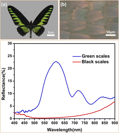
The macroscopic morphology of the butterfly wings and the reflectance spectroscopy of black and green region of the butterfly wings. a Photograph of butterfly Trogonoptera brookiana. b Optical microscopic image of the black butterfly wing scales. c The lower reflectance of the black wing scales was confirmed in the entire wave range
Microscopic Morphology Observations of Black Butterfly Wing Scales
In order to obtain more detailed information about the black scales’ nanostructures, FESEM was employed for the characterization of the morphology and distribution of the black scales, as shown in Fig. 2. It was clearly found that these scales ranged in good order on the substrate under low magnification. The scales with an area of about 160 μm in length and 70 μm in width had sharp serrate ends. The root of each scale was embedded on the substrate, as shown in Fig. 2a. More exquisite nanostructures of the black scales were observed under medium magnification as shown in Fig. 2b, c. It could be observed that the scales had longitudinal ridges, which run through the scales. The distance between two adjacent ridges was approximately 1.6 μm. This surface of the scale comprised a set of raised longitudinal quasi-parallel lamellae (ridges). The space between adjacent ridges was filled with a netlike reticulum composed of pores. As discussed further in this paper, the reticulum and lamellae were both the optical elements that endowed the wing scales a super light trapping effect. It was called a quasi-honeycomb-like structure. Figure 2d shows the high-magnification images of the quasi-honeycomb-like structure. It could be observed clearly that there were parallel fold stripes on both sides of the ridges. These fold stripes could be the proof if whether the original nanostructures in bio-templates were faithfully inherited by the replica or not.
Fig. 2.
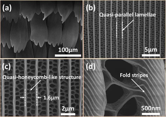
FESEM images of original butterfly scales in the black region with different magnifications. a Lower magnification image. It was found clearly that these scales ranged in good order on the substrate. b, c Medium magnification image. This surface of the scale comprised a set of raised longitudinal quasi-parallel lamellae (ridges), the space between which was filled with a quasi-honeycomb-like structure. d The high-magnification images of the quasi-honeycomb-like structure
The Formation Mechanism of the SiO2 Negative Replica
As shown in Fig. 3, 3D CATIA models were built according to the overall process of the preparation using bio-templates from butterfly Trogonoptera brookiana wings: (a) the clean original templates (3 cm × 2 cm), which was pretreated with diethyl ether and ethanol absolute for 10 min, respectively, was sandwiched between two glass slides; (b) a suitable amount of the precursor solution (~8 μL) was added to the edge of the assembly using a micropipette; (c) the assembly was heated at 125 °C for 25 min in an electric vacuum-drying oven to solidify the precursor solution; (d) the original template was removed through the process of selective etching in a mixture of concentrated nitric acid and perchloric acid (1:1 in volume) at 130 °C for another 40 min. Then, the whole assembly was washed by ultrasonic oscillation for 5 min in deionized water to get rid of the residue. Finally, the whole assembly was washed with deionized water for 15 min. After drying at room temperature, the SiO2 negative replica was fabricated.
Fig. 3.
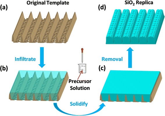
Fabrication process from the original templates of butterfly wings to the SiO2 negative replica. a 3D nanostructured model of the original butterfly wings. b The precursor solution filled the space left between the micro-ridges with a micropipettor. c The precursor solution became solidified through heating process. d The solid negative replica was obtained after the original bio-templates were etched away and cooled at room temperature
Microscopic Morphology Observations and EDS Analysis of the SiO2 Negative Replica
The morphology of the SiO2 negative replica was investigated by FESEM, and the result is illustrated in Fig. 4a. Some scale-like structures were distributed on the surface of the replica, the shapes of which were similar to those of the black scales shown in Fig. 1b. From the aspect of the arrangement, the scaly structures of the replica also had good periodicity and arranged regularly from the front to the end of the replica in the same sequence just like tiles on the roof, which was also analogous with the bio-templates. The structural details of the SiO2 negative replica of the single scale were illustrated in Fig. 4b under medium magnification. It could be observed that notches were lying in parallel, with humps of different shapes between them. These notches were formed from the ridges on the scales of the black wings. The sol–gel precursor filled the space left between the ridges and became solidified, making the places that used to be ridges became notches, and the pores between ridges became humps between notches. Figure 4c is a high-magnification image of the negative replica of the black wing scales. The period of the negative replica nanostructures was about 1.5 μm. The size and shape of the humps were in conformity with those of the pores shown in Fig. 2c, which confirmed that these humps were the reverse structures of those pores. It was worth to mention that parallel nanostructures were obtained on both sides of the notches. These nanostructures were formed from fold stripes on both sides of the ridges mentioned when illustrating the morphology of black wing scales (Fig. 2d). In a word, after comparing Fig. 4 with Fig. 2 from a variety of angles, such as appearance, arrangement, size of scales, notches, and bumps, it was obvious that the original super light trapping architectures of bio-template were well inherited by the SiO2 negative replica. What is more, the appearance and comparison of the fold stripes on both sides of the ridges also draw the conclusion that the original architectures in bio-template were faithfully inherited by the SiO2 negative replica. To the best of our knowledge, this is the first time that a SiO2 negative replica of the original black butterfly Trogonoptera brookiana wing scales has been synthesized. However, the size of the negative replica was a bit different from the original scales. The element types and content analysis of the surface of the SiO2 negative replica were characterized with the help of an energy dispersive spectrometer (EDS). The EDS microanalyses (Fig. 4d) of the SiO2 replica indicated that the SiO2 replica was mainly composed of silicon and oxygenium. Peaks of silicon and oxygenium could be observed clearly, and the weight percentages of these two elements were 31.32 and 45.31 %, respectively. And the element enrichment regions of both silicon and oxygenium were consistent with the shape of the SiO2 negative replicas as the area scanning maps shown in Fig. 4e, f, which further indicated that highly purified SiO2 replicas were obtained.
Fig. 4.
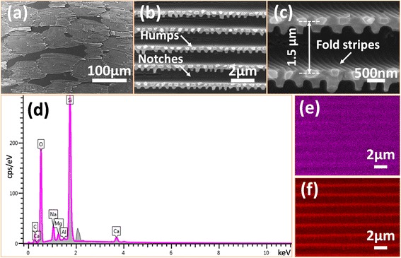
FESEM images and EDS spectrum of the SiO2 negative replica. a Lower magnification image. It could be found that the scales were still arranged in rows. However, they were no longer overlapped with each other. b Medium magnification image of the SiO2 negative replica. It can be observed that notches are lying in parallel, with humps of different shapes between them. c High-magnification images of the replica surface. The sizes and shapes of the humps were in conformity with those of the pores shown in Fig. 2c. d The EDS spectrum showed the main elements constituting the SiO2 negative replicas. e, f The scanning maps of silicon and oxygenium demonstrated the distribution of silicon and oxygenium, which was consistent with the structures shape of the subwavelength antireflective nanoditches arrays
Analysis of the Light Trapping Mechanism of the SiO2 Negative Replicas
The simplified model of the SiO2 negative replicas was built as shown in Fig. 5a. When incident light traveled through air to the SiO2 material, reflection and refraction would happen on the interface at the same time. After multiple reflections and refractions, incident light traveled for a longer distance. Only a small part of the solar energy was reflected back to the air, resulting in most of the incident light being effectively adsorbed within the super light trapping architectures eventually. What is more, the humps on the top of the ridges and the fold stripes on both sides of the ridges enhanced the scattering of incident light within super light trapping architectures of the SiO2 negative replicas, which also reduced the solar energy loss (SEL) due to the reflectance from the SiO2 negative replica.
Fig. 5.
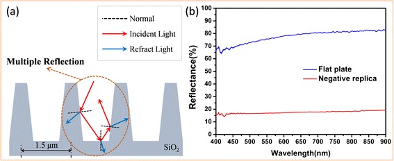
Schematic illustration of the multiple reflection and refraction occurred in the SiO2 negative replicas and the reflectance spectroscopy analysis of the SiO2 negative replica. a After multiple reflections and refractions, incident light traveled for a longer distance, and only a small part of the solar energy was reflected back to the air. b The average reflection of the SiO2 negative replicas was about 20 % which was just 1/4 of the reflection of the flat plate without the negative quasi-honeycomb-like structure
Measured reflectance spectroscopy in Fig. 5b shows that the applying of the negative quasi-honeycomb-like structure on flat plate played the key role in producing the super light trapping property. The red curve shows that the average reflection of the SiO2 negative replica was about 20 %, and the blue curve shows that the average reflection of the flat plate was about 80 %. It was obtained that the average reflection of the SiO2 negative replica was just 1/4 of the reflection of the flat plate without the negative quasi-honeycomb-like structure. To further clarify the influence of the negative quasi-honeycomb-like structure on the super light trapping property of the SiO2 negative replica, the SEL caused by the reflection of the SiO2 negative replica and the flat plate was calculated according to the equation below, where I (λ) is the solar energy intensity as a function of the wavelength λ at AM1.5 and α (λ) is the measured reflectance of the SiO2 negative replica, and the flat plate is a function of the wavelength λ [37–40].
The SEL calculated from the measured reflectance spectroscopy (seen in Fig. 5b) of the flat plate and the SiO2 negative replica were 491.34 and 110.98 W/m2, respectively. Apparently, the SEL of the flat plate was more than four times the SEL of the SiO2 negative replica, which confirmed that the SiO2 negative replica had the better light trapping characteristics. Given that they shared uniform intensity of the light and material, it could be inferred from the results that the negative quasi-honeycomb-like structure which was borrowed from the black wing scales of butterfly acted as super light trapping architectures. Thus, it could be concluded that the SiO2 negative replica inherited not only the original super light trapping architectures, but also the super light trapping characteristics of bio-template.
Conclusions
In summary, the reflectivity of the black wings of butterfly Trogonoptera brookiana was carefully examined, and the wings showed good optical absorption in the visible spectrum, which confirmed that the black wings of this butterfly possessed structure-based super light trapping property. So, a super light trapping material was then fabricated templated from these black wings by transferring the quasi-honeycomb-like structure from the black wing scales to flat plate. The structural replication fidelity of the process is demonstrated on both the macro- and microscale, and even down to the nanoscale, as evidenced by FESEM and energy dispersive spectroscopy (EDS). The comparison results of feature size between the SiO2 negative replica and original bio-templates were in concordance to some extent. Considering of the final observation result, the obtained SiO2 negative replica preserved the super light trapping architectures successfully from the aspects of morphology, dimensions, and distributions of the scales. At last, the reflectance spectroscopy of the SiO2 negative replica and a flat plate was measured. It was found that the SiO2 negative replica obtained 20 % reflection in visible light spectrum. Reflection of the SiO2 negative replica with the negative quasi-honeycomb-like structure was merely 1/4 of that in the flat plate. Thus, it was obvious that the obtained SiO2 negative replica possessed super light trapping property. From the analysis above, it was proved that the super light trapping surface of original bio-templates was also inherited by the SiO2 negative replica faithfully in terms of structure and function. Thus, it could be concluded that the SiO2 negative replica inherited not only the original super light trapping architectures, but also the super light trapping characteristics of bio-template.
The manufacture of the super light trapping architectures of butterfly Trogonoptera brookiana black wing scales is meaningful. The SiO2 negative replica was not only stable but also corrosion resistant due to the complex super light trapping architectures and inert SiO2 material, which made it a promising light trapping surface for various fields such as photoelectrical devices, photo-induced sensors, and solar cells. Moreover, this work sets up a strategy for the design and fabrication of super light trapping materials and may encourage people to look for more super light trapping architectures in nature.
Acknowledgements
This work is supported by the National Natural Science Foundation of China (Nos. 51175220, 51505183, 51325501, and 51290292), and China Postdoctoral Science Foundation Funded Project (Project No. 2015 M571360).
Abbreviations
- SEM
scanning electron microscope
- FESEM
field emission scanning electron microscope
- EDS
energy dispersive spectroscopy
- ALD
atomic layer deposition
Footnotes
Competing Interests
The authors declare that they have no competing interests.
Authors’ Contributions
ZWH and SCN designed the study. BL and ZZM performed the experiments with help from MY. BL and SCN contributed in drafting the manuscript. All the authors provided technical and scientific insight and contributed to the editing of the manuscript. All authors read and approved the final manuscript.
Contributor Information
Zhiwu Han, Email: zwhan@jlu.edu.cn.
Bo Li, Email: libo14@mails.jlu.edu.cn.
Zhengzhi Mu, Email: muzz13@mails.jlu.edu.cn.
Meng Yang, Email: yangmeng12@mails.jlu.edu.cn.
Shichao Niu, Email: niushichao@jlu.edu.cn.
Junqiu Zhang, Email: zhangjunqiu2005@163.com.
Luquan Ren, Email: lqren@jlu.edu.cn.
References
- 1.Zhu DF, Li X, Zhang G, Zhang X, Zhang X, Wang TQ, et al. Mimicking the rice leaf-from ordered binary structures to anisotropic wettability. Langmuir. 2010;26:14276–83. doi: 10.1021/la102243c. [DOI] [PubMed] [Google Scholar]
- 2.Anjusree GS, Bhupathi A, Balakrishnan A, Vadukumpully S, Subramanian KRV, Sivakumar N, et al. Fabricating fiber, rice and leaf-shaped TiO2 by tuning the chemistry between TiO2 and the polymer during electrospinning. RSC Adv. 2013;3:16720–7. doi: 10.1039/c3ra42250j. [DOI] [Google Scholar]
- 3.Nishimoto S, Bhushan B. Bioinspired self-cleaning surfaces with superhydrophobicity, superoleophobicity, and superhydrophilicity. RSC Adv. 2013;3:671–90. doi: 10.1039/C2RA21260A. [DOI] [Google Scholar]
- 4.Liu Y, Li SY, Zhang JJ, Wang YM, Han ZW, Ren LQ. Fabrication of biomimetic superhydrophobic surface with controlled adhesion by electrodeposition. Chem Eng J. 2014;248:440–7. doi: 10.1016/j.cej.2014.03.046. [DOI] [Google Scholar]
- 5.Koch K, Bhushan B, Jung YC, Barthlott W. Fabrication of artificial lotus leaves and significance of hierarchical structure for superhydrophobicity and low adhesion. Soft Matter. 2009;5:1386–93. doi: 10.1039/b818940d. [DOI] [Google Scholar]
- 6.Park H, Shin D, Kang G, Baek S, Kim K, Padilla WJ. Broadband optical antireflection enhancement by integrating antireflective nanoislands with silicon nanoconical-frustum arrays. Adv Mater. 2011;23:5796–800. doi: 10.1002/adma.201103399. [DOI] [PubMed] [Google Scholar]
- 7.Liu KS, Jiang L. Multifunctional integration: from biological to bio-inspired materials. ACS Nano. 2011;5:6786–90. doi: 10.1021/nn203250y. [DOI] [PubMed] [Google Scholar]
- 8.Chen Q, Hubbard G, Shields PA, Liu C, Allsopp DWE, Wang WN, et al. Broadband moth-eye antireflection coatings fabricated by low-cost nanoimprinting. Appl Phys Lett. 2009;94:263118. doi: 10.1063/1.3171930. [DOI] [Google Scholar]
- 9.Ju J, Yao X, Yang S, Wang L, Sun RZ, He YX, et al. Cactus stem inspired cone-arrayed surfaces for efficient fog collection. Adv Funct Mater. 2014;24:6933–8. doi: 10.1002/adfm.201402229. [DOI] [Google Scholar]
- 10.Cao MY, Ju J, Li K, Dou SX, Liu KS, Jiang L. Facile and large-scale fabrication of a cactus-inspired continuous fog collector. Adv Funct Mater. 2014;24:3235–40. doi: 10.1002/adfm.201303661. [DOI] [Google Scholar]
- 11.Wang ZZ, Gu P, Wu XP. A gecko-inspired double-sided adhesive. Phys Chem Chem Phys. 2013;15:20764–70. doi: 10.1039/c3cp53961j. [DOI] [PubMed] [Google Scholar]
- 12.Su YW, Ji BH, Huang YG, Hwang K. Effects of contact shape on biological wet adhesion. J Mater Sci. 2007;42:8885–93. doi: 10.1007/s10853-007-1759-7. [DOI] [Google Scholar]
- 13.Liu KS, Tian Y, Jiang L. Bio-inspired superoleophobic and smart materials: design, fabrication, and application. Prog Mater Sci. 2013;58:503–64. doi: 10.1016/j.pmatsci.2012.11.001. [DOI] [Google Scholar]
- 14.Ji XY, Wang JW, Feng XQ. Role of flexibility in the water repellency of water strider legs: theory and experiment. Phys Rev E. 2012;85:021607. doi: 10.1103/PhysRevE.85.021607. [DOI] [PubMed] [Google Scholar]
- 15.Niu SC, Li B, Mu ZZ, Yang M, Zhang JQ, Han ZW, et al. Excellent structure-based multifunction of Morpho butterfly wings: a review. J Bionic Eng. 2015;12:170–89. doi: 10.1016/S1672-6529(14)60111-6. [DOI] [Google Scholar]
- 16.Zhang D, Zhang W, Gu JJ, Fan TX, Liu QL, Su HL, et al. Butterfly effects: novel functional materials inspired from the wings scales. Prog Mater Sci. 2015;68:67–96. doi: 10.1016/j.pmatsci.2014.10.003. [DOI] [PubMed] [Google Scholar]
- 17.England G, Kolle M, Kim P, Khan M, Muñoz P, Mazur E, et al. Bioinspired micrograting arrays mimicking the reverse color diffraction elements evolved by the butterfly Pierella luna. Proc Natl Acad Sci. 2014;111:15630–4. doi: 10.1073/pnas.1412240111. [DOI] [PMC free article] [PubMed] [Google Scholar]
- 18.Pris AD, Utturkar Y, Surman C, Morris WG, Vert A, Zalyubovskiy S, et al. Towards high-speed imaging of infrared photons with bio-inspired nanoarchitectures. Nat Photonics. 2012;6:195–200. doi: 10.1038/nphoton.2011.355. [DOI] [Google Scholar]
- 19.Zhang W, Tian JL, Wang YA, Fang XT, Huang YQ, Chen WX, et al. Single porous SnO2 microtubes templated from Papilio maacki bristles: new structure towards superior gas sensing. J Mater Chem A. 2014;2:4543–50. doi: 10.1039/C3TA13845C. [DOI] [Google Scholar]
- 20.Han ZW, Niu SC, Yang M, Mu ZZ, Li B, Zhang JQ, et al. Unparalleled sensitivity of photonic structures in butterfly wings. RSC Adv. 2014;4:45214–9. doi: 10.1039/C4RA06117A. [DOI] [Google Scholar]
- 21.Han ZW, Niu SC, Zhang LF, Liu ZN, Ren LQ. Light trapping effect in wing scales of butterfly Papilio peranthus and its simulations. J Bionic Eng. 2013;10:162–9. doi: 10.1016/S1672-6529(13)60211-5. [DOI] [Google Scholar]
- 22.Han ZW, Niu SC, Shang CH, Liu ZN, Ren LQ. Light trapping structures in wing scales of butterfly Trogonoptera brookiana. Nanoscale. 2012;4:2879–83. doi: 10.1039/c2nr12059c. [DOI] [PubMed] [Google Scholar]
- 23.Martín-Palma RJ, Pantano CG, Lakhtakia A. Biomimetization of butterfly wings by the conformal-evaporated-film-by-rotation technique for photonics. Appl Phys Lett. 2008;93:083901. doi: 10.1063/1.2973167. [DOI] [PubMed] [Google Scholar]
- 24.Zhao QB, Fan TX, Ding J, Zhang D, Guo QX, Kamada M. Super black and ultrathin amorphous carbon film inspired by anti-reflection architecture in butterfly wing. Carbon. 2011;49:877–83. doi: 10.1016/j.carbon.2010.10.048. [DOI] [Google Scholar]
- 25.Zhao QB, Guo XM, Fan TX, Ding J, Zhang D, Guo QX. Art of blackness in butterfly wings as natural solar collector. Soft Matter. 2011;7:11433. doi: 10.1039/c1sm06167d. [DOI] [Google Scholar]
- 26.Zhang FY, Shen QC, Shi XD, Li SP, Wang WL, Luo Z, et al. Infrared detection based on localized modification of Morpho butterfly wings. Adv Mater. 2015;27:1077–82. doi: 10.1002/adma.201404534. [DOI] [PubMed] [Google Scholar]
- 27.Potyrailo RA, Ghiradella H, Vertiatchikh A, Dovidenko K, Cournoyer JR, Olson E. Morpho butterfly wing scales demonstrate highly selective vapour response. Nat Photonics. 2007;1:123–8. doi: 10.1038/nphoton.2007.2. [DOI] [Google Scholar]
- 28.Kang SH, Tai TY, Fang TH. Replication of butterfly wing microstructures using molding lithography. Curr Appl Phys. 2010;10:625–30. doi: 10.1016/j.cap.2009.08.007. [DOI] [Google Scholar]
- 29.Liu F, Liu YP, Huang L, Hu XH, Dong BQ, Shi WZ, et al. Replication of homologous optical and hydrophobic features by templating wings of butterflies Morpho menelaus. Opt Commun. 2011;284:2376–81. doi: 10.1016/j.optcom.2011.01.017. [DOI] [Google Scholar]
- 30.Kolle M, Salgard-Cunha PM, Scherer MR, Huang F, Vukusic P, Mahajan S, et al. Mimicking the colourful wing scale structure of the Papilio blumei butterfly. Nat Nanotechnol. 2010;5:511–5. doi: 10.1038/nnano.2010.101. [DOI] [PubMed] [Google Scholar]
- 31.Hiller J, Mendelsohn JD, Rubner MF. Reversibly erasable nanoporous anti-reflection coatings from polyelectrolyte multilayers. Nat Nanotechnol. 2002;1:59–63. doi: 10.1038/nmat719. [DOI] [PubMed] [Google Scholar]
- 32.Miyako E, Sugino T, Okazaki T, Bianco A, Yudasaka M, Iijima S. Self-assembled carbon nanotube honeycomb networks using a butterfly wing template as a multifunctional nanobiohybrid. ACS Nano. 2013;7:8736–42. doi: 10.1021/nn403083v. [DOI] [PubMed] [Google Scholar]
- 33.Tang J, Zhu SM, Chen ZX, Feng CL, Shen YJ, Yao F, et al. Replication of polypyrrole with photonic structures from butterfly wings as biosensor. Mater Chem Phys. 2012;131:706–13. doi: 10.1016/j.matchemphys.2011.10.038. [DOI] [Google Scholar]
- 34.Ding L, Zhou H, Lou S, Ding J, Zhang D, Zhu HX, et al. Butterfly wing architecture assisted CdS/Au/TiO2 Z-scheme type photocatalytic water splitting. Int J Hydrogen Energ. 2013;38:8244–53. doi: 10.1016/j.ijhydene.2013.04.093. [DOI] [Google Scholar]
- 35.Yao F, Yang QQ, Yin C, Zhu SM, Zhang D, Moon WJ, et al. Biomimetic Bi2WO6 with hierarchical structures from butterfly wings for visible light absorption. Mater Lett. 2012;77:21–4. doi: 10.1016/j.matlet.2012.02.116. [DOI] [Google Scholar]
- 36.Peng WH, Zhu SM, Zhang W, Yang QQ, Zhang D, Chen ZX. Spectral selectivity of 3D magnetophotonic crystal film fabricated from single butterfly wing scales. Nanoscale. 2014;6:6133–40. doi: 10.1039/c4nr00477a. [DOI] [PubMed] [Google Scholar]
- 37.Wang ZY, Zhang RJ, Wang SY, Lu M, Chen X, Zheng YX, et al. Broadband optical absorption by tunable Mie resonances in silicon nanocone arrays. Sci Rep. 2015;5:7810. doi: 10.1038/srep07810. [DOI] [PMC free article] [PubMed] [Google Scholar]
- 38.Wu ZL, Wang ZY, Wang SY, Zhong ZY. Substantial influence on solar energy harnessing ability by geometries of ordered Si nanowire array. Nanoscale Res Lett. 2014;9:495. doi: 10.1186/1556-276X-9-495. [DOI] [PMC free article] [PubMed] [Google Scholar]
- 39.Spinelli P, Verschuuren MA, Polman A. Broadband omnidirectional antireflection coating based on subwavelength surface Mie resonators. Nat Commun. 2012;3:692. doi: 10.1038/ncomms1691. [DOI] [PMC free article] [PubMed] [Google Scholar]
- 40.Xi JQ, Schubert MF, Kim JK, Schubert EF, Chen MF, Lin SY, et al. Optical thin-film materials with low refractive index for broadband elimination of Fresnel reflection. Nat Photonics. 2007;1:176–9. [Google Scholar]


