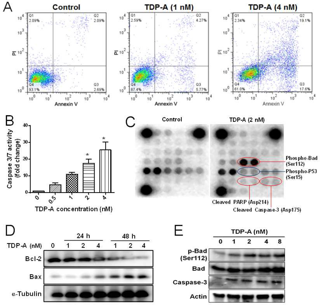
Figure 2. TDP-A induced apoptosis in MDA-MB-231 breast cancer cells.
(A) Flow cytometric analysis of apoptosis by annexin V/PI dual staining assay after TDP-A treatment (1 and 4 nM) for 48 h. (B) The activities of caspase 3/7 in MDA-MB-231 cells after treatment with various concentrations of TDP-A (0, 0.5, 1, 2 and 4 nM) for 24 h. (C) Chemiluminescent array images of the PathScan Intracellular Signaling array kit in MDA-MB-231 cells treated with TDP-A (2 nM) for 24 h. (D) Western blot of Bcl-2 and Bax expression after TDP-A treatment for 24 h or 48 h. (E) Western blot of phosphor-Bad (Ser112), Bad, caspase-3 protein expression after TDP-A treatment (24 h).

