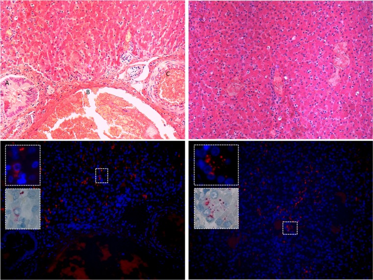
FIG 4 .
Harbor seal infected liver tissue. (Top) Two liver sections from animal NEAQ-11-295-Pv, stained with hematoxylin; left-hand image includes bile duct (A), hepatic vein (B), and hepatic artery (C). No inflammation is observed in the infected tissue. (Bottom) Distribution of phopivirus in liver using fluorescent in situ hybridization (FISH), with higher-magnification insets demonstrating clear cytoplasmic infection.

