Abstract
Background:
Lead poisoning has been an old but perpetual public health problem in developing countries. Lead has an adverse effect on fertility, and this study aimed to examine the effect of consuming green tea extract (GTE) on fertility parameters in rats exposed to lead.
Materials and Methods:
In this experimental study, 70 rats have been classified, as it is described later, into 4 groups of 10 and were studied over 2 months. Group 1: Normal diet and tap water; Group 2: 10 mg/kg intraperitoneal lead acetate weekly over 8 weeks; Group 3: Lead acetate + 100 mg/kg green tea, Group 4: Extract green tea. Distal epididymal sperm samples were collected to assess the sperm counts, motility, and morphology. Testicular tissue and blood level of testosterone were also studies. Data were analyzed by SPSS-17 software using ANOVA and independent t-test with a significant level of 0.05.
Results:
The rats exposed to lead acetate had the lowest weight, and green tea had the highest weight. Green tea consumption in rats exposed to lead, reduced the effect of lead and the difference in mean body weight in these rats, compared to other groups, was minimized (P < 0.05). The group exposed to lead acetate had the highest sperm abnormalities, and the lowest sperm abnormalities were observed in groups taking green tea.
Conclusion:
Consumption of green tea can reduce the adverse effects of lead, and also can effectively prevent fertility reduction.
Keywords: Detoxification, green tea, lead acetate
INTRODUCTION
About 15% of couples naturally and without any problem are not fertilized in the 1st year of their common life. The causes are 40% male infertility, 40% female infertility, 10% both sexes, and 10% is unknown problems. Male infertility can be caused by different factors such as environment and exposure to toxic chemicals (e.g., lead), and preventing the risks of exposure to these substances and identification of appropriate and simple treatments are of most importance.[1]
Lead (Pb), a ubiquitous and potent toxic agent, induces several physiological and behavioral changes while Pb alters the function of multiple organs and systems; it primarily affects the reproductive system. Pb can produce oxidative stress, barrier and alter several Ca2+ -dependent processes, including physiological processes that involve nitric oxide synthesis in development and adult animals.[2,3]
Recent studies have shown that lead (Pb) could disrupt tissue prooxidant/antioxidant balance which lead to physiological dysfunction. Natural antioxidants are particularly useful in such situation. In a study, the effect of GTE on lead-induced toxicity was studied in Sprague-Dawley rats. Results of their study showed that lead exposure was found to attenuate the antioxidant potential of liver, which was, however, augmented when supplemented with GTE. Liver enzymes alanine transaminase, aspartate aminotransferase and alkaline phosphatase and serum protein determinations indicated the protective effects of GTE. Histopathological studies of liver revealed that supplementation of GTE resulted in mild degeneration and congestion of the blood vessels and an enhanced regenerative capacity.[4,5,6]
Lead poisoning has been an old but perpetual public health problem in developing countries. Lead has an adverse effect on fertility, and this study aimed to examine the effect of consuming GTE on fertility parameters in rats exposed to lead.
According to the lead exposure in general population and the side effects on their reproductive system and the properties of green tea and the availability and acceptance by the people to use it, this study was conducted to determine the effect of GTE on fertility of rats exposed to lead. The aim of the study was investigated the detoxification effect of GTE in rats exposed to lead acetate. We surveyed the detoxification effect of green tea in reproductive system and infertility due to lead poisoning.
MATERIALS AND METHODS
This is an in vitro experimental study, in which 70 Wistar male rats about 200 ± 250 in weight, randomly classified into 4 groups of 10, as described later, were studied in 12 h light and 12 h dark cycle with enough access to food and water during 2 months.
Group 1: Normal diet and tap water
Group 2: 10 mg/kg intraperitoneal lead acetate weekly administered over 8 weeks
Group 3: Lead acetate + green tea 100 mg/kg[5]
Group 4: GTE.
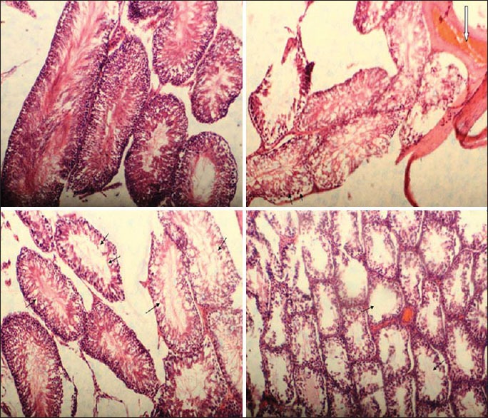
The results of testicular tissue slices from four groups are showed in Figure 1.
Figure 1.

The results of testicular tissue slices from four groups. Rat testis. Normal tubules and interstitial tissue (H and E *400) (group 1). Normal: Seminiferous tubules contain 3-5 cell layered spermatogenic epithelium with normal spermatid, Germinal epithelium detachment from basement membrane of a seminiferous tubule (black arrows) and Hyperaemia (white arrow) in testicular interstitium and sever reduction in number luminal spermatozoa in areas in a rat exposed to (group 2). (H and E *40), Rat testis. Atrophy, tubular, segmental and Vacuolar degeneration with necrosis in the germinal epithelium of a seminiferous tubule (arrows) (arrow) (group 3) (H and E *100), Reduction in number luminal spermatozoa and Atrophy, tubular, diffuse (arrow) (H and E *100) (group 4)
Blood sampling
Blood was taken from ventricles of animals under ether anesthesia; then blood samples were centrifuged at 300 rpm for 5 min and serum was separated. Hormones were measured using routine laboratory methods as well as using radioimmunoassay and Kentron gamma counter apparatus which was made in Switzerland. Mice semen samples were taken for chromatin DNA analysis, and each sample was kept in incubator with 37°C for 30 min in order for liquefaction. After this, seminal liquid analysis involved the determination of sperm concentration, motility, normal morphology and other microscopic and macroscopic parameters which were conducted according to WHO guidelines, and the remaining sample was used for sperm chromatin tests. Histology was conducted by an expert with equipment in the laboratory.
Data collection methods and tools
Plant extraction
In order to prepare GTE, its dry leaves are grinded by an electronic mill, and then passed through a sieve number 10; the obtained powder is mixed with ethanol-80 with the ratio of 1:1, and then the soaked powder is transferred to a percolator, and then extracted with percolation method according to German Pharmacopoeia 10. Then, the resulting extract will be distilled with vacuum rotary evaporator to the withdrawal of alcohol, and the produced extract of green tea will be used in later stages of the study.
Evaluation of antioxidant activities of the plant
The system of linoleat beta-carotene model is used for the evaluation of antioxidant activities.
The measurement of total phenol
The amount of total phenolic compounds is measured according to folin-ciocalteu colorimetric method and based on Gallic acid.[7]
Fertility evaluation
After 2 months of the above regimen, the mice were killed by cervical dislocation and their epididymis was removed. Spermatozoa of the epididymis were released in the culture medium of Gibco Ham's F10 Company, and the motility of sperms was examined. Then, the percentage of alive sperms was measured by eosin staining, and sperms containing DNA were examined by acridine orange staining. The ratio of motile to nonmotile sperms (motility) was expressed as a percentage, and it was determined by direct microscopic examination. Sperm live-dead counting (viability) was conducted by the permeability property of cell membrane with vital eosine-nigrosin staining,[8] and finally, distal epididymal sperm samples were collected to assess count, motility and morphology of sperms.[9]
Histological evaluation of testicle
In order for histological evaluation, the right testicle was cut into small pieces and by fixing it into Bouin solution and washing with lithium carbonate, 6-micron transverse sections were prepared, and hematoxylin and eosin staining were performed. Then, spermatogenesis process was examined through the optical microscope.[10]
DATA ANALYSIS METHOD
The results were obtained through a computer with SPSS-18 software and ANOVA, and significance level was considered to be P ≤ 0.05.
Ethical considerations
Permission from the deputy of research and technology.
Observance of all issues related to animal rights.
RESULTS
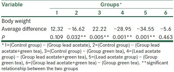
The results of this study revealed that those rats exposed to lead acetate had the lowest weight, and rats taking green tea found to have the highest weight, respectively (213.57 ± 11.43 g vs. 235.8 ± 9.02 g, P < 0.05), which showed a significant difference between all groups and lead acetate group in a way that it seems green tea had an increasing and lead acetate had a reducing effect on the rats’ weight [Table 1].
Table 1.
Comparison between the average ranges of body weight in the four groups

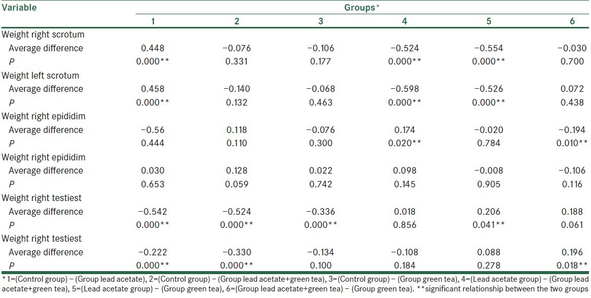
Also, the mean weight of left and right testicles in control group was approximately equal, but in other groups, there was slight difference between the weight of right and left testicles and after that the group of rats exposed to lead acetate had the lowest mean weight of testicles (right: 0.71 ± 0.15 g and left: 0.69 ± 0.18 g) and the green tea consumed group had the highest weight of testicle (right: 1.26 ± 0.11 g and left: 1.32 ± 0.13 g). There was a significant difference between the lead acetate and control groups and also between the lead acetate and green tea groups (P < 0.05).
On the other hand, lead acetate has decreased the weight of the right and left epididymis but the green tea had a desired effect with an increase with no significant difference (P > 0.05).
The mean weight of the right and the left kidneys in the lead-exposed rats used green tea had no significant change in comparison with lead acetate group, but it was significant in the lead acetate and green tea groups (P < 0.05) [Table 2].
Table 2.
Comparison between the average ranges of scrotum and epididim and testiest weight in the four groups

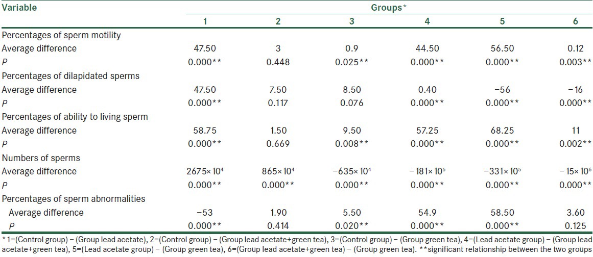
In addition, based on the results of the present study, in the lead acetate exposed group the percentage of sperm motility (22.5% ±0.08%), sperm progression (15% ±0.07%), sperm viability (13.8 ± 0.03) and mean count of sperm (105 × 105 ± 3,915,780) was significantly lower than in other groups (P < 0.05) but in green tea group these values were higher with these amounts respectively 79 ± 0.03, 71 ± 0.06, 82 ± 0.04 and 436 × 105 ± 5,189,733 with significant difference (P < 0.05). The percentage of sperm disorders was more in lead acetate group rather than green tea group (72.5 ± 203 vs. 14 ± 3) [Table 3].
Table 3.
Comparison between the percentages of sperm parametric in the four groups

DISCUSSION
The aim of this study was to determine the effects of GTE consumption on fertility parameters of male rats which had been exposed to lead.
The results of this study revealed that those rats exposed to lead acetate had the lowest weight, and rats taking green tea found to have the highest weight, respectively. Also, the addition of green tea consumption to those rats which were exposed to lead, reduced the effect of lead, and the difference in mean body weight in rats, compared to other groups, was minimized (P < 0.05).
In a study conducted by Khalaf et al. in 2012, the rats exposed to lead acetate also had lost weight. Moreover, glutathione in the brain tissues of these rats was significantly reduced. In addition, the results of the above study indicated that GTE protects against lead neurotoxicity in rats exposed to lead acetate, and GTE can prevent DNA damage in neural tissues.[10] The study of Kirchgatterer et al. also indicated that lead poisoning can be an independent risk factor for weight loss.[11] The results of the above study are consistent with the present study.
The adverse impacts of lead on DNA, especially on proliferating cells, are more evident. Testicular tissues and spermatozoid progenitor cells are from factors which are at risk.
In a study carried out by Graça et al. in 2004, it was observed that the testes weight, seminiferous tubule diameter and sperm count were significantly decreased by lead administration, however, these effects are reversible over time.[12] Also, Anjum et al., in their study in 2011, added lead acetate to the water of rats for 45 days, and they observed that genitalia weight, count, motility and viability of sperms were dramatically reduced in the group exposed to lead.[13]
The results of this study also indicated that the mean weight of left and right testicles in control group was approximately equal, but in other groups, there was slight difference between the weight of right and left testicles; and the group of rats exposed to lead acetate had the lowest mean weight of testicles. In other words, those rats exposed to lead acetate that took GTE had significantly lower mean weight of testicles than the group that hadn’t taken the GTE.
On the other hand, the previous studies indicated that antioxidants can have a protective effect on adverse impacts of lead in sperm count and motility.
In a study in 2009, Shan et al. examined the effect of acid ascorbic and thiamin on rats which simultaneously took lead intragastrically within 6 weeks, and compared to control group they observed sperm count and motility reduction in lead group, although, these parameters were higher in ascorbic group. Therefore, regarding the results of the above study, acid ascorbic with thiamin, due to their antioxidant characteristics, can play a protective role on genitals’ exposure to lead.[7]
In most of the studies, cellular protective properties of green tea are attributed to its antioxidant effects.[14,15]
By examining the therapeutic effect of green tea on allergic vernal conjunctivitis inflammation, Attarzadeh et al. showed that green tea has health benefits, such as prevention of cellular proliferation by inducing apoptosis, and also it has antioxidant properties, which seems that, due to its lack of noticeable complications in topical and systemic applications (based on previous studies), can be used for treatment of this disease.[16]
In addition to lead acetate effects on body weight, and based on the results of the present study, this substance can have potential effects on sperm parameters; so that the percentage of sperm motility in rats exposed to lead acetate was significantly lower than in other groups it means that lead acetate had dramatic effect on the percentage of sperm motility. Also, sperm count in rats exposed to lead acetate was significantly low; on the contrary, the group of rats which received green tea had the highest sperm count.
Unlike the present study, Murthy et al. and also Johansson and Wide in separate studies, found no significant changes in epididymal sperm count and motility after exposing rats to lead.[17,18]
The research results of the above study, with respect to protective property of green tea, are consistent with the results of the present study.
In another recent study which was carried out by Dias et al. in 2014, antioxidant effect of GTE was examined. The research results showed that due to antioxidant properties, green tea also prevents changes in and destruction of sperm cells stored in glass in room temperature.[19]
Most of the studies indicate the adverse effects of lead on spermatozoa performance and production, and subsequently resulting in infertility.[20,21]
The results of the present study also revealed that the group exposed to lead acetate had the highest sperm abnormality; in contrast, minimum sperm abnormalities were observed in groups taking green tea. So, it probably can be said that green tea can be effective in reducing sperm abnormalities.
Along with the present study, Graça et al. examined low or moderate dose of lead acetate on rats over 2–4 weeks. The results showed that sperm velocity and the rate of abnormal sperms decreased at both doses, but with changes found in luteinizing hormone and follicle-stimulating hormone rates. Testosterone levels increased and this indicated that lead has a selective effect on performance of testicles.[12]
Also, another study showed that abnormal sperm counts increase in Algerian rats with the administration of sequential doses of lead.[22]
RECOMMENDATIONS
Due to the growing environmental pollution with heavy metals, especially lead, and the adverse effects of lead on fertility, and also due to increasing prevalence of infertility among men, the results of this study indicates that the consumption of green tea can reduce the adverse effects of lead, and also it can effectively prevent fertility reduction. Considering the low price and availability of green tea in Iran, it is recommended that:
Future studies would examine its beneficial effects, especially on human model.
A comparative study concerning the effects of green tea and Vitamin E, each of which conducted separately and also in comparison to the group which is exposed to lead.
Footnotes
Source of Support: Nil
Conflict of Interest: None declared.
REFERENCES
- 1.Macleod J, Gold RZ. The male factor in fertility and infertility. II. Spermatozoon counts in 1000 men of known fertility and in 1000 cases of infertile marriage. J Urol. 1951;66:436–49. doi: 10.1016/S0022-5347(17)74358-3. [DOI] [PubMed] [Google Scholar]
- 2.Sigman M, Jarow JP. Male infertility. In: Wein AJ, Kavoussi LR, Novick AC, Partin AW, Peters CA, editors. Campbell-Wash Urology. 9th ed. Philadelphia: Saunders Elseviers; 2007. pp. 609–53. [Google Scholar]
- 3.American Urological Association Education and Research. Linthicum (MD): American Urological Association Education and Research, Inc; 2010. The OptimAal Evaluation of the Infertile Male: AUA Best Practice Statement; p. 30. [Google Scholar]
- 4.Showell MG, Brown J, Yazdani A, Stankiewicz MT, Hart RJ. Antioxidants for male subfertility. Cochrane Database Syst Rev. 2011;19:CD007411. doi: 10.1002/14651858.CD007411.pub2. [DOI] [PubMed] [Google Scholar]
- 5.Tapisso JT, Marques CC, Mathias Mda L, Ramalhinho Mda G. Induction of micronuclei and sister chromatid exchange in bone-marrow cells and abnormalities in sperm of Algerian mice (Mus spretus) exposed to cadmium, lead and zinc. Mutat Res. 2009;678:59–64. doi: 10.1016/j.mrgentox.2009.07.001. [DOI] [PubMed] [Google Scholar]
- 6.Abshenas J, Babaei H, Zare MH, Allahbakhshi A, Sharififar F. The effects of green tea (Camellia sinensis) extract on mouse semen quality after scrotal heat stress. Vet Res Forum. 2011;2:242–7. [Google Scholar]
- 7.Shan G, Tang T, Zhang X. The protective effect of ascorbic acid and thiamine supplementation against damage caused by lead in the testes of mice. J Huazhong Univ Sci Technolog Med Sci. 2009;29:68–72. doi: 10.1007/s11596-009-0114-4. [DOI] [PubMed] [Google Scholar]
- 8.Abdolahi M, Khordandi L, Ahrar K. The protective effect of green tea extract on acetaminophen induced nephro-toxicity in mice. Arak Med Univ J (Rahavard Danesh) 2010;13:90–96. [Google Scholar]
- 9.Narayana K, Prashanthi N, Nayanatara A, Kumar HH, Abhilash K, Bairy KL. Effects of methyl parathion (o, o-dimethyl o-4-nitrophenyl phosphorothioate) on rat sperm morphology and sperm count, but not fertility, are associated with decreased ascorbic acid level in the testis. Mutat Res. 2005;588:28–34. doi: 10.1016/j.mrgentox.2005.08.012. [DOI] [PubMed] [Google Scholar]
- 10.Khalaf AA, Moselhy WA, Abdel-Hamed MI. The protective effect of green tea extract on lead induced oxidative and DNA damage on rat brain. Neuro toxicol. 2012;33:280–9. doi: 10.1016/j.neuro.2012.02.003. [DOI] [PubMed] [Google Scholar]
- 11.Kirchgatterer A, Rammer M, Knoflach P. Weight loss, abdominal pain and anemia after a holiday abroad – Case report of lead poisoning. Dtsch Med Wochenschr. 2005;130:2253–6. doi: 10.1055/s-2005-918557. [DOI] [PubMed] [Google Scholar]
- 12.Graça A, Ramalho-Santos J, de Lourdes Pereira M. Effect of lead chloride on spermatogenesis and sperm parameters in mice. Asian J Androl. 2004;6:237–41. [PubMed] [Google Scholar]
- 13.Anjum MR, Sainath SB, Suneetha Y, Reddy PS. Lead acetate induced reproductive and paternal mediated developmental toxicity in rats. Ecotoxicol Environ Saf. 2011;74:793–9. doi: 10.1016/j.ecoenv.2010.10.044. [DOI] [PubMed] [Google Scholar]
- 14.Newsome BJ, Petriello MC, Han SG, Murphy MO, Eske KE, Sunkara M, et al. Green tea diet decreases PCB 126-induced oxidative stress in mice by up-regulating antioxidant enzymes. J Nutr Biochem. 2014;25:126–35. doi: 10.1016/j.jnutbio.2013.10.003. [DOI] [PMC free article] [PubMed] [Google Scholar]
- 15.Kim HS, Quon MJ, Kim JA. New insights into the mechanisms of polyphenols beyond antioxidant properties; lessons from the green tea polyphenol, epigallocatechin 3-gallate. Redox Biol. 2014;2:187–95. doi: 10.1016/j.redox.2013.12.022. [DOI] [PMC free article] [PubMed] [Google Scholar]
- 16.Attarzadeh A, Khalili MR, Mosallaei M. The potential therapeutic effect of green tea in treatment of vernal keratoconjunctivitis. Iran J Med Hypotheses Ideas. 2008;2:4–7. [Google Scholar]
- 17.Murthy RC, Gupta SK, Saxena DK. Nuclear alterations during acrosomal cap formation in spermatids of lead-treated rats. Reprod Toxicol. 1995;9:483–9. doi: 10.1016/0890-6238(95)00040-h. [DOI] [PubMed] [Google Scholar]
- 18.Johansson L, Wide M. Long-term exposure of the male mouse to lead: Effects on fertility. Environ Res. 1986;41:481–7. doi: 10.1016/s0013-9351(86)80142-6. [DOI] [PubMed] [Google Scholar]
- 19.Dias TR, Alves MG, Tomás GD, Socorro S, Silva BM, Oliveira PF. White tea as a promising antioxidant medium additive for sperm storage at room temperature: A comparative study with green tea. J Agric Food Chem. 2014;62:608–17. doi: 10.1021/jf4049462. [DOI] [PubMed] [Google Scholar]
- 20.Hosni H, Selim O, Abbas M, Fathy A. Semen quality and reproductive endocrinal function related to blood lead levels in infertile painters. Andrologia. 2013;45:120–7. doi: 10.1111/j.1439-0272.2012.01322.x. [DOI] [PubMed] [Google Scholar]
- 21.Al-Saleh I, Coskun S, Mashhour A, Shinwari N, El-Doush I, Billedo G, et al. Exposure to heavy metals (lead, cadmium and mercury) and its effect on the outcome of in-vitro fertilization treatment. Int J Hyg Environ Health. 2008;211:560–79. doi: 10.1016/j.ijheh.2007.09.005. [DOI] [PubMed] [Google Scholar]
- 22.Tariq SA. Role of ascorbic acid in scavenging free radicals and lead toxicity from biosystems. Mol Biotechnol. 2007;37:62–5. doi: 10.1007/s12033-007-0045-x. [DOI] [PubMed] [Google Scholar]


