Abstract
A glandular odontogenic cyst (GOC) is a developmental cyst that is a clinically rare and histopathologically unusual type of odontogenic cyst. GOCs are now relatively well-known entities; their importance relates to the fact that they exhibit a propensity for recurrence rates from 21% to 55%, similar to odontogenic keratocysts, and may be confused microscopically with central mucoepidermoid carcinoma. Furthermore, some microscopic features of GOCs may also be found in dentigerous, botryoid, radicular and surgical ciliated cysts. The present case report aims to describe a typical case of GOC, throwing light on its epidemiology and origin, as well as on its clinical, radiographic and microscopic features, which may be helpful for diagnosis in problematic cases, long-term follow-up and to determine the most appropriate treatment.
Background
Glandular odontogenic cysts (GOCs) are rare odontogenic lesions with bizarre histopathological features. It will be our great privilege to share this case with the oral and maxillofacial fraternity and standardise histopathology for diagnosing these types of odontogenic cysts.
Case presentation
A 63-year-old woman presented with pain and swelling over the lower right third region of her face of 4 months duration. The swelling was associated with mild, dull aching and non-radiating pain. There was a history of trauma over the chin region 15 years earlier. On extraoral examination (figure 1), a single diffuse swelling measuring approximately 3×2.5 cm was noted on the right lower third region of the patient's face from the corner of the mouth to the body of the mandible. Paraesthesia was present on the right side of the body of the mandible.
Figure 1.
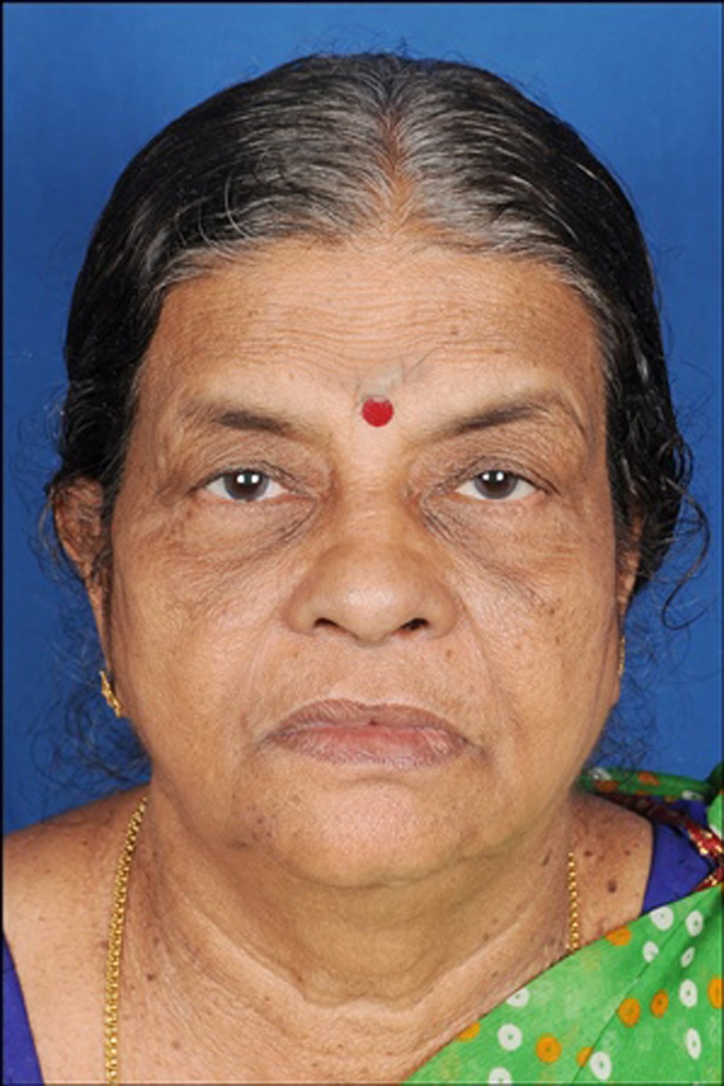
Frontal view showing diffuse swelling present on lower right third region of the face, involving the chin.
Intraoral examination (figure 2) revealed a single, diffuse swelling over the right labial mucosa extending from 31 to 45, crossing the midline and obliterating the right buccal vestibule. The overlying mucosa appeared to be blanched. Grade I mobility was evident in relation to teeth 31–45. Investigation of these teeth showed absence of tenderness on percussion; they displayed a delayed response to electric pulp test (EPT). Fine needle aspiration was carried out and a brown coloured fluid was obtained from the affected area. The panoramic view (figure 3) showed a well-defined unilocular radiolucency with corticated margins in the periapical region extending from the mesial surface of root of 31 to distal surface of root of 45. Based on clinical and radiographic findings, a provisional diagnosis of radicular cyst was performed.
Figure 2.
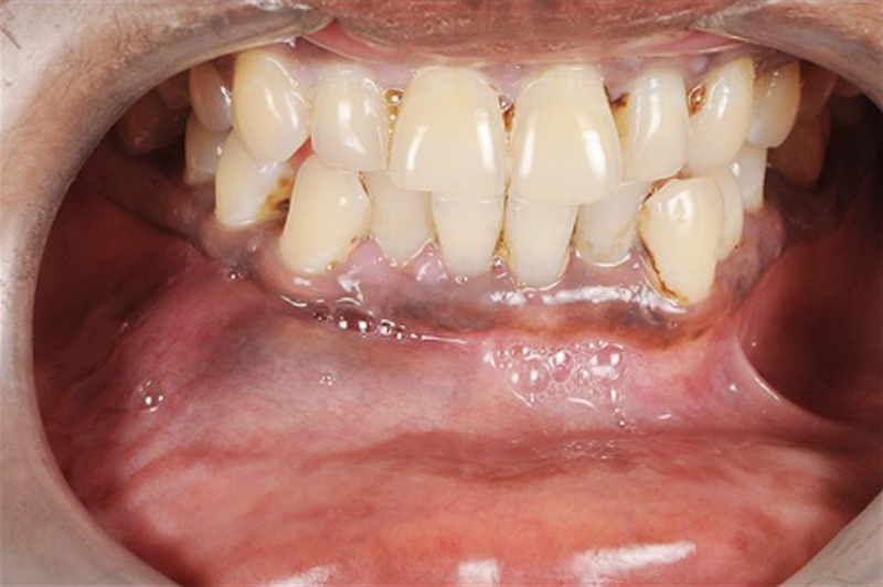
Diffuse intraoral soft tissue swelling extending from 31 to 45 region.
Figure 3.
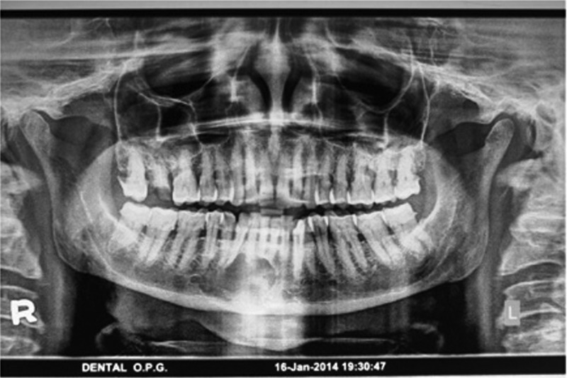
Panoramic radiograph showing unilocular radiolucency with corticated borders involving periapical/rootapex region from mesial surface of 31 to mesial surface of 45.
All preoperative investigations were made and a medical fitness report was obtained from the patient's physician. The patient was operated under GA. Nasotracheal intubation was performed from the left external nare with cuffed portex endotracheal tube number 7. A crevicular incision was made extending from the mesial of 33 to the mesial of 47. Careful dissection was inevitable to maintain the patency of the cystic lining as far as possible and the residual cavity was debrided thoroughly to eliminate fragile residual lining, if any. In anticipation of the high recurrence rate of this pathology, it is absolutely essential to ensure no residual cystic lining is left over (figure 4). Closure of the surgical site was accomplished using 3-0 vicryl (2328), and a pressure bandage was applied extraorally.
Figure 4.
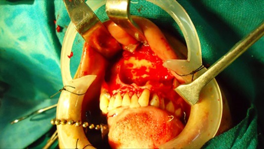
Cystic cavity involving periapical region from 31 to 46.
The pathological specimen obtained (figure 5) was soft in consistency and creamish-brown in colour, with irregular borders.
Figure 5.
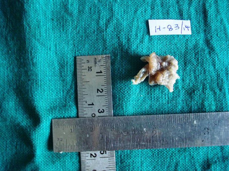
Gross appearance of the lesion.
The histopathological examination of the specimen (figure 6) showed non-keratinised pseudostratified epithelium with superficial layer of epithelium depicting ciliated and columnar cells. The epithelium (figure 7) showed variation in thickness, intraepithelial crypts and clear and hobnail cells. The underlying connective tissue showed epithelium-like structures, nerve bundles and inflammatory infiltrate composed of lymphocytes. The final diagnosis of GOC was performed. Apart from this there was a strong positivity in all layers of epithelium for cytokeratin 19 (CK19) immunohistochemical staining (figure 8), which strongly points towards the odontogenic nature of epithelium. Follow-up was carried out clinically and radiographically at 1, 3 and 6 months, and at 1 year, showing no recurrence, and the patient remained asymptomatic.
Figure 6.
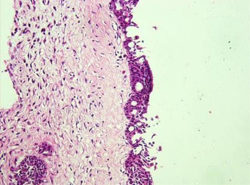
(H&E stain) ×10 image showing cystic lining epithelium with pseudocysts and connective tissue wall with loosely arranged collagen fibre bundles and epithelial islands in connective tissue wall.
Figure 7.
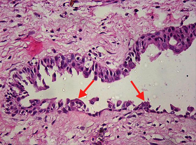
(H&E stain) ×40 image showing cystic lining of pseudostratified epithelium (with red arrow indicating) hobnail cells in the superficial epithelium and connective tissue wall, with loosely arranged collagen fibre bundles and fibroblasts.
Figure 8.
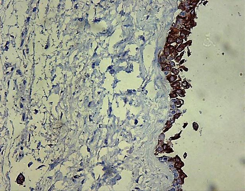
Cystic epithelium with CK19 immunohistochemical staining showing strong positivity. CK19, cytokeratin19.
Investigations
FNAC (fine needle aspiration cytology)
OPG (orthopantomogram)
Immunohistochemical staining with CK19
Cystic swelling commonly contains fluid, and for that, FNAC was the right choice; in addition, for better localising position of the cystic swelling, an OPG radiographic image was taken. In order to confirm the odontogenic nature of epithelium, CK19 immunohistochemical staining was carried out, which showed strong positivity.
As the cytology slide was made from the FNAC, no units were required. The cytology slide showed extravasated RBC's intermixed with sparse inflammatory cells.
Differential diagnosis
Because of delayed response to EPT and history of trauma 15 years before with respect to 31–35, a working diagnosis of radicular cyst was given, although a differential diagnosis of midline radiolucencies of the mandible may be possible, which were excluded.
Traumatic cyst: commonly seen in young patients and in the posterior mandible (molar ramus) region.
Central giant cell granuloma: more common in the anterior segment of the jaws and, not uncommonly, across the midline, as seen in our case. It is more common in patients aged under 30 years. These radiographically show smooth or ragged borders.
Central ossifying fibroma: may occur at any age, but young adults are most commonly involved. Clinically, growth produces a noticeable swelling and mild deformity—displacement or mobility of teeth may be an early feature, as seen in our case. Radiographically, early lesions show uniform radiolucency without internal ossifications, also seen in our case.
Keratocystic odontogenic tumour: affect bimodal age group (young and old) with anterior mandible less commonly seen. Among the more common features are pain, soft-tissue swelling and expansion of bone with various neurological manifestations such as paraesthesia, which was seen in our patient. More commonly, keratocystic odontogenic tumours are seen radiographically with unilocular radiolucency with scalloping border, as seen in our case.
Ameloblastoma: most cases cluster between ages 20–60 years. They are seen less commonly within the mandible premolar incisor area compared to molar-angle-ramus area. Rarely, unilocular radiolucency is also seen radiographically, as in our case.
All these findings were apparently negative for the present case, hence ruled out.
Treatment
In this case, assessment of the pathological lesion clinically and radiographically revealed the complete intrabony nature of this lesion. Hence, precise enucleation was the treatment of choice. Had it been a more aggressive lesion, more radical treatment would have been required. Surgical intervention resulted in complete resolution of the symptoms and transient neuropraxia was corrected within 2 weeks.
Definitive diagnosis is usually based on postoperative histological findings correlating with clinical and radiological findings. Success was assessed by complete resolution of subjective symptoms, and immediate clinical and radiographic postoperative assessment with subsequent follow-up at regular intervals. We performed surgical enucleation and, during postoperative follow-up, the patient was asymptomatic.
Outcome and follow-up
Postoperatively, the surgical site healed uneventfully with no complications. The patient recovered well with no signs of persistent neurosensory deficit. Follow-up of 1, 3 and 6 months, and 1 year, showed no recurrence, and the patient remained asymptomatic.
Discussion
A case of GOC, a rare development of cyst of the jaws, has been presented.1 It has a frequency of occurrence ranging from 0.012% to 1.3% of all jaw cysts, and its prevalence is 0.17%.2 Similar to previous case reports, our case had mandibular involvement, with swelling and pain as symptoms.1 In addition, the radiological features showed similarities with previous reports, such as well-defined radiolucency with distinct sclerotic borders and root displacement. Histopathological features were also suggestive of a cystic cavity lined with pseudostratified, ciliated columnar epithelial lining and fibrous vascular connective tissue.3 The disagreement was related to gender predilection and age: the literature showed a predilection toward males and a mean age of 49.5 years, with the anterior mandible being the most commonly affected site, whereas the present case was reported in a 63-year-old woman, however, it was supporting the most commonly affected site, the anterior mandible region.
Literature review shows that GOC may mimic a wide spectrum of lesions ranging from a lateral periodontal cyst to a destructive malignant neoplasm such as central mucoepidermoid carcinoma.4
The GOC has two clinically important attributes: it has a high recurrence rate and it displays an aggressive growth potential. This is because of GOC's complex and frequently non-specific histopathology.5
Our case was considered to be GOC because it fulfilled all major and minor microscopic criteria specified by Kaplan et al and Brannon and colleagues6 for diagnosis of GOC, as mentioned in (table 1).
Table 1.
Diagnostic histopathological criteria for GOC given by Kaplan et al7
| Major criteria | Minor criteria |
|---|---|
|
|
GOC, glandular odontogenic cyst.
Apart from this there was a strong positivity in all layers of epithelium for CK19 (figure 8), which strongly points towards the odontogenic nature of epithelium. Many other molecular markers have been investigated as new tools to diagnose GOC and differentiate it from other lesions (table 2).2 7
Table 2.
Diagnostic molecular markers to differentiate glandular odontogenic cyst (GOC) from other lesions7
| GOC | Central mucoepidermoid carcinoma (CMEC) and other lesions |
|---|---|
| ▸ MASPIN (mammary serine protease inhibitor)—negative ▸ EMA (epithelial membrane antigen)—negative ▸ Ki67 lesion index—more ▸ p53=3% ▸ CK18=30%, (cytokeratin 18) ▸ CK19=50% | ▸ MASPIN (Mammary Serine Protease Inhibitor)—positive ▸ EMA (epithelial membrane antigen)—positive ▸ Ki67 lesion index—less ▸ p53=4.9% ▸ Radicular cyst p53=0.4% ▸ CK18=100% in low mucoepidermoid carcinoma (MEC) ▸ Odontogenic cyst CK18=7% ▸ CK19=100% |
Various treatment modalities have been recommended for GOC patients, depending on patient status, location of the lesion and clinicians’ views. Several authors have advised conservative approaches, such as enucleation and curettage with or without applying Carnoy's solution. However, due to its high recurrence rate, others recommend marginal resection or en bloc excision.3 8
Patient's perspective.
I came to hospital because of pain and swelling in the lower part of my face for 4 months. After the operation I have been fine, the swelling and pain have completely subsided.
Learning points.
The glandular odontogenic cyst is a well-known clinical entity and important to recognise and diagnose due to its aggressive behaviour and tendency to recur.
This cyst is often misdiagnosed because of its overlapping histopathological features with that of other odontogenic cysts such as lateral periodontal cyst, dentigerous cyst, radicular cyst and central mucoepidermoid carcinoma.
There is no consensus on how many histopathological features are necessary for diagnosis of glandular odontogenic cysts. To help with diagnosis, a clear definition of criteria is necessary, as well as a search for specific markers that would support the diagnosis.
Footnotes
Acknowledgements: The authors specially thank Dr Swapan Goswami, MD, Pathology, Professor in the Department of Pathology, SBKS Medical College Piparia, Vadodara, Gujarat, India, and Dr Raju Sugankar (MDS Oral and Maxillofacial Pathology, Kamineni Institute of Dental Sciences, Nalgonda, Telangana, India), for their technical help.
Competing interests: None declared.
Patient consent: Obtained.
Provenance and peer review: Not commissioned; externally peer reviewed.
References
- 1.Oliveira JX, Santos KC, Nunes FD et al. Odontogenic glandular cyst: a case report. J Oral Sci 2009;51:467–70. 10.2334/josnusd.51.467 [DOI] [PubMed] [Google Scholar]
- 2.Mascitti M, Santarelli A, Sabatucci A et al. Glandular odontogenic cyst: review of literature and report of a new case with cytokeratin-19 expression. Open Dent J 2014;8:1–12. 10.2174/1874210601408010001 [DOI] [PMC free article] [PubMed] [Google Scholar]
- 3.Tambawala SS, Karjodkar FR, Yadav A et al. Glandular odontogenic cyst: a case report. Imaging Sci Dent 2014;44:75–9. 10.5624/isd.2014.44.1.75 [DOI] [PMC free article] [PubMed] [Google Scholar]
- 4.Krishnamurthy A, Sherlin HJ, Ramalingam K et al. Glandular odontogenic cyst: report of two cases and review of literature. Head Neck Pathol 2009;3:153–8. 10.1007/s12105-009-0117-2 [DOI] [PMC free article] [PubMed] [Google Scholar]
- 5.Jain V, Ahuja S, Gupta VS et al. Glandular odontogenic cyst: a rare case in maxilla. IJDPMS 2013;1:9–15. [Google Scholar]
- 6.Fowler CB, Brannon RB, Kessler HP et al. Glandular odontogenic cyst: analysis of 46 cases with special emphasis on microscopic criteria for diagnosis. Head Neck Pathol 2011;5:364–75. 10.1007/s12105-011-0298-3 [DOI] [PMC free article] [PubMed] [Google Scholar]
- 7.Kaplan I, Anavi Y, Hirshberg A. Glandular odontogenic cyst: a challenge in diagnosis and treatment. Oral Dis 2008;14:575–81. 10.1111/j.1601-0825.2007.01428.x [DOI] [PubMed] [Google Scholar]
- 8.Boffano P, Cassarino E, Zavattero E et al. Surgical treatment of glandular odontogenic cysts. J Craniofac Surg 2010;21:776–80. 10.1097/SCS.0b013e3181d7a3e6 [DOI] [PubMed] [Google Scholar]


