Abstract
Background
The Scratch Collapse Test (SCT) is used to assist in the clinical evaluation of patients with ulnar nerve compression. The purpose of this study is to introduce the hierarchical SCT as a physical examination tool for identifying multilevel nerve compression in patients with cubital tunnel syndrome.
Methods
A prospective cohort study (2010–2011) was conducted of patients referred with primary cubital tunnel syndrome. Five ulnar nerve compression sites were evaluated with the SCT. Each site generating a positive SCT was sequentially “frozen out” with a topical anesthetic to allow determination of both primary and secondary ulnar nerve entrapment points. The order or “hierarchy” of compression sites was recorded.
Results
Twenty-five patients (mean age 49.6 ± 12.3 years; 64 % female) were eligible for inclusion. The primary entrapment point was identified as Osborne’s band in 80 % and the cubital tunnel retinaculum in 20 % of patients. Secondary entrapment points were also identified in the following order in all patients: (1) volar antebrachial fascia, (2) Guyon’s canal, and (3) arcade of Struthers.
Conclusion
The SCT is useful in localizing the site of primary compression of the ulnar nerve in patients with cubital tunnel syndrome. It is also sensitive enough to detect secondary compression points when primary sites are sequentially frozen out with a topical anesthetic, termed the hierarchical SCT. The findings of the hierarchical SCT are in keeping with the double crush hypothesis described by Upton and McComas in 1973 and the hypothesis of multilevel nerve compression proposed by Mackinnon and Novak in 1994.
Electronic supplementary material
The online version of this article (doi:10.1007/s11552-014-9721-z) contains supplementary material, which is available to authorized users.
Keywords: Scratch collapse test, Cubital tunnel syndrome, Multilevel nerve compression
Introduction
We have previously described the Scratch Collapse Test (SCT) as a provocative test to assist in the clinical evaluation of patients with ulnar nerve symptomatology [2, 3]. This clinical test has been shown to have a higher sensitivity for cubital tunnel syndrome than other provocative tests, including Tinel’s sign and the elbow flexion test, with an accuracy of 89 % [3]. Using the SCT, we identified Osborne’s band as the point of maximal compression in cubital tunnel syndrome in the majority of patients [2]. Additionally, several refinements have been made to the SCT since its publication, which have allowed us to further characterize sites of ulnar nerve compression in each individual patient. Specifically, we have noted that application of a topical cold spray to a compression site with a positive SCT can unmask other sites of entrapment along the ulnar nerve. We have termed this the “hierarchical” SCT.
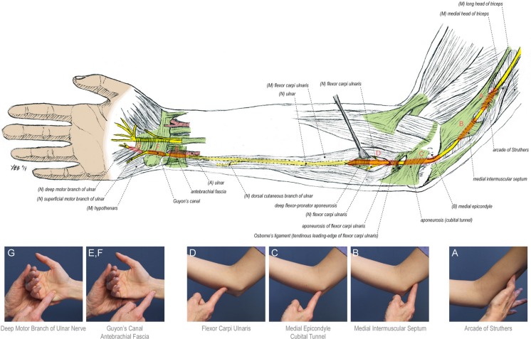
Cubital tunnel syndrome, or compression of the ulnar nerve at the elbow, is the second most common upper extremity compression neuropathy after carpal tunnel syndrome [22]. Other sites of ulnar nerve compression, such as Guyon’s canal or the arcade of Struthers, are less frequently recognized but can result in functional impairment. The accepted sites of potential ulnar nerve compression are depicted in Fig. 1 and include (1) the arcade of Struthers [36], (2) the medial intermuscular septum [31], (3) the bony retrocondylar groove and the overlying cubital tunnel retinaculum [24], (4) Osborne’s band [8, 25], (5) the volar antebrachial fascia just proximal to the wrist crease (proximal Guyon’s canal), and (6) the leading edge of the hypothenar musculature overlying the deep motor branch of the ulnar nerve (distal Guyon’s canal) [12, 13].
Fig. 1.
Ulnar nerve anatomy, course, and sources of compression in the upper extremity. Clinical photographs indicate the surface landmarks for the sites of ulnar nerve compression tested in this study, which include the following: (1) the arcade of Struthers (a), a transverse attachment of the deep fascia of the distal triceps to the medial intermuscular septum, located approximately 8 cm proximal to the medial epicondyle (2.22–24); (2) the medial intermuscular septum (b); (3) the bony retrocondylar groove and the overlying cubital tunnel retinaculum spanning the olecranon and medial epicondyle (c); (4) Osborne’s band (d), a thickened confluence of fascia between the two heads of the flexor carpi ulnaris muscle (5–6); (5) the volar antebrachial fascia just proximal to the wrist crease (e, f); and (6) the tendinous leading edge of the hypothenar musculature overlying the deep motor branch of the ulnar nerve [8] (g)
Management of ulnar nerve compression depends on an accurate diagnosis, yet localizing the site of nerve compression can be challenging. This is compounded by other conditions that may mimic ulnar nerve compression neuropathy, such as cervical disc disease and motor neuropathies. Furthermore, patients presenting with multilevel ulnar nerve symptomatology and patients with electrodiagnostic studies that are normal or demonstrate non-localizable ulnar neuropathy remain a diagnostic challenge.
The objective of this study is to demonstrate this new examination technique—the hierarchical SCT—in the clinical evaluation of patients with compression neuropathy of the ulnar nerve and specifically in the management of patients with complex and multilevel symptomatology.
Materials and Methods
Patients
A prospective cohort study was performed of patients referred to the senior author with a provisional diagnosis of compression neuropathy of the ulnar nerve over a 1-year period (2010–2011). Patients with a definitive diagnosis of ulnar nerve transection, ulnar nerve tumor, cervical radiculopathy, motor neuron disease, and a non-ulnar compression neuropathy were subsequently excluded. Patients with major shoulder pathology were also excluded owing to difficulty participating in the SCT. Ethics approval for this study was obtained from the Institutional Review Board, and written informed consent was obtained from all individual study participants.
Procedures
All patients underwent routine evaluation with history, physical examination, and electrodiagnostic studies. Nerve compression was evaluated with sensory and motor testing, Tinel’s sign, the SCT, and provocative maneuvers. The hierarchical SCT was performed to evaluate five sites of ulnar nerve compression (Fig. 1): (1) the arcade of Struthers, (2) the cubital tunnel retinaculum, (3) Osborne’s band, (4) the volar antebrachial fascia (proximal Guyon’s canal), and (5) the leading edge of the hypothenar muscles (deep motor branch). All examinations were performed by one individual (GG) experienced in performance of the SCT.
The Scratch Collapse Test
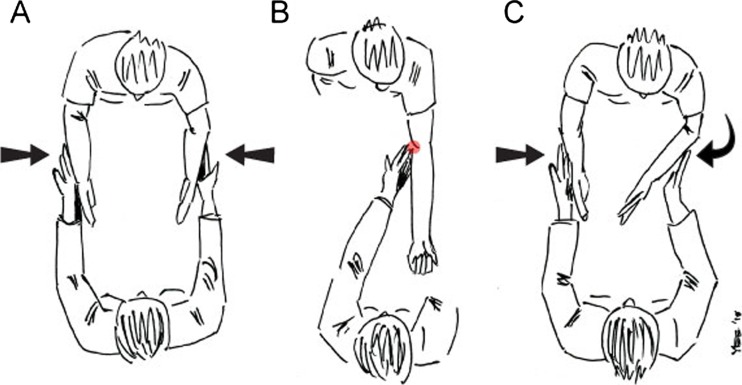
The basic technique for performing the Scratch Collapse Test has been described elsewhere [2, 3]. In brief, the patient is seated in front of the examiner, with their back free from the back of the chair, feet on the ground, shoulders in neutral position, elbows bent at 90°, forearms and wrists in neutral, and fingers in full extension. The upper arms are parallel to each other by the sides of the chest, with no forward flexion or abduction of the shoulders (Figs. 2 and 3). A force is then applied to the dorsal forearms in an effort to internally rotate the shoulders. The patient provides a baseline resistance against the initial force, and equilibrium is established between the examiner and the patient. We emphasize to the patient that it is not a “fight” between examiner and patient, but rather that we are trying to achieve a balance in resistance at the forearms. In the absence of significant shoulder pathology or other confounding injuries to the upper extremity, the arms will not collapse. With the baseline resistance set, the skin overlying the suspected site of nerve compression is gently scratched, and the examiner again exerts an internal rotation force to the dorsal forearms. A positive test will result in a significant change in resistance, and the arm on the affected side will “collapse” in toward the patient’s abdomen.
Fig. 2.
Illustration of the Scratch Collapse Test technique, which was modified after the initial technique, was published by Cheng et al. in 2008 [3]. a The initial resistance applied by the examiner to the dorsal surface of the patient’s forearms. b Scratching the patient’s arm at the cubital tunnel in the affected extremity. c Reapplying resistance to the dorsal forearms of the patient after the scratch provocation, with subsequent collapse of the affected extremity
Fig. 3.
Clinical photographs demonstrating the steps for performing the Scratch Collapse Test. a Patient positioning for the SCT: The patient is seated facing the examiner, back free from the chair, feet on the ground, shoulders adducted and in neutral position, elbows bent at 90°, forearms and wrists in neutral, and fingers in full extension. b The initial resistance applied by the examiner to the dorsal surface of the patient’s forearms. c Scratching the patient’s right (affected) arm just posterior to the medial epicondyle. d Reapplication of resistance to the dorsal forearms and collapse of the patient’s right arm (a positive SCT)
The senior author has made important refinements to the SCT since its initial publication [3], which are outlined here:
Ensure that the patient’s elbows are kept tight against their flanks, as the shoulder abductors can be used to “cheat” and overcome the collapse. If the patient starts to abduct their arms, they are trying too hard and the concept of balance is reemphasized. Indeed, their arms must stay against their flanks with no forward flexion of the shoulders.
Scratching the skin overlying the site of nerve compression is not the only option for a stimulus. The site can also be rubbed gently, or the examiner can simply blow on the skin of the region. With nerves that are submuscular or located deep in the extremity (i.e., deep motor branch of the ulnar nerve, median nerve at the pronator teres, and tibial nerve at the soleus arch), or if the patient is morbidly obese, the site should be “pushed” with the fingertip deep against the nerve.
Sustain the second resisted test force of internal rotation for at least 2–3 s, and pause as you do this, as it may take this long to identify a collapse and avoid a false negative test.
The SCT is always done over a “control area” first (a site where no nerve or compression point is present; i.e., the lateral deltoid) to establish a negative control.
This test requires a strong knowledge of surface anatomy of the nerve and will be most useful for surgeons who are more familiar with this than other specialists. The examiner can use a broader “swipe” or “blow” over the area of interest if they are less familiar with the underlying anatomy.
The SCT has a definite learning curve, and we suggest for those interested in incorporating it into their clinical examination, that they start with patients with electrically positive carpal and cubital tunnel syndrome and see how it compares with the Tinel and pressure provocative tests.
In patients with underlying shoulder pathology or other confounding upper extremity injuries, the feet can be used in a similar fashion with the forearms. In this version of the SCT, the patient is seated in front of the examiner with the hips flexed at 90° and knees flexed at 60°, ankles dorsiflexed and everted, and heels resting on the ground. Force is then applied to the lateral aspects of the feet in an effort to invert them against resistance (resistance on the legs yet scratching the arms). The remainder of the test is the same as previously described. Of note, patients with shoulder pathology were excluded from this study.
The Hierarchical SCT: Ulnar Nerve
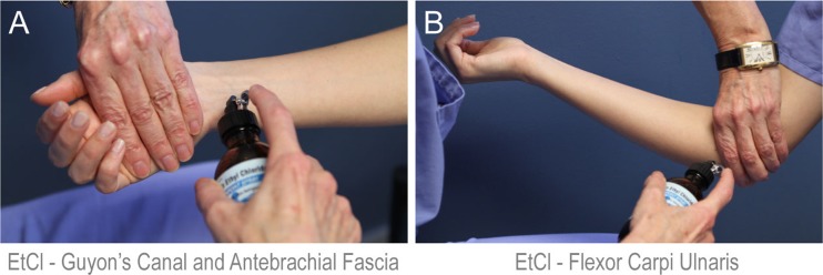
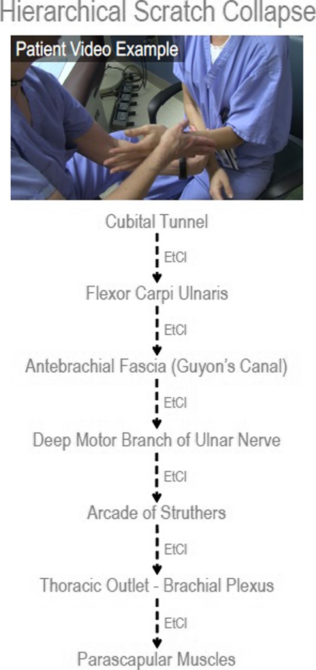
The examiner begins by performing the SCT at the five compression sites along the ulnar nerve. The site of compression generating a positive SCT is noted. This primary site is then temporarily “frozen out” (Fig. 4) with the application of topical ethyl chloride (Gebauer’s Ethyl Chloride®, Gebauer Company, Cleveland, OH). The primary site is then retested with the SCT to confirm that the patient no longer collapses at this site. The remaining four ulnar nerve entrapment points are once again evaluated with the SCT. The next compression point with a positive SCT (if any) is noted, and topical ethyl chloride is utilized to freeze this second area out. The above process is then repeated until no further entrapment points give a positive SCT (Fig. 5 and Supplementary Digital Content 1, https://vimeo.com/59943112). The order (“hierarchy”) in which the five entrapment points generate a positive SCT is recorded for each patient.
Fig. 4.
Demonstration of the use of ethyl chloride (EtCl), a topical anesthetic, to “freeze out” a site of ulnar nerve compression that has yielded a positive SCT. In order to very specifically anesthetize the desired anatomic site, it is important to shield the areas where you do not want the ethyl chloride to reach. a Application of ethyl chloride to the volar antebrachial fascia proximal to the wrist crease, while shielding Guyon’s canal distally. b Application of ethyl chloride to Osborne’s band between the two heads of the flexor carpi ulnaris, while shielding the retrocondylar groove and the medial intermuscular septum
Fig. 5.
Example of the hierarchical SCT in cubital tunnel syndrome. This figure illustrates the sequence of collapse of the five anatomic sites of ulnar nerve compression tested in this study, in a patient with mild cubital tunnel syndrome (corresponds to Video, Supplementary Digital Content 1, https://vimeo.com/59943112). In this patient, the primary site of collapse was the cubital tunnel retinaculum just posterior to the medial epicondyle, and the remaining sequence was identical to that found in all study patients. Additionally, this patient demonstrated collapse at the thoracic outlet and parascapular muscles once the initial five compression sites in the arm, forearm, and hand had been “frozen out” with ethyl chloride
Patient demographic data, including age, gender, symptom duration, and comorbid disease (diabetes, hypothyroidism), were also collected for demographic purposes. If patients have coexisting significant neuromuscular issues, then the most significant point will collapse first.
Results
Twenty-five patients with cubital tunnel syndrome were identified as eligible for inclusion. Patient characteristics are presented in Table 1. In all patients, cubital tunnel syndrome was their primary or only extremity clinical diagnosis.
Table 1.
Patient characteristics
| Characteristics | N = 25 |
|---|---|
| Patient age (years), mean ± SD (range) | 49.6 ± 12.3 (22.0–72.3) |
| Sex | |
| Female | 16 (64.0 %) |
| Male | 9 (36.0 %) |
| Symptom duration (months), mean ± SD (range) | 16.1 ± 8.9 (4.0–36.0) |
| Comorbid conditions | |
| Diabetes mellitus | 3 (12.0 %) |
| Obesitya | 7 (28.0 %) |
aObesity was defined as a body mass index ≥30
All patients demonstrated a positive SCT around the elbow in the first iteration of testing. The primary site of ulnar nerve entrapment was identified as Osborne’s band in 20 patients (80 %) and the cubital tunnel retinaculum posterior to the medial epicondyle in 5 patients (20 %).
After freezing out these primary compression sites with ethyl chloride, a consistent sequence of secondary entrapment points was identified. In patients where the primary site was Osborne’s band, subsequent entrapment points in order of collapse with SCT were the cubital tunnel retinaculum, the volar antebrachial fascia proximal to the wrist crease, the leading edge of the hypothenar muscles (deep motor branch), and the arcade of Struthers. In patients where the primary site of collapse was the cubital tunnel retinaculum, the hierarchy of subsequent compression sites was Osborne’s band, followed by the volar antebrachial fascia, the leading edge of the hypothenar muscles (deep motor branch), and finally the arcade of Struthers (Fig. 5; see also Video, Supplementary Digital Content 1, https://vimeo.com/59943112). All patients in this study demonstrated a positive SCT at all five compression sites evaluated with sequential “freezing out” of each previous site yielding a positive collapse.
Discussion
Provocative tests, such as Tinel’s sign and the elbow flexion test, are useful in making the clinical diagnosis of cubital tunnel syndrome. The recently introduced SCT, a sensitive test for nerve compression, is an additional provocative test that can assist the surgeon in rendering a diagnosis, particularly when other provocative tests and electrodiagnostic testing are negative [2, 3, 10]. The senior author was first introduced to the SCT in the early 1990s by Bronson and Beck and has spent the last 2 decades refining the test, understanding its nuances, and recognizing its consequent utility to clinical practice [3]. Without presuming to know all the pathophysiology of this test [15, 16, 18, 34], we believe that it has important utility in the management of the most challenging nerve entrapment populations.
Using the SCT, this study identified Osborne’s band as the most common primary site of ulnar nerve compression in cubital tunnel syndrome, consistent with the findings of Brown et al. [2]. Importantly, however, these patients all exhibited a secondary positive SCT at the cubital tunnel retinaculum overlying the retrocondylar groove just posterior to the medial epicondyle. The fibro-osseous tunnel was also the second most common primary entrapment point in this study. These findings make sense considering the known pathophysiology of cubital tunnel syndrome. Indeed, whereas the ulnar nerve may be compressed by the thickened confluence of fascia between the two heads of the FCU (and therefore more likely to generate a positive SCT), it is the dynamic tension on the nerve with elbow flexion behind the medial epicondyle that may also contribute to symptomatology [9, 11, 28, 33, 37].
The use of a topical anesthetic to freeze out entrapment points generating a positive SCT has not been previously described in the literature. The idea to use ethyl chloride was in fact sparked by a review of a manuscript noting increased Substance P levels in the carpal canal in patients with carpal tunnel syndrome [26]. We hypothesized that Substance P release in an area of nerve constriction could be the source of the pain stimulus resulting in the reflex “give way” seen in the SCT. Thus, if we could temporarily “freeze out” this stimulus with a topical anesthetic, the SCT would be temporarily blocked and indeed this bore out in clinical use. The idea to blow as an obnoxious stimulus, rather than scratch, on the entrapped area also derived from this Substance P hypothesis. The hypothesis that the “give away/collapse” sign was a spinal reflex also stimulated the idea to test the give away/collapse in external rotation at the shoulders or eversion at the ankles.
The ability to freeze out sequential sites of compression generating a positive SCT (what we have termed the hierarchical SCT) has importantly allowed us to identify secondary entrapment points, such as in the current study along the ulnar nerve. We were surprised by our finding that all patients demonstrated a secondary positive SCT at the arcade of Struthers. Scratching higher toward the axilla did not result in a collapse response. The arcade of Struthers has been demonstrated in cadaver dissections and is accepted as a potential site of kinking of the ulnar nerve following anterior transposition for cubital tunnel syndrome proximally [19, 26, 29, 30]. Indeed, since we began using sterile tourniquets to perform our ulnar nerve transpositions, we have been increasingly impressed by the presence of a tendinous band coursing below the ulnar nerve, from the triceps to the medial intermuscular septum. The senior author believes that in the past, she greatly underestimated the frequency of this arcade of Struthers because the nonsterile tourniquet prevented digital evaluation to a level proximal enough to evaluate for its presence.
The fact that all patients in this study ultimately collapsed at the arcade of Struthers lends further support to its role as a subclinical entrapment point in primary cubital tunnel syndrome. On the other hand, the fact that the majority of patients do well surgically without addressing the arcade of Struthers suggests that the SCT is sensitive enough to elicit subclinical entrapment points. In effect, the arcade of Struthers was always the last point to collapse in this patient cohort.
Ulnar nerve compression points around the wrist yielded positive SCTs in patients with cubital tunnel syndrome after sequentially freezing out Osborne’s band and the cubital tunnel retinaculum. Among the wrist entrapment points evaluated in this study, the volar antebrachial fascia was found to be a more significant site of compression than the tendinous leading edge of the hypothenar musculature in Guyon’s canal. This concept differs from our traditional thinking and stresses the importance of releasing this proximal fascia during a Guyon’s canal decompression. In our experience, an incision across the wrist is required to completely release this volar antebrachial fascia overlying the ulnar neurovascular bundle.
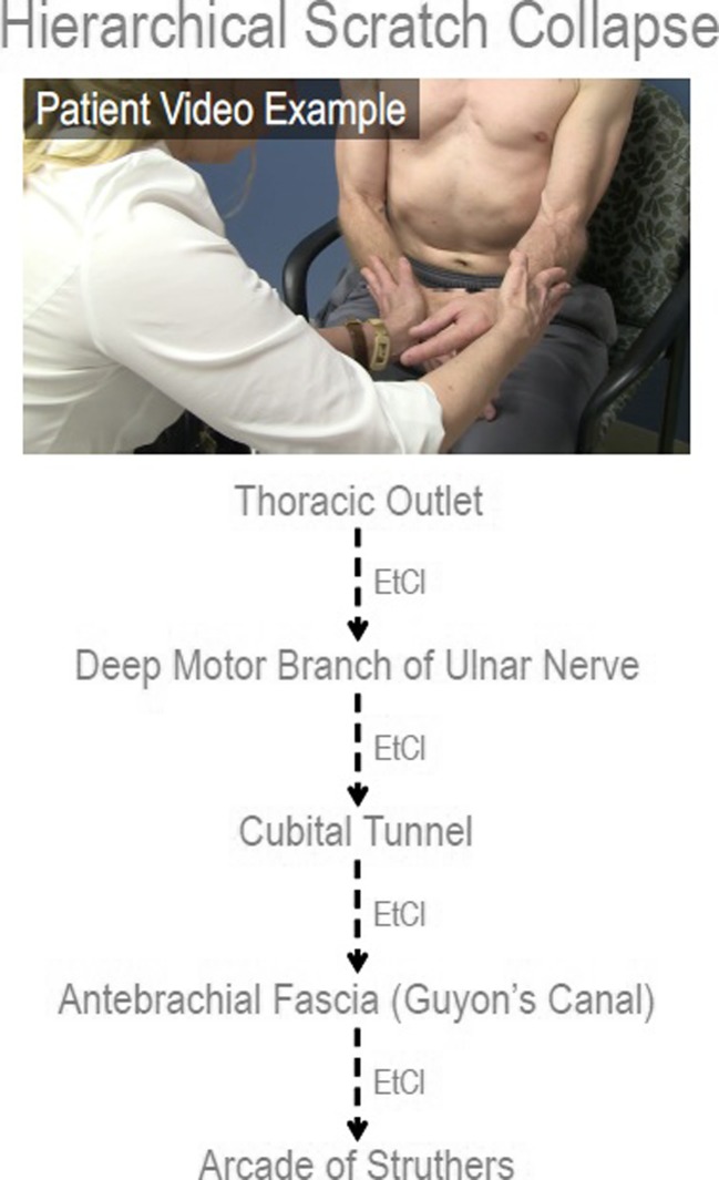
We have found the addition of ethyl chloride to the examination, and the ability to identify other sites of secondary nerve compression to be extremely valuable in clinical practice (Fig. 6; see also Video, Supplementary Digital Content 2, https://vimeo.com/59943111). The concept of multiple points of entrapment along the same nerve is not new and is in keeping with the double crush hypothesis of Upton and McComas [35]. They postulated that a proximal nerve compression (i.e., cervical disc disease) renders the nerve more susceptible to distal nerve compression (i.e., carpal tunnel syndrome). Lundborg also suggested the reverse effect, in that a distal nerve entrapment sensitized a nerve to proximal areas of compression [4, 5]. The double crush hypothesis also suggests that serial constraints to axoplasmic flow, while individually are insufficient to result in clinically appreciable nerve dysfunction, can be an additive in causing ultimate nerve dysfunction. There exists both clinical and experimental evidence to support multiple compression sites in peripheral nerves [1, 6, 7, 14, 17, 21, 23, 27, 32]. Other potential etiologies include medical conditions which increase the susceptibility of compression (e.g., diabetic neuropathy), neurologic conditions (e.g., syringomyelia), and genetic conditions such as hereditary neuropathy with liability to pressure palsies.
Fig. 6.
Application of the hierarchical SCT to brachial plexus injury (corresponds to Video, Supplementary Digital Content 2, https://vimeo.com/59943111). This patient had a right medial cord injury and on physical examination had significantly more wasting of the ulnar-innervated intrinsic muscles as compared to the median-innervated intrinsics. A multilevel injury was suspected, and a hierarchical SCT was performed to determine secondary entrapment points on the ulnar nerve. The sequence of collapse in this patient was the right thoracic outlet (consistent with the medial cord injury), followed by the deep motor branch of the ulnar nerve, cubital tunnel, volar antebrachial fascia, and the arcade of Struthers
The results of this study suggest that the hierarchical SCT can identify concomitant compression of the ulnar nerve at the elbow and wrist. Clinical complaints, physical examination, and electrical studies will combine to determine patient management. In patients with significant intrinsic atrophy, the consideration of a concomitant decompression of the nerve at both levels can be made. Secondary ulnar nerve compression at the leading edge of the hypothenar musculature and antebrachial fascia may also explain persistent patient symptoms following surgical decompression or transposition of the ulnar nerve at the elbow alone. Consequently, we will utilize the SCT to evaluate for secondary compression of the ulnar nerve at the wrist in patients with cubital tunnel syndrome who do not recover excellent function after primary surgical treatment at the elbow. Particularly in secondary or tertiary cases, clear demonstration of a positive SCT test at Guyon’s canal and not at the elbow indicates that these patients may respond well to a distal release without the need to perform revision cubital tunnel surgery.
Finally, the hierarchical SCT has allowed for the identification of multiple concomitant neuromuscular problems and reinforces the idea that multilevel nerve compression and muscle imbalance often act in concert [20]. Indeed, we use this in our practice not only for nerve entrapments but also for muscle imbalance. For example, in patients with concomitant thoracic outlet syndrome, muscle imbalance, poor posture, and cubital tunnel syndrome, we find this test very helpful when it prioritizes periscapular muscle imbalance and scapular dyskinesis as a more significant component of the clinical presentation than the thoracic outlet issues. In patients with muscle imbalance and a positive SCT, the SCT will become negative if the patient corrects and maintains their posture by adducting and depressing their scapula. We use this demonstration to motivate such patients to work toward improving their abducted scapular posture, with physical therapy and on their own. In 1994, when the hypothesis was presented to explain to patients with significant upper extremity neuromuscular complaints, especially those that failed to respond to a simple carpal tunnel release or had normal electrodiagnostic findings, several vocal commentators generally criticized the hypothesis [20]. The hierarchical SCT mirrors that 1994 hypothesis and is critical in our successful management of this very challenging patient population.
Identifying multiple clinical problems with the hierarchical SCT does not imply surgical intervention at these sites, but it does allow for a greater awareness of the significance of these issues. Wherever possible, these issues can be addressed with physical therapy or conservative management, utilizing surgery judiciously. Specifically, in the study population, the majority of patients are first treated conservatively, and surgery at the cubital tunnel is only suggested with failure of conservative management; surgery at Guyon’s canal is suggested when ulnar intrinsic atrophy is present.
While this study is limited by its small sample size and focus on patients with primary cubital tunnel syndrome, it highlights the utility of the hierarchical SCT in the clinical assessment of patients with ulnar nerve compression. In particular, it demonstrates the ability of the SCT to preoperatively identify all recognized sites of compression along the ulnar nerve in a hierarchical fashion. We do not mean to imply that all of these points are clinically significant; rather, it is the primary compression site generating a positive SCT that is the most clinically relevant. However, it is likely that in some patients with cubital tunnel syndrome and intrinsic atrophy, release of the ulnar nerve at the wrist may be indicated. In patients with persistent, new, or recurrent symptoms after cubital tunnel surgery, we have seen the SCT localize symptom etiology to primary compression of the ulnar nerve at the wrist or arcade of Struthers, directing surgery away from the elbow and toward these other sites (thereby avoiding revision cubital tunnel surgery). This information may have a role in clarifying and guiding the surgical decision-making process, indicating which procedures are most appropriate to meet the specific needs of each patient. Future studies will continue to define and enhance the diagnostic properties of the hierarchical SCT and its utility in the evaluation of patients with multilevel nerve compression.
In summary, we use the population of patients with primary cubital tunnel syndrome as a way to introduce this concept of the hierarchical SCT as a new test to evaluate complex patients with nerve compression problems when other clinical tests fail to satisfactorily identify the etiology of patients’ subjective complaints.
Electronic supplementary material
This video demonstrates performance of the hierarchical SCT in a patient with cubital tunnel syndrome, as delineated in this study. This video can also be accessed through the following link: https://vimeo.com/59943112. (WMV 90.9 MB)
This video demonstrates application of the hierarchical SCT in a patient with medial cord injury to identify and prioritize distal ulnar nerve entrapment points. This video can also be accessed through the following link: https://vimeo.com/59943112. (WMV 72.8 MB)
Acknowledgments
Conflict of Interest
Kristen M. Davidge has no conflicts of interest to declare.
Gil Gontre has no conflicts of interest to declare.
David Tang has no conflicts of interest to declare.
Kirsty U. Boyd has no conflicts of interest to declare.
Andrew Yee has no conflicts of interest to declare.
Marci S. Damiano has no conflicts of interest to declare.
Susan E. Mackinnon has no conflicts of interest to declare.
Statement of Human and Animal Rights
All procedures followed were in accordance with the ethical standards of the responsible committee on human experimentation (institutional and national) and with the Helsinki Declaration of 1975, as revised in 2008.
Statement of Informed Consent
Written informed consent was obtained from all individual participants included in the study.
Funding
None
Contributor Information
Kristen M. Davidge, Phone: 647-292-9070, Email: kristen.davidge@utoronto.ca
Susan E. Mackinnon, Phone: 314-362-4586, Email: mackinnon@wudosis.wustl.edu
References
- 1.Baba H, Maezawa Y, Uchida K, et al. Cervical myeloradiculopathy with entrapment neuropathy: a study based on the double-crush concept. Spinal Cord. 1998;36(6):399–404. doi: 10.1038/sj.sc.3100539. [DOI] [PubMed] [Google Scholar]
- 2.Brown JM, Mohktee D, Evangelista MS, Mackinnon SE. Scratch collapse test localizes Osborne’s band as the point of maximal nerve compression in cubital tunnel syndrome. Hand. 2010;5:141–147. doi: 10.1007/s11552-009-9225-4. [DOI] [PMC free article] [PubMed] [Google Scholar]
- 3.Cheng CJ, Mackinnon-Patterson B, Beck JL, Mackinnon SE. Scratch collapse test for evaluation of carpal and cubital tunnel syndrome. J Hand Surs. 2008;33A:1518–1524. doi: 10.1016/j.jhsa.2008.05.022. [DOI] [PubMed] [Google Scholar]
- 4.Dahlin LB, Lundborg G. The neurone and its response to peripheral nerve compression. J Hand Surg (Br) 1990;15:5–10. doi: 10.1016/0266-7681(90)90040-B. [DOI] [PubMed] [Google Scholar]
- 5.Dahlin LB, Nordborg C, Lundborg G. Morphologic changes in nerve cell bodies induced by experimental graded nerve compression. Exp Neurol. 1987;95:611–621. doi: 10.1016/0014-4886(87)90303-7. [DOI] [PubMed] [Google Scholar]
- 6.Dahlin LB. Aspects on pathophysiology of nerve entrapments and nerve compression injuries. Neurosurg Clin N Am. 1991;2:21–29. [PubMed] [Google Scholar]
- 7.Dellon AL, Mackinnon SE. Chronic nerve compression model for the double crush hypothesis. Ann Plast Surg. 1991;26:259–264. doi: 10.1097/00000637-199103000-00008. [DOI] [PubMed] [Google Scholar]
- 8.Dellon AL. Musculotendinous variations about the medial humeral epicondyle. J Hand Surg. 1986;11B:175–181. doi: 10.1016/0266-7681(86)90254-8. [DOI] [PubMed] [Google Scholar]
- 9.Gelberman RH, Yamaguchi K, Hollstien SB, et al. Changes in interstitial pressure and cross-sectional area of the cubital tunnel and of the ulnar nerve with flexion of the elbow. J Bone Joint Surg. 1998;80A:492–501. doi: 10.2106/00004623-199804000-00005. [DOI] [PubMed] [Google Scholar]
- 10.Gillenwater J, Cheng J, Mackinnon SE. Evaluation of the scratch collapse test in peroneal nerve compression. Plast Reconstr Surg. 2011;128:933–939. doi: 10.1097/PRS.0b013e3181f95c36. [DOI] [PubMed] [Google Scholar]
- 11.Grewal R, Varitimidis SE, Vardakas DG, et al. Ulnar nerve elongation and excursion in the cubital tunnel after decompression and anterior transposition. J Hand Surg. 2000;25B:457–460. doi: 10.1016/S0266-7681(00)80012-1. [DOI] [PubMed] [Google Scholar]
- 12.Gross MS, Gelberman RH. Anatomy of the distal ulnar tunnel. Clin Orthop. 1985;196:238–247. [PubMed] [Google Scholar]
- 13.Guyon F. Note sur une disposition anatomique propre a la face anterieure de la region du poignet et non encore decrite par le docteur. Bull Soc Anat Paris. 1861;6:184–186. [Google Scholar]
- 14.Hurst LC, Weissberg D, Carroll RE. The relationship of the double crush to carpal tunnel syndrome (an analysis of 1,000 cases of carpal tunnel syndrome) J Hand Surg (Br) 1985;10:202–204. doi: 10.1016/0266-7681(85)90018-X. [DOI] [PubMed] [Google Scholar]
- 15.Kofler M. Functional organization of exteroceptive inhibition following nociceptive electrical fingertip stimulation in humans. Clin Neurophysiol. 2003;114:973–980. doi: 10.1016/S1388-2457(03)00060-9. [DOI] [PubMed] [Google Scholar]
- 16.Leis AA, Stokic DS, Fuhr P, et al. Nociceptive fingertip stimulation inhibits synergistic motoneuron pools in the human upper limb. Neurology. 2000;55:1305–1309. doi: 10.1212/WNL.55.9.1305. [DOI] [PubMed] [Google Scholar]
- 17.Lo SF, Chou LW, Meng NH, et al. Clinical characteristics and electrodiagnostic features in patients with carpal tunnel syndrome, double crush syndrome, and cervical radiculopathy. Rheumatol Int. 2012;32:1257–1263. doi: 10.1007/s00296-010-1746-1. [DOI] [PubMed] [Google Scholar]
- 18.Logigian EL, Plotkin GM, Shefner JM. The cutaneous silent period is mediated by spinal inhibitory reflex. Muscle Nerve. 1999;22:467–472. doi: 10.1002/(SICI)1097-4598(199904)22:4<467::AID-MUS7>3.0.CO;2-Y. [DOI] [PubMed] [Google Scholar]
- 19.Mackinnon SE, Dellon AL. Surgery of the peripheral nerve. New York: Thieme Medical Publishers; 1988. pp. 65–85. [Google Scholar]
- 20.Mackinnon SE, Novak CB. Clinical commentary: pathogenesis of cumulative trauma disorder. J Hand Surg [Am] 1994;19:873–883. doi: 10.1016/0363-5023(94)90205-4. [DOI] [PubMed] [Google Scholar]
- 21.Mackinnon SE. Double and multiple crush syndromes. Hand Clin. 1992;8:369–80. [PubMed] [Google Scholar]
- 22.Maggi SP, Lowe JB, III, Mackinnon SE. Pathophysiology of nerve injury. Clin Plast Surg. 2003;30:109–126. doi: 10.1016/S0094-1298(02)00101-3. [DOI] [PubMed] [Google Scholar]
- 23.Nemoto K, Matsumoto N, Tazaki K, et al. An experimental study on the “double crush” hypothesis. J Hand Surg Am. 1987;12:552–559. doi: 10.1016/S0363-5023(87)80207-1. [DOI] [PubMed] [Google Scholar]
- 24.O’Driscoll SW, Horii E, Carmichael SW, Morrey BF. The cubital tunnel and ulnar neuropathy. J Bone Joint Surg. 1991;73B:613–617. doi: 10.1302/0301-620X.73B4.2071645. [DOI] [PubMed] [Google Scholar]
- 25.Osborne GV. Surgical treatment of tardy ulnar neuritis. J Bone Joint Surg. 1957;39B:782. [Google Scholar]
- 26.Ozturk N, Erin N, Tuzuner S. Changes in tissue substance P levels in patients with carpal tunnel syndrome. Neurosurgery. 2010;67:1655–1660. doi: 10.1227/NEU.0b013e3181fa7032. [DOI] [PubMed] [Google Scholar]
- 27.Schmid AB, Coppieters MW. The double crush syndrome revisited—a Delphi study to reveal current expert views on mechanisms underlying dual nerve disorders. Man Ther. 2011;16:557–562. doi: 10.1016/j.math.2011.05.005. [DOI] [PubMed] [Google Scholar]
- 28.Schuind FA, Goldschmidt D, Bastin C, Burny F. A biomechanical study of the ulnar nerve at the elbow. J Hand Surg. 1995;20B:623–627. doi: 10.1016/S0266-7681(05)80124-X. [DOI] [PubMed] [Google Scholar]
- 29.Siquera MG, Martins RS. The controversial arcade of Struthers. Surg Neurol. 2005;64(S1):17–20. doi: 10.1016/j.surneu.2005.04.017. [DOI] [PubMed] [Google Scholar]
- 30.Spinner M, Kaplan EB. The relationship of the ulnar nerve to the medial intermuscular septum in the arm and its clinical significance. Hand. 1976;8:239–242. doi: 10.1016/0072-968X(76)90008-5. [DOI] [PubMed] [Google Scholar]
- 31.Stewart JD. Focal peripheral neuropathies. 2. New York: Thieme Medical Publishers; 1991. [Google Scholar]
- 32.Suzuki Y, Shirai Y. Motor nerve conduction analysis of double crush syndrome in a rabbit model. J Orthop Sci. 2003;8:69–74. doi: 10.1007/s007760300011. [DOI] [PubMed] [Google Scholar]
- 33.Toby EB, Hanesworth D. Ulnar nerve strains at the elbow. J Hand Surg. 1998;23A:992–997. doi: 10.1016/S0363-5023(98)80005-1. [DOI] [PubMed] [Google Scholar]
- 34.Uncini A, Kujirai T, Gluck B, Pullman S. Silent period induced by cutaneous stimulation. Electroencephalogr Clin Neurophysiol. 1991;81:344–352. doi: 10.1016/0168-5597(91)90023-Q. [DOI] [PubMed] [Google Scholar]
- 35.Upton AR, McComas AJ. The double crush in nerve entrapment syndromes. Lancet. 1973;2:359–362. doi: 10.1016/S0140-6736(73)93196-6. [DOI] [PubMed] [Google Scholar]
- 36.Wehrli L, Oberlin C. The internal brachial ligament versus the arcade of Struthers: an anatomical study. Plast Reconstr Surg. 2005;115(2):471–477. doi: 10.1097/01.PRS.0000150144.73603.24. [DOI] [PubMed] [Google Scholar]
- 37.Wright TW, Glowczewskie F, Cowin D, Wheeler DL. Ulnar nerve excursion and strain at the elbow and wrist associated with upper extremity motion. J Hand Surg. 2001;26A:655–662. doi: 10.1053/jhsu.2001.26140. [DOI] [PubMed] [Google Scholar]
Associated Data
This section collects any data citations, data availability statements, or supplementary materials included in this article.
Supplementary Materials
This video demonstrates performance of the hierarchical SCT in a patient with cubital tunnel syndrome, as delineated in this study. This video can also be accessed through the following link: https://vimeo.com/59943112. (WMV 90.9 MB)
This video demonstrates application of the hierarchical SCT in a patient with medial cord injury to identify and prioritize distal ulnar nerve entrapment points. This video can also be accessed through the following link: https://vimeo.com/59943112. (WMV 72.8 MB)


