Abstract
Di-2-ethylhexyl adipate (DEHA) is a common plasticizer used in food packaging. At high doses, DEHA can cause adverse health effects in rats. Although the potential for human exposure to DEHA is high, no DEHA specific biomarkers are identified for human biomonitoring. Using human liver microsomes, we investigated the in vitro phase I metabolism of DEHA and its hydrolytic metabolite mono-2-ethylhexyl adipate (MEHA) and, for comparison purposes, of the analogous di-2-ethylhexyl phthalate (DEHP) and its hydrolytic metabolite mono-2-ethylhexyl phthalate. We unequivocally identified MEHA, a DEHA specific biomarker, and adipic acid, a nonspecific biomarker, using authentic standards. On the basis of their mass spectrometric fragmentation patterns, we tentatively identified two other DEHA specific metabolites: mono-2-ethylhydroxyhexyl adipate (MEHHA) and mono-2-ethyloxohexyl adipate (MEOHA), analogous to the oxidative metabolites of DEHP. Interestingly, although adipic acid was the major in vitro metabolite of DEHA, the analogous phthalic acid was not the major in vitro metabolite of DEHP. Our preliminary data for 144 adults with no known exposure to DEHA suggests that adipic acid is also the main in vivo urinary metabolite, while MEHA, MEHHA, and MEOHA are only minor metabolites. Therefore, the use of these specific metabolites for assessing the exposure of DEHA may be limited to highly exposed populations.

INTRODUCTION
Di-2-ethylhexyl adipate (DEHA; hexanedioic acid, di-2-ethylhexyl ester) is used extensively as a plasticizer in flexible polyvinyl chloride (PVC) and food contact films.1 The migration of DEHA from PVC film into food content2,3 is thought to be a major source of human exposure among the general population.4 Results from a recent study5 based on duplicate diet samples suggest that median dietary intake of DEHA among a group of German adults is 0.67 μg/kg bw, well below the tolerable daily intake of 280 μg/kg bw.5
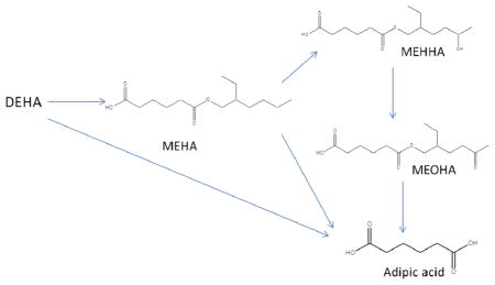
In humans and rats, after oral administration, DEHA is hydrolyzed first to mono-2-ethylhexyl adipate (MEHA), which can be further metabolized and rapidly excreted in urine, mainly as adipic acid.4,6 Exposure of Harlan/ICR albino Swiss mice after a single intraperitoneal dose of 10 mL/kg DEHA before the 8 week mating period caused a reduced percentage of pregnancies and an increased number of fetal deaths.7 At doses at or above 1,000 mg/kg, DEHA disturbed ovulation and follicle growth in rats.8 Animal carcinogenicity data are limited,9 and DEHA is not classifiable as to its carcinogenicity to humans.1
No human data are available on any potential toxicity associated with DEHA exposure, but in order to study the potential health effects of human exposure to DEHA at environmental levels, identification of sensitive and specific biomarkers is necessary. Previously, five metabolites of DEHA, namely, 2-ethylhexanoic acid, 2-ethylhexanol, 2-ethyl-5-hydroxyhexanoic acid, 2-ethylhexane dioic acid, and 2-ethyl-5-ketohexanoic acid were identified in the urine of six adult volunteers orally administered with 46 mg of deuterium labeled-DEHA10 and accounted for 12.1% of the administered DEHA dose.10 However, these metabolites are unsuitable for human exposure assessment because they are nonspecific biomarkers of DEHA that can be formed from any esters containing a 2-ethylhexyl side chain, including di-2-ethylhexyl phthalate (DEHP), another widely used plasticizer. Metabolism of DEHP formed mono-2-ethylhexyl phthalate (MEHP) and several DEHP specific metabolic products that can be used as biomarkers of DEHP exposure, namely, mono-2-ethyl-5-oxohexyl phthalate (MEOHP), mono-2-ethyl-5-hydroxyhexyl phthalate (MEHHP), and mono-2-ethyl-5-carboxypentyl phthalate (MECPP).11–13 We hypothesized that DEHA may produce similar metabolites and that they could be used as DEHA specific exposure biomarkers.
For the present study, we used online solid phase extraction (SPE) followed by high performance liquid chromatography (HPLC) and mass spectrometry to investigate in vitro phase I metabolism of DEHA and DEHP by human liver microsomes in order to identify potential DEHA specific exposure biomarkers for human biomonitoring and compare them to the known DEHP metabolites. Because DEHA is rapidly hydrolyzed to MEHA that may be further metabolized before its elimination in urine, much like MEHP, we also investigated the in vitro metabolism of MEHA and MEHP.
EXPERIMENTAL PROCEDURES
Reagents and Standards
MEHA was purchased from CanSyn (Ontario, Canada). All phthalate metabolites and their stable-isotope labeled analogues, 13C6-DEHA and 13C6-MEHA, were purchased from Cambridge Isotope Laboratories (Andover, MA, USA). Adipic acid, DEHA, and DEHP were purchased from SigmaAldrich (St. Louis, MO, USA). All reagents, solvents, and standard materials were used without further purification.
Human Samples
The urine samples analyzed for this study (N = 144) were archived samples (stored at −70 °C) collected anonymously in Atlanta, GA between 2000 and 2013 from a demographically diverse group of U.S. male and female adults with no known DEHA exposure. No personal information from the subjects was available. Samples were collected between 8:00 a.m. and 5:00 p.m. and were not necessarily first-morning voids. The Centers for Disease Control and Prevention (CDC) Institutional Review Board approved the collection of the urine for the development and validation of analytical methods at the CDC. A waiver of informed consent was requested under 45 CFR 46.116(d).
In Vitro Metabolism
We incubated DEHA (273 μg/mL), 13C6-DEHA (200 μg/mL), MEHA (234 μg/mL), or 13C6-MEHA (200 μg/mL) separately with human liver microsome homogenates (BD Gentest, Woburn, MA, USA). Each standard solution (50 μL) was mixed with pH 7.4 phosphate buffer (0.1M, 8 mL), water (1 mL), NADPH regenerating solution A (500 μL, BD Gentest), NADPH regenerating solution B (100 μL, BD Gentest), and male human liver microsomes (200 μL, 50 donor pooled-20 mg/mL, BD Gentest) in a glass beaker. The contents were gently mixed and placed on a rotary shaker in an incubator (Fisher Scientific, Hampton, NH, USA) at 37 °C for 5 h. After incubation, each microsomal suspension was transferred into a microcentrifuge tube and centrifuged at 12,500 rpm for 20 min on an Avanti high performance centrifuge (Beckman Coulter, Inc., Brea, CA, USA). The supernatant was then transferred into an autosampler vial for metabolite identification following the procedure described below.
For the comparison study with DEHP and MEHP, we used the above for solutions containing (A) DEHP (100 μM) and DEHA (100 μM), and (B) MEHP (10 μM) and MEHA (10 μM) incubated with human liver microsome homogenates for 24 h for solution A and 12 h for solution B. Controls did not contain either microsomes or DEHA/MEHA (or DEHP/MEHP). At several time intervals, 100 μL aliquots (N = 3) were withdrawn into microcentrifuge tubes containing acetonitrile (200 μL) and an internal standard solution (100 μL) prepared with 13C2-phthalic acid, 13C4-MEHP, D4-mono-2-ethyl-5-oxohexyl phthalate (D4-MEOHP), D4-mono-2-ethyl-5-hydrox-yhexyl phthalate (D4-MEHHP), D4-mono-2-ethyl-5-carboxypentyl phthalate (D4-MECPP), and 13C6-MEHA in 10% aqueous acetonitrile. The contents in the microcentrifuge tubes were vortex mixed and centrifuged at 12,500 rpm for 20 min on an Avanti high performance centrifuge. The supernatant was transferred into autosampler vials for the quantification of DEHP and DEHA metabolites. All stock standard solutions were prepared in acetonitrile. The dilutions of stock solutions were made in deionized water.
Identification of in Vitro Metabolites
Details on the HPLC gradient for the separation of DEHA and DEHP metabolites and the method for online SPE are presented elsewhere.14,15 Briefly, metabolites in the supernatant of the human liver microsomal homogenate (500 μL), obtained after incubating for 5 h with DEHA and MEHA, were extracted using online SPE on a Chromolith RP-18 precolumn (Merck KGaA, Darmstadt, Germany), resolved on a Betasil phenyl HPLC column (3 μM, 2.1 mm × 25 mm, ThermoFisher Scientific, San Jose, CA, USA) using a water/acetonitrile gradient program and detected by mass spectrometry on a TSQ Vantage AM triple quadrupole mass spectrometer (ThermoFisher Scientific, San Jose, CA, USA).
All ions on Q1 were scanned from m/z = 125 to m/z = 400 in negative ion mode. The fragmentation patterns of electrospray ionization (ESI) mass spectra of the major peaks were analyzed to identify potential DEHA metabolites (Table 1). ESI Q1 full scan produced multiple peaks. Metabolites unique to DEHA were identified by comparing the mass transitions of the peaks resulting from DEHA or MEHA to their isotopically labeled analogues. Product ion scans were performed only for the peaks with a mass difference (Δm) of 6 between the DEHA or MEHA metabolites and their 13C6-analogues. All common peaks with similar m/z values were excluded from further evaluation. The phthalate metabolites phthalic acid, MEHP, MEHHP, MECPP, and MEOHP were identified using authentic standards.
Table 1.
Mass Spectrometric Specifications Used for Measuring the Metabolites of Di-2-ethylhexyl Adipate (DEHA)a

| ||||
|---|---|---|---|---|
| DEHA metabolite |
m/z
|
CE (eV) | S-Lens (V) | |
| precursor | productb | |||
| adipic acid | 145 | 83 | 13 | 53 |
| mono-2-ethylhexyl adipate (MEHA) | 257 | 83 | 15 | 72 |
| mono-2-ethylhydroxyhexyl adipate (MEHHA)c | 273 | 83 | 15 | 72 |
| mono-2-ethyloxohexyl adipate (MEOHA)c | 271 | 83 | 15 | 72 |
Structures shown are for only one of the potential isomers.
Optimized for the most abundant peak.
Multiple isomeric metabolites.
Quantification of DEHP and DEHA Metabolites
Adipic acid, MEHA, MEHP, MEHHP, MECPP, and MEOHP were quantified using authentic standards; mono-2-ethyloxohexyl adipate (MEOHA) and mono-2-ethylhydroxyhexyl adipate (MEHHA) were quantified using the MEHA calibration curve. Isotope-dilution quantification was used for all phthalate metabolites. 13C6-MEHA was used as the internal standard for all adipate metabolites (Table 1). Because all coeluting isomeric metabolites of DEHA produced similar fragmentation patterns, no attempts were made to characterize the individual isomers of MEHHA and MEOHA. Instead, all coeluting isomers were quantified together. The limits of detection (LOD) for adipic acid, MEHHA, MEOHA, and MEHA were set as the lowest detectable standard (0.5 ng/mL); for the phthalate metabolites, the LODs were 0.5 ng/mL (MEHP) and 0.2 ng/mL (MEHHP and MEOHP).
To determine the urinary concentrations of DEHA metabolites in the human samples (N = 144), 100 μL of urine was spiked with the internal standard solution containing 13C6-MEHA. The target metabolites, after enzymatic hydrolysis with β-glucuronidase, were extracted by online solid phase extraction using a Chromolith RP-18 precolumn, chromatographically resolved using a gradient program, and detected by ESI-tandem mass spectrometry as described.14 The previous method was modified to quantify DEHA metabolites (Table 1). The mobile phase contained 0.1% acetic acid in water and 0.1% acetic acid in acetonitrile.
RESULTS AND DISCUSSION
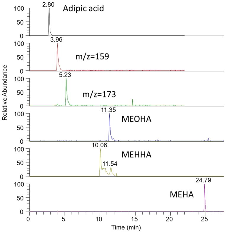
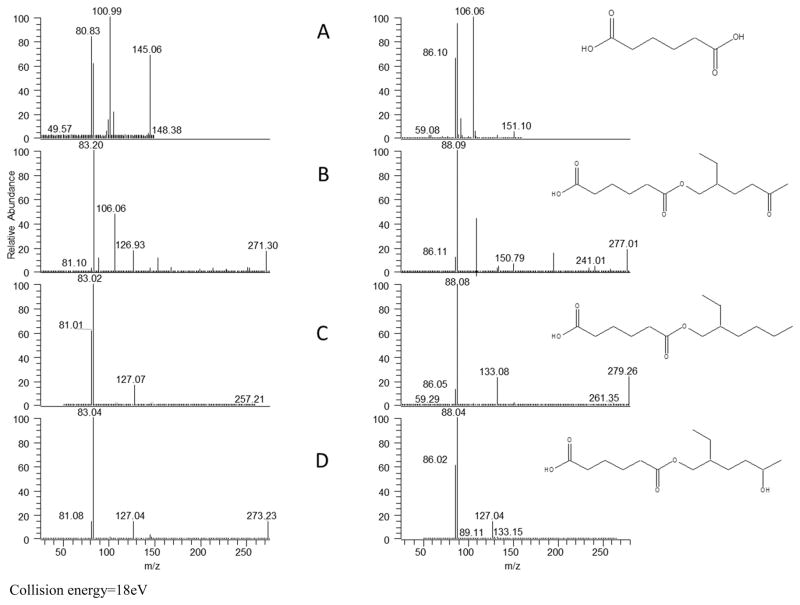
Five nonspecific metabolites of DEHA have been identified previously in the urine of six adult volunteers administered with deuterium labeled DEHA.4 We used in vitro metabolism to identify specific biomarkers of DEHA. Full scan analysis in negative ion mode from m/z = 125 to m/z = 400 of the human liver microsomal supernatant after 5 h of incubation with DEHA resulted in six unique peaks at different retention times (Figure 1). Two of these metabolites, adipic acid (m/z = 145, RT = 3.9 min) and MEHA (m/z = 257, RT = 24.2 min), were positively identified using authentic standards. Attempts to identify the two metabolites with m/z = 159 (RT = 3.96 min) and 173 (RT = 5.3 min) were not made because they are nonspecific metabolites of DEHA derived from chemical modifications of the adipic acid backbone. In the absence of authentic standards and based on their fragmentation patterns, we tentatively identified MEOHA (RT = 11.2 min, m/z = 271) and MEHHA (RT = 10–12 min, m/z = 273). In vitro metabolism of 13C6-DEHA and 13C6-MEHA formed analogous metabolites, 13C6-MEHHA and 13C6-MEOHA, further supporting the identity of these oxidative products (Figure 2). Interestingly, MEHHA and MEOHA eluted as two separate clusters of peaks, likely due to multiple oxidation sites in the 2-ethyl hexyl side chain and the adipic acid backbone that produced similar fragmentation patterns (Figure 1). We previously observed similar metabolite clusters for other plasticizers, including di-isononyl phthalate and 1,2-cyclohexane dicarboxylic acid, diisononyl ester.15–18 Because all coeluting isomers of MEHHA and MEOHA are valid biomarkers of exposure to DEHA and similar in structure with different oxidation sites, we did not attempt to separate the isomers and included them together to facilitate their detection.
Figure 1.
Chromatographic separation of DEHA metabolites detected in a human liver microsomes suspension of DEHA after 5 h of incubation at 37 °C. MEOHA, mono-2-ethyl oxohexyl adipate; MEHHA, mono-2-ethylhydroxyhexyl adipate; MEHA, mono-2-ethyl-hexyl adipate.
Figure 2.
Mass spectrometric fragmentation of (A) adipic acid, (B) MEOHA, (C) MEHA, and (D) MEHHA formed after in vitro phase I metabolism of DEHA (left) and 13C6-DEHA (right) using human liver microsomes. Structures shown are for only one of the potential isomers.
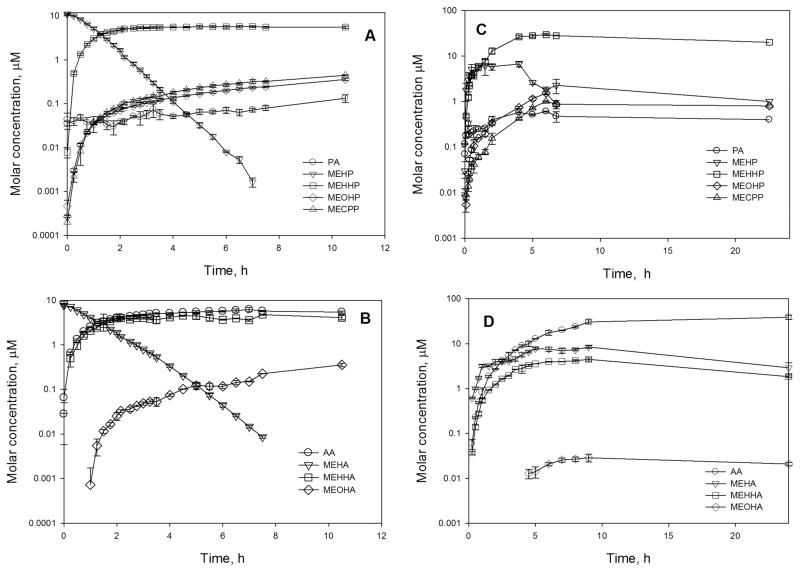
MEHHA and MEOHA are analogous to MEHHP and MEOHP, oxidative metabolites of DEHP;11,13 therefore, we also evaluated the in vitro metabolism of DEHP. Our data suggest that the first products formed, the hydrolytic monoesters MEHA (from DEHA) and MEHP (from DEHP), are further metabolized to the diacids (adipic acid or phthalic acid, respectively) and other oxidative products (Figure 3). Adipic acid was the major metabolite of DEHA/MEHA after 24 h/12 h of incubation with human liver microsomes, but phthalic acid was only a minor product of the DEHP/MEHP metabolism (Figure 3). In vitro metabolism of MEHP by human liver microsomes was faster (t1/2 = 0.56 h) compared to MEHA (t1/2 = 0.77 h). We did not detect MEHP and MEHA after 8 h of incubation with human liver microsomes (Figure 3). Also, both DEHA and DEHP formed the corresponding oxo metabolite (MEOHA and MEOHP, respectively); MEOHA was not detectable until 4 h after incubation. By contrast, we detected MEOHP immediately upon the incubation of DEHP and MEHP with human liver microsomes. Although MECPP is present as a major urinary oxidative metabolite of DEHP in humans,12,19 under our experimental conditions, MECPP was a minor in vitro metabolite (Figure 3). Similarly, we did not detect mono-2-ethyl-5-carboxypentyl adipate from the in vitro metabolism of DEHA. These findings suggest important differences in the in vitro metabolism of DEHA/MEHA and DEHP/MEHP.
Figure 3.
Time dependent formation of phase I in vitro metabolites of MEHP (A), MEHA (B), DEHP (C), and DEHA (D) with human liver microsomes. Error bars represent the standard deviation (N = 3). MEHHA and MEOHA were quantified using MEHA. DEHA and DEHP levels were not monitored.
In 144 urine samples from a group of US adults with no known exposure to DEHA, we detected adipic acid in all of the samples (median 313 nM (45.7 ng/mL); the maximum was 77,664 nM (11,333 ng/mL), whereas MEHHA, MEOHA, and MEHA were detectable in fewer than 20% of the samples and at lower concentration ranges: MEHA, <LOD-165 nM (LOD-42.5 ng/mL); MEHHA, <LOD-87 nM (LOD-23.9 ng/mL); and MEOHA, <LOD-38 nM (LOD-10.4 ng/mL). The urinary concentrations of MEHHA and MEOHA in these samples correlated well (r = 0.83, p < 0.01); however, these concentrations did not correlate with the concentrations of adipic acid. Adipic acid is not a unique biomarker for DEHA but the final hydrolytic metabolite of all adipates, including dibutyl adipate and diisononyl adipate. Further, adipic acid is used as a food additive.20 The lack of correlation between the urinary concentrations of adipic acid and the specific DEHA metabolites suggest additional sources for urinary adipic acid besides DEHA in the group of adults examined.
In summary, our study suggests that measuring the urinary concentrations of DEHA specific metabolites would be a suitable approach for assessing DEHA background exposure in humans. However, in contrast to the phthalate plasticizer DEHP, because DEHA appears to be metabolized mainly to the nonspecific metabolite adipic acid, MEHA, MEHHA, and MEHOA may only serve as sensitive exposure biomarkers of DEHA at high exposure levels.
Acknowledgments
Funding
This work was supported by the Centers for Disease Control and Prevention, U.S. Department of Health and Human Services.
ABBREVIATIONS
- DEHA
di-2-ethylhexyl adipate
- MEHA
mono-2-ethylhexyl adipate
- DEHP
di-2-ethylhexyl phthalate
- MEHP
mono-2-ethylhexyl phthalate
- MEOHP
mono-2-ethyl-5-oxohexyl phthalate
- MEHHP
mono-2-ethyl-5-hydroxyhexyl phthalate
- MECPP
mono-2-ethyl-5-carboxypentyl phthalate
- MEOHA
mono-2-ethyloxohexyl adipate
- MEHHA
mono-2-ethylhydroxyhexyl adipate
Footnotes
The authors declare no competing financial interest.
The findings and conclusions in this report are those of the authors and do not necessarily represent the official position of the Centers for Disease Control and Prevention.
References
- 1.IARC. WHO IARC Monographs. Vol. 77. IARC; Lyon, France: 2000. Di(2-ethylhexyl) Adipate; pp. 149–175. [PMC free article] [PubMed] [Google Scholar]
- 2.Fasano E, Bono-Blay F, Cirillo T, Montuori P, Lacorte S. Migration of phthalates, alkylphenols, bisphenol A and di(2-ethylhexyl)adipate from food packaging. Food Control. 2012;27:132–138. [Google Scholar]
- 3.Goulas AE, Salpea E, Kontominas MG. Di(2-ethylhexyl) adipate migration from PVC-cling film into packaged sea bream (Sparus aurata) and rainbow trout (Oncorhynchus mykiss) fillets: kinetic study and control of compliance with EU specifications. Eur Food Res Technol. 2008;226:915–923. [Google Scholar]
- 4.Loftus NJ, Woollen BH, Steel GT, Wilks MF, Castle L. An assessment of the dietary uptake of di-2-(ethylhexyl) adipate (DEHA) in a limited population study. Food Chem Toxicol. 1994;32:1–5. doi: 10.1016/0278-6915(84)90029-2. [DOI] [PubMed] [Google Scholar]
- 5.Fromme H, Gruber L, Schlurnmer M, Wz G, Bohmer S, Angerer J, Mayer R, Liebl B, Bolte G. Intake of phthalates and di(2-ethylhexyl)adipate: results of the integrated exposure assessment survey based on duplicate diet samples and biomonitoring data. Environ Int. 2007;33:1012–1020. doi: 10.1016/j.envint.2007.05.006. [DOI] [PubMed] [Google Scholar]
- 6.Takahashi T, Tanaka A, Yamaha T. Elimination, distribution and metabolism of di-(2-ethylhexyl)adipate (Deha) in rats. Toxicology. 1981;22:223–233. doi: 10.1016/0300-483x(81)90085-8. [DOI] [PubMed] [Google Scholar]
- 7.Singh AR, Lawrence WH, Autian J. Dominant lethal mutations and antifertility effects of di-2-ethylhexyl adipate and diethyl adipate in male mice. Toxicol Appl Pharmacol. 1975;32:566–576. doi: 10.1016/0041-008x(75)90121-0. [DOI] [PubMed] [Google Scholar]
- 8.Wato E, Asahiyama M, Suzuki A, Funyu S, Amano Y. Collaborative work on evaluation of ovarian toxicity 9) Effects of 2-or 4-week repeated dose studies and fertility study of di(2-ethylhexyl)adipate (DEHA) in female rats. J Toxicol Sci. 2009;34:SP101–SP109. doi: 10.2131/jts.34.s101. [DOI] [PubMed] [Google Scholar]
- 9.EPA. [accessed Sep 05, 2013];Integrated Risk Information System (IRIS), Di(2-ethylhexyl)adipate (CASRN 103-23-1) 2012 available at http://www.epa.gov/iris/subst/0420.htm.
- 10.Loftus NJ, Laird WJD, Steel GT, Wilks MF, Woollen BH. Metabolism and pharmacokinetics of deuterium-labeled di-2-(ethylhexyl) adipate (Deha) in humans. Food Chem Toxicol. 1993;31:609–614. doi: 10.1016/0278-6915(93)90042-w. [DOI] [PubMed] [Google Scholar]
- 11.Koch HM, Bolt HM, Angerer J. Di(2-ethylhexyl)phthalate (DEHP) metabolites in human urine and serum after a single oral dose of deuterium-labelled DEHP. Arch Toxicol. 2004;78:123–130. doi: 10.1007/s00204-003-0522-3. [DOI] [PubMed] [Google Scholar]
- 12.Koch HM, Preuss R, Angerer J. Di(2-ethylhexyl)phthalate (DEHP): human metabolism and internal exposure - an update and latest results. Int J Androl. 2006;29:155–165. doi: 10.1111/j.1365-2605.2005.00607.x. [DOI] [PubMed] [Google Scholar]
- 13.Silva MJ, Reidy A, Preau JL, Samandar E, Needham LL, Calafat AM. Measurement of eight urinary metabolites of di(2-ethylhexyl) phthalate as biomarkers for human exposure assessment. Biomarkers. 2006;11:1–13. doi: 10.1080/13547500500382868. [DOI] [PubMed] [Google Scholar]
- 14.Silva MJ, Samandar E, Preau JL, Reidy JA, Needham LL, Calafat AM. Quantification of 22 phthalate metabolites in human urine. J Chromatogr B. 2007;860:106–112. doi: 10.1016/j.jchromb.2007.10.023. [DOI] [PubMed] [Google Scholar]
- 15.Silva MJ, Samandar E, Preau JL, Calafat AM. Environmental exposure to the plasticizer 1,2-cyclohexan dicarboxylic acid, diisononly ester (DINCH) in US adults (2000–2012) [accessed Sep 6, 2013];Environ Res. 2013 doi: 10.1016/j.envres.2013.05.007. Online early access, published online Jun 15, 2013. [DOI] [PMC free article] [PubMed] [Google Scholar]
- 16.Silva MJ, Reidy JA, Preau JL, Needham LL, Calafat AM. Oxidative metabolites of diisononyl phthalate as biomarkers for human exposure assessment. Environ Health Perspect. 2006;114:1158–1161. doi: 10.1289/ehp.8865. [DOI] [PMC free article] [PubMed] [Google Scholar]
- 17.Koch HM, Angerer J. Di-iso-nonylphthalate (DINP) metabolites in human urine after a single oral dose of deuterium-labelled DINP. Int J Hyg Environ Health. 2007;210:9–19. doi: 10.1016/j.ijheh.2006.11.008. [DOI] [PubMed] [Google Scholar]
- 18.Schutze A, Palmke C, Angerer J, Weiss T, Bruning T, Koch HM. Quantification of biomarkers of environmental exposure to di(isononyl)cyclohexane-1,2-dicarboxylate (DINCH) in urine via HPLC-MS/MS. J Chromatogr, B. 2012;895:123–130. doi: 10.1016/j.jchromb.2012.03.030. [DOI] [PubMed] [Google Scholar]
- 19.Silva MJ, Reidy A, Preau JL, Samandar E, Needham LL, Calafat AM. Measurement of eight urinary metabolites of di(2-ethylhexyl) phthalate as biomarkers for human exposure assessment. Biomarkers. 2006;11:1–13. doi: 10.1080/13547500500382868. [DOI] [PubMed] [Google Scholar]
- 20.Organization for Economic Co-operation and Development. [accessed Sep 05, 2013];SIDS Initial Assessment Report for Adipic Acid. 2004 available at http://www.epa.gov/chemrtk/hpvis/rbp/103-23-1_Adipic%20acid,%20bis[2-ethylhexyl]%20ester_Web_SuppDocs_Sept08.pdf.


