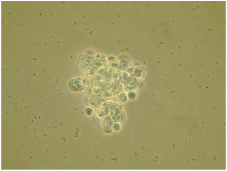
Figure 1.
A microaggregate of hemocytes (approximately 10–12 μm) formed at 1 h after injecting S. marcescens into the hemocoel of a tobacco hornworm. For this microphotograph (taken 1 h after injection), hemolymph was withdrawn, diluted with buffer and placed on a microscope slide for observation and photography. The cells in these photographs range from 10–12 microns. Photo by JSM.

