Abstract
Glucocorticoid (GC) therapy is associated with the risk of life-threatening adverse events in patients with autoimmune disease. To determine accurately the incidence and predictors of GC-related adverse events during initial GC treatment, we conducted a cohort study. Patients with autoimmune disease who were initially treated with GCs in Japan National Hospital Organization (NHO) hospitals were enrolled. Cox proportional hazard regression was used to determine the independent risks for GC-related serious adverse events and mortality. Survival was analyzed according to the Kaplan-Meier method and was assessed with the log-rank test.
The 604 patients had a total follow-up of 1105.8 person-years (mean, 1.9 year per patient). One hundred thirty-six patients had at least 1 infection with objective confirmation, and 71 patients had serious infections. Twenty-two cardiovascular events, 55 cases of diabetes, 30 fractures, 23 steroid psychosis events, and 4 avascular bone necrosis events occurred during the follow-up period. The incidence of serious infections was 114.8 (95% confidence interval, 95.7–136.6) per 1000 person-years. After adjustment for covariates, the following independent risk factors for serious infection were found: elderly age (hazard ratio [HR], 1.25/10-yr age increment; p = 0.016), presence of interstitial lung disease (HR, 2.01; p = 0.011), high-dose GC use (≥29.9 mg/d) (HR, 1.71; p = 0.047), and low performance status (Karnofsky score, HR, 0.98/1-score increment; p = 0.002). During the follow-up period, 73 patients died, 35 of whom died of infection. Similarly, elderly age, the presence of interstitial lung disease, and high-dose GC use were found to be significant independent risk factors for mortality. The incidence of serious and life-threatening infection was higher in patients with autoimmune disease who were initially treated with GCs. Although the primary diseases are important confounding factors, elderly age, male sex, the presence of interstitial lung diseases, high-dose GCs, and low performance status were shown to be risk factors for serious infection and mortality.
INTRODUCTION
Glucocorticoids (GCs) are commonly used as antiinflammatory and immunosuppressive therapies in autoimmune diseases.3,6 Despite the considerable benefits of GCs in controlling autoimmune diseases, serious adverse events (AEs) have been observed.29 These AEs vary from mild and self limited to major or life threatening.21 It is generally considered that GC-related AEs are dose related, thus, the higher the initial and cumulative doses, the longer the course, and the greater the likelihood of significant side effects.8,12 However, there remains considerable debate regarding the true incidence of these AEs.7,23 The inability to differentiate AEs that are attributable to GCs from those occurring due to underlying diseases or other comorbidities confounds the identification of potential associations. Although there is emerging evidence concerning the AEs of lower- to moderate-dose GCs,8,10,16,17,26,27 clear guidelines on the use of GCs are lacking. Comorbid conditions and risk factors for serious AEs of GCs should be identified before initiating GC therapy. To better define the toxicity of GCs, we addressed the question of whether the dose of GCs can cause an increased incidence of serious GC-associated AEs in patients with autoimmune disease. We undertook a multicenter cohort study of GC treatments of patients with newly diagnosed autoimmune disease, and investigated the incidence of GC-related AEs in the Japan National Hospital Organization (NHO)-EBM study group.
METHODS
Patients and Study Design
We conducted a multicenter cohort study following patients with newly diagnosed autoimmune diseases in NHO hospitals (total 55 hospitals). Patients were eligible if they were initially treated with GCs against the following autoimmune diseases, which were newly diagnosed (within the 4 weeks prior to entry) by the established criteria. The cohort start date was defined as the time of initiation of the first GC prescription. The autoimmune diseases registered in this study were rheumatic disease, systemic lupus erythematous (SLE), mixed connective tissue disease, polymyositis, dermatomyositis, vasculitis, Behçet disease, systemic scleroderma, adult-onset Still disease, Sjögren syndrome, rheumatoid arthritis, autoimmune bullous diseases, and anaphylactoid purpura. Neurologic diseases included multiple sclerosis, myasthenia gravis, and chronic inflammatory demyelinating polyradiculoneuropathy. The following gastro-hepatobiliary diseases were included: ulcerative colitis, autoimmune hepatitis, autoimmune pancreatitis, and primary biliary cirrhosis. Interstitial lung diseases included idiopathic interstitial pneumonia and collagen vascular disease preceded by interstitial pneumonia. The primary glomerular diseases included were rapidly progressive glomerulonephritis, chronic glomerulonephritis, and nephrotic syndrome. Patients were excluded from the study if they fit any of the following criteria: 1) clinically unstable cardiovascular disease, 2) age <16 years at study entry, 3) previous use of steroids to treat other diseases <6 months before cohort entry, or 4) other pre-existing autoimmune diseases. A total of 604 patients with newly diagnosed autoimmune disease were enrolled. These patients were enrolled between April 1, 2007, and March 31, 2008, and were regularly followed for the observation period that ended on March 31, 2009. The study was approved by the ethical committees of the NHO central institutional review board.
Data Collection
Data from all participating physicians were entered into the J-NHOSAC database at the data center of International Medical Center of Japan (Shinjuku, Tokyo) via the Hospnet Internet system. Information was collected during the study follow-up period regarding the development of the following AEs: fracture, avascular bone necrosis, diabetes, hyperlipidemia, infections, cardiovascular events (myocardial infarction, pulmonary emboli, heart failure, arrhythmia, and cerebrovascular accidents], gastrointestinal (GI) bleeding, and death. Physicians assessed AEs and serious AEs according to accepted guidelines. Diagnosis of pulmonary emboli required that a contrast-enhanced computed tomography (CT) scan of the chest or ventilation/perfusion scintigraphy indicated the presence of an embolism. Diagnosis of avascular osteonecrosis was based on the 2011 revised criteria for classification of osteonecrosis from the Japanese Ministry of Health, Labor, and Welfare.24 Osteonecrosis due to primary joint diseases, such as osteoarthritis or septic arthritis, was excluded. For quality assurance, the participating physicians were provided with the definitions of AEs and serious AEs. These definitions were included in the case report form at each visit.
Data on Study Entry
The past comorbid conditions of each of the patients were reviewed by each of the principal physicians. In addition, the incidences of specific conditions, including pre-existing pulmonary tuberculosis, hepatitis viral infection (hepatitis B virus, hepatitis C virus), diabetes, hyperlipidemia, arrhythmia, and performance status (Karnofsky score) were assessed. The physicians also provided information on smoking or drinking habits and history of tuberculosis. At entry, patients underwent chest X-ray and were screened for hepatitis B surface antigen (HBsAg) and anti-hepatitis C virus antibodies.
Medications
Every day of steroid use in each patient was recorded in the J-NHOSAC database, and average and cumulative steroid doses were calculated. Information on concomitant medications was recorded and hospitalizations for any reason were documented. Details of GCs, immunosuppressive agents, and biological agents were recorded at each visit, including the route of administration and the dose. In addition, we recorded the use of other medications, including prophylaxis agents, antibiotics, cardiovascular drugs, immunomodulating drugs, disease-modifying antirheumatic drugs (DMARDs), anti-osteoporosis agents, and nonsteroidal antiinflammatory drugs (NSAIDs). We categorized GC exposure according to the average daily dose throughout the follow-up period for each patient. We calculated “dose equivalents” of prednisolone as follows: 1 mg of prednisolone = 5 mg of cortisone = 4 mg of hydrocortisone = 1 mg of prednisone = 0.8 mg of triamcinolone = 0.8 mg of methylprednisolone = 0.15 mg of dexamethasone = 0.15 mg of betamethasone.5
Outcome Variables
At the start of the study, standardized lists were used to document AEs, which were classified using the System Organ Class (SOC) of the Medical Dictionary for Regulatory Activities (MedDRA, v. 11.1; International Conference on Harmonisation of Technical Requirements for Registration of Pharmaceuticals for Human Use [ICH], Geneva, Switzerland). All physicians documented episodes of infection requiring medical care and death certificates and the causes of deaths that occurred during the follow-up period. Serious infections occurring during the observation periods were counted. Serious infections (≥grade 3, defined by Common Terminology Criteria for Adverse Events v. 3.0; National Cancer Institute, National Institutes of Health, Bethesda, MD) were defined as life threatening, requiring hospitalization and/or intravenous antibiotic therapy, or leading to significant disability/incapacity.2 Fungal infection was defined as a “proven” or “possible” invasive fungal infection according to the criteria of the European Organization for Research and Treatment of Cancer/Mycoses Study Group.1 Cytomegalovirus (CMV) infection was defined as CMV end-organ disease,20 in which the signs and symptoms of affected organs (pneumonia, GI disease, hepatitis) plus CMV antigenemia were present. Diagnosis of CMV antigenemia was defined by a positive CMV PP65 antigenemia assay. If a serious infection was identified, verification was sought by the patient’s physician. If not already provided, additional information about all serious infections was required, including causative organism, treatment, and outcomes. Telephone interviews concerning the health assessment and presence of GC-related AEs were conducted with a few patients who were moved or transferred to another hospital at the end of the cohort study. The overall outcome was not available for 17 patients (2.8%) at the end of the study. In statistical analyses, we excluded the participants without final outcome data.
Statistical Analysis
The chi-square test for categorical variables and the Mann-Whitney test for continuous variables were used to calculate statistical differences between the 2 groups. The incidence rate (IR) (per 1000 person-years) was calculated with 95% confidence interval (CI).13 Cox proportional hazard models were used to estimate the risk of serious infections and mortality. In Cox proportional hazard models, we identified the best subset of explanatory variables by all combinations as variable selection in terms of score statistics. The variables included in the analysis were age, sex, types of primary autoimmune disease, comorbidities (diabetes, renal diseases, cardiovascular diseases, and interstitial lung diseases), medications (average dose of GC, use of immunosuppressive agents), and performance status or laboratory data on entry (Karnofsky score, serum albumin, serum IgG, lymphocyte counts). The analysis was conducted using SAS v. 9.1 software (SAS Institute, Cary, NC). Two-sided p values < 0.05 were considered statistically significant.
Propensity analysis was also performed regarding the probability of high-dose GC use (≥30 mg/d). The propensity for high-dose corticosteroid use was determined without regard to outcome. For each patient, a propensity score indicating the likelihood of having high-dose GCs prescribed was calculated by logistic regression analysis and included 25 covariates: age, sex and primary autoimmune diseases, previous cerebrovascular disease, ischemic heart disease, diabetes, hyperlipidemia, proteinuria, macrohematuria, previous autoimmune diseases, and performance status (Karnofsky score). The patients were stratified into 5 groups according to the propensity score, and the effect of high-dose GC (≥30 mg/d) on mortality was analyzed using the log-rank test.
RESULTS
Demographic Data
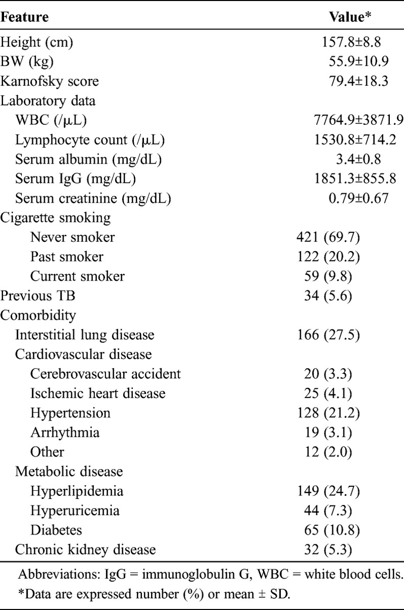
This cohort study comprised 604 patients, of whom 59.3% were female. The mean age at entry, which was within 4 weeks of the first diagnosis of autoimmune diseases, was 59.1 ± 16.9 years. The total follow-up time was 1105.8 person-years, and the mean follow-up was 22.4 months. The distribution of primary autoimmune diseases and general characteristics are shown in Table 1. All patients received GCs at entry, and the mean GC dose for the first month was 50.4 ± 63.1 mg/d. Table 2 lists variables examined at the time of cohort initiation, including performance status, laboratory data, and comorbidities. During the follow-up period, 46.9% (283) of patients had been treated with either an immunosuppressive or biological agent (see Table 1).
TABLE 1.
Characteristics of 604 Patients With Autoimmune Diseases
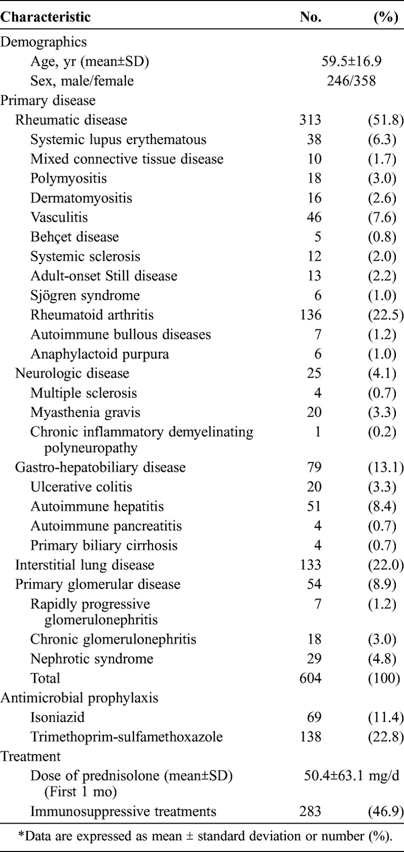
TABLE 2.
Clinical and Laboratory Findings on Entry
Types and Incidence of AEs
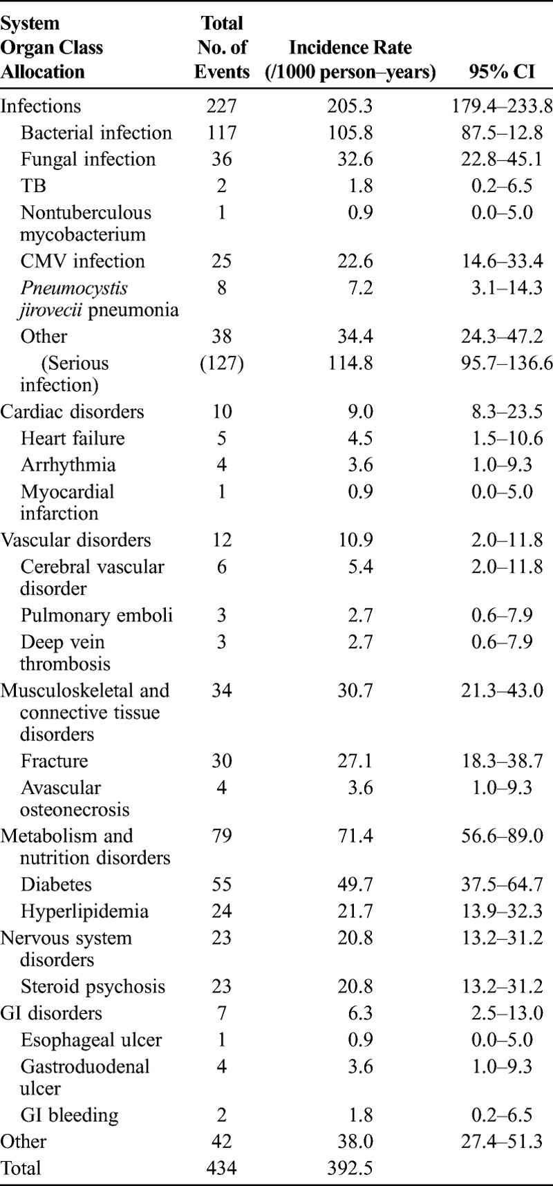
Overall, 434 AEs occurred (Table 3). Based on the AE categories classified using the SOC allocation, infections were the most common, followed by metabolic disease, fractures, steroid psychosis, and cardiovascular events.
TABLE 3.
Categories of Adverse Events (AE) by System Organ Class Allocation
Predictors of Serious Infections
A total of 136 patients experienced at least 1 infectious AE, 43 of whom experienced 2 or more infections. A significant number of the infections were opportunistic infections, including Pneumocystis jirovecii pneumonia (IR, 7.2 events/1000 person-years), fungal infection (IR, 32.6 events/1000 person-years), and CMV infection (IR, 22.6 events/1000 person-years), in addition to bacterial infections. There were 2 cases of Mycobacterium tuberculosis and 1 case of nontuberculosis mycobacterium (see Table 3). The crude IR for tuberculosis was 1.8 events/1000 person-years, and for nontuberculosis mycobacterium it was 0.9 events/1000 person-years. A total of 71 patients had serious infections; the crude IR of serious infections was 114.8 events/1000 person-years. The cumulative incidence curve of infectious AEs increased steadily from the cohort start, and close to 30% of patients experienced their first AEs within 3 months after the start of GC treatment. Average daily doses of GCs were time-dependent categorical variables. Therefore, the average daily dose for the first 1 month was used for analysis.
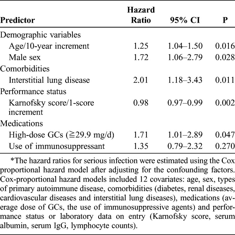
Univariate analysis revealed several statistically significant predictors for increased risk of infection (Table 4). The factors that were independently associated with an increase in the risk of serious infections in multivariate models are shown in Table 5. Factors associated with serious infections included increasing age (hazard ratio [HR], 1.25/10-yr age increment; 95% CI, 1.04–1.50), male sex (HR, 1.72; 95% CI, 1.06–2.79), the presence of interstitial lung disease (HR, 2.01; 95% CI, 1.18–2.89), low performance status (Karnofsky score, HR, 0.98/1-score increment; 95% CI, 0.97–0.99; p = 0.002), and high-dose GCs (≥29.88 mg/d) (HR, 1.71; 95% CI, 1.01–2.89).
TABLE 4.
Risk Factors for Serious Infection in Patients Treated With GCs
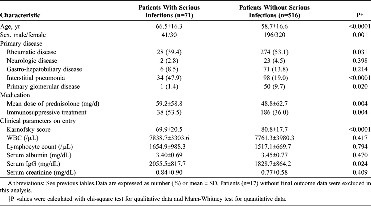
TABLE 5.
Predictors of Serious Infection Identified in the Multivariate Model*
Fatal Outcome
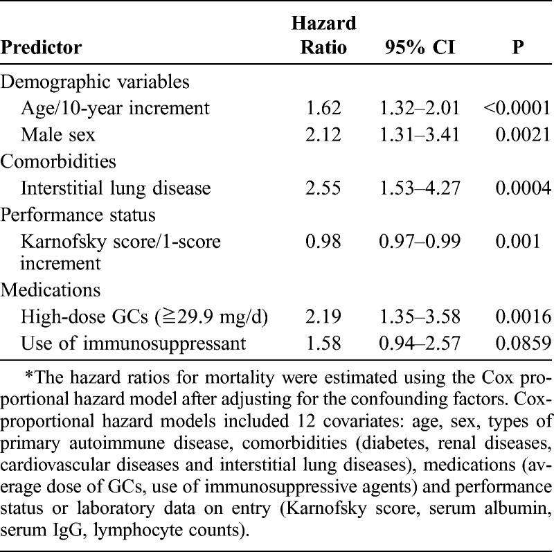
During the follow-up period, 73 patients died. The most common cause of death was infection, followed by interstitial pneumonia, respiratory failure, and cardiovascular events (Table 6). Factors that were independently associated with risk for mortality in Cox multivariate regression analysis are shown in Table 7. Factors associated with mortality included increasing age (HR, 1.62/10-yr age increment; 95% CI, 1.31–3.41), male sex (HR, 2.12; 95% CI, 1.31–3.41), presence of interstitial lung disease (HR, 2.55; 95% CI, 1.53–4.27), low performance status (Karnofsky score, HR, 0.98/1-score increment; 95% CI, 0.97–0.99; p = 0.001), and high-dose GCs (HR, 2.19; 95% CI, 1.35–3.58).
TABLE 6.
Causes of Death
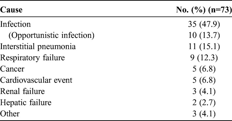
TABLE 7.
Predictors of Mortality Identified in the Multivariate Model*
In Kaplan-Meier survival curves stratified by the categorical ranges of the average prednisolone dose, a statistically significant difference was observed between patients receiving high-dose GCs (≥29.9 mg/d) and those receiving low-dose GCs (<12.7 mg/d) (p < 0.0001, log-rank test, Figure 1A). There was a significant difference in survival between patients receiving steroid pulse therapy (>500 mg of intravenous methylprednisolone) and those without steroid pulse therapy (Figure 1B). The overall survival was estimated for each category of autoimmune disease. The survival rate was comparable among patients with rheumatic disease and those with neurologic disease, primary glomerular disease, and gastro-hepatobiliary diseases. However, there was a significant difference in survival between patients with rheumatic disease and interstitial lung disease (Figure 2A). Similarly, the presence of interstitial lung disease significantly affected patient survival (p < 0.0001, log-rank test) (Figure 2B).
FIGURE 1.
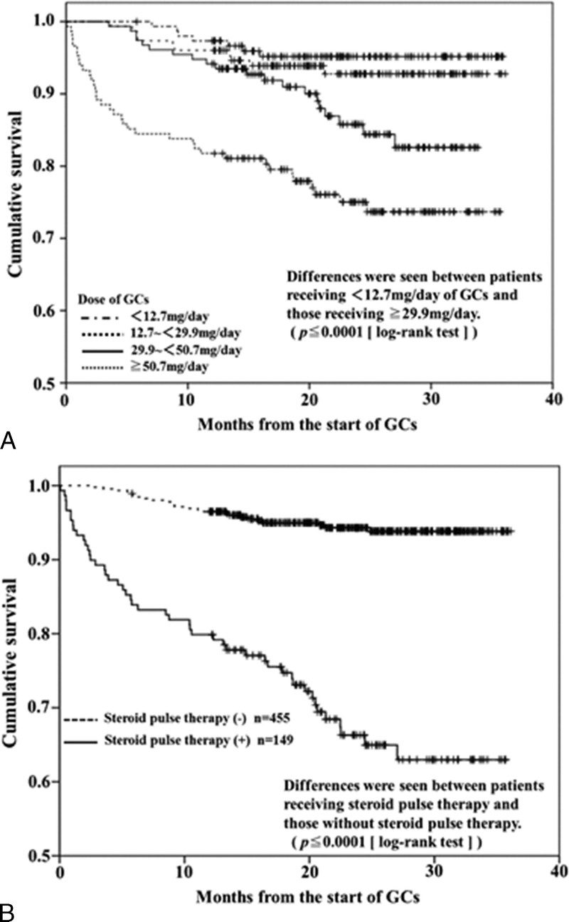
A, Kaplan-Meier survival curves illustrating survival distribution. Curves are stratified by average prednisolone dose (ranges shown in lower left corner). Statistically significant differences were observed between patients receiving high-dose GCs (≥29.88 mg/d) and those receiving low-dose GCs (<12.7 mg/d). B, Kaplan-Meier survival curves stratified by the presence or absence of steroid pulse therapy (>500 mg/d of intravenous methylprednisolone). There was a significant difference in survival between patients receiving and those not receiving steroid pulse therapy (p < 0.0001, log-rank test).
FIGURE 2.
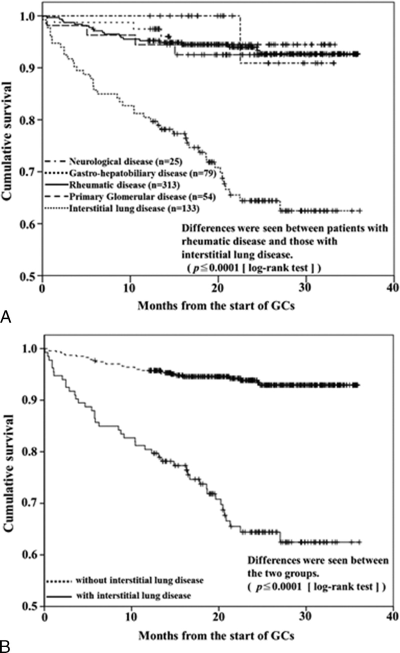
A, Kaplan-Meier survival curves stratified by category of autoimmune disease. The survival rate was comparable among patients with rheumatic disease and those with neurologic disease, renal disease, and gastro-hepatobiliary disease. However, there was a significant difference in survival between patients with rheumatic disease and those with interstitial lung disease (p < 0.0001, log-rank test). B, Kaplan-Meier survival curves stratified by the presence or absence of interstitial lung disease. Statistically significant differences were observed between patients with or without interstitial lung disease (p < 0.0001, log-rank test).
Propensity Score Analysis
Because high-dose GC treatment is reserved for severe autoimmune diseases, incomplete adjustment for underlying diseases or their disease activity would bias these results toward higher mortality rates, assuming that severe autoimmune diseases are associated with a higher mortality rate. Therefore, we used propensity score analysis30 to address the effect of covariate imbalance between patients receiving high-dose GCs (≥30 mg/d) and those not receiving high-dose GCs (<30 mg/d). In the propensity analysis, variables that correlated strongly with prescription of high-dose GCs were underlying primary autoimmune diseases (rheumatoid arthritis, systemic sclerosis, SLE, vasculitis, primary Sjögren syndrome, dermatomyositis, polymyositis, ulcerative colitis, and myasthenia gravis), presence of comorbidities (interstitial lung disease, diabetes, hyperlipidemia, and ischemic heart disease), and presence of proteinuria and macrohematuria. The likelihood of receiving high-dose GCs for each patient was modeled using logistic regression conditioned on covariate values for each individual. Effect of high-dose GCs on survival in each of 5 strata of equal size was analyzed on the basis of the propensity score (Table 8). In the log-rank test, the difference in time to death was statistically significant (HR, 2.07; 95% CI, 1.07–4.00; p = 0.029) between patients receiving or not receiving high-dose GCs, suggesting that patients receiving high-dose GCs had a mortality risk even after adjustment for treatment selection bias. However, in the individual propensity score quintiles, the adjusted relative risk (RR) for mortality did not reach statistical significance except in the lowest propensity score group (quintile 1).
TABLE 8.
Effects of High-Dose Corticosteroid on Mortality, Stratification by Propensity Score*

DISCUSSION
Glucocorticoids (GCs) are among the most indispensable therapeutic agents used against autoimmune diseases.4 Despite the considerable benefits of GCs in controlling autoimmune diseases, various AEs are well established.21 However, there is little evidence concerning their incidence, dose-dependence, and true impact on survival. Several large retrospective reviews have reported that long-term GC use was a significant predictor of potentially serious AEs.15 Considering the diversity of their mechanisms and sites of action, GCs can cause a wide array of AEs.18 One population-based study showed that patients who were exposed to dosages of GCs greater than the equivalent of 7.5 mg of prednisolone per day for 1–5 years had substantially higher rates of all cardiovascular diseases.35 The incidence of cardiovascular events was reported to be 23.9 per 1000 person-years in the group exposed to GCs.35 In the current cohort study with a mean follow-up of 1.9 years, the incidence of cardiovascular events in patients with autoimmune disease treated with GCs was 19.9 per 1000 person-years, which is consistent with that previous study.35 However, the overall incidence of infections was 205.3 per 1000 person-years, which was substantially higher compared with the overall incidence of cardiovascular events. Furthermore, some of these infections, including opportunistic infections, were serious enough to contribute to a fatal outcome. Opportunistic infections have been reported with corticosteroid use in conjunction with other risk factors, including immunosuppressive therapy.14,19,31,32 Data from the current study indicated that the actual incidence of fungal infections was 32.6 persons/1000 person-years in patients treated initially with GCs; CMV was 22.6 persons; Mycobacterium tuberculosis was 1.8 persons; nontuberculosis mycobacterium was 0.9 persons; and Pneumocystis jirovecii pneumonia was 7.2 persons/1000 person-years in patients treated initially with GCs. The identified risk factors for serious infection included male sex, increasing age, interstitial lung disease, low performance status, and an initial high dose of GCs (≥29.9 mg/d). Some of these factors (sex, increasing age, and performance status) have already been demonstrated to be risks for infection or mortality in patients treated with GCs.11,25,28
Moderate- to high-dose GC therapy leads to an increased risk of infection.9 The risk of infection increases with the dose and duration of treatment, and tends to remain low in patients exposed to low doses, even with a high cumulative dosage.22,34 The strongest evidence for increased risk of infection from GCs is meta-analysis of 71 controlled clinical trials in which patients were treated with corticosteroid or placebo.33 Stuck et al33 found a significant risk of lethal and nonlethal infections in patients receiving systemic corticosteroid (RR, 1.6; 95% CI, 1.3–1.9). This association was dose-dependent with no increased risk observed in patients receiving ≤10 mg of prednisolone a day or a cumulative dosage ≤700 mg. The RR of infection increased with a mean daily dosage of steroid (RR, 1.3; 95% CI, 1.0–1.6) of <20 mg prednisolone, RR of 2.1 (95% CI, 1.3–3.6) with a daily dose of 20–40 mg prednisolone, and RR of 2.1 (95% CI, 1.6–2.9) with a daily dose >40 mg prednisolone.33 Our results are in overall agreement with this previous meta-analysis in that risk of serious infections increased in a dose-dependent manner related to GC usage. When underlying disease was considered, Stuck et al33 detected no significant increase in the incidence of infection in those patients with pulmonary diseases, which were mainly chronic obstructive pulmonary diseases. These results are in contrast with results from our study, in which the presence of interstitial lung disease was an independent risk factor for serious infections and mortality. It is likely that differences between the types of pulmonary disease may contribute to the differential outcome between our study and the previous meta-analysis.
Although high-dose GCs have been demonstrated to be a causal risk factor for mortality, a key question of our cohort study was whether the association of high-dose GC use with mortality reflects the effects of GCs or an association with the underlying diseases for which high-dose GCs were prescribed. The difference in mortality by high-dose GC treatments needs to be analyzed by including all available confounding factors into the models. Statistical adjustment through propensity scoring, including types of diseases and activity, may partly resolve these problems.30 We found an association between high-dose GCs and mortality in propensity score-based stratification groups. However, our results also indicated that the association between high-dose GC exposure and mortality tends to be weak in high propensity scoring groups. These data suggest that the association between high-dose GCs and mortality may not be prominent in patients with autoimmune disease for which high-dose GCs are necessary.
One major limitation of the present study is that nonrandom allocation of subjects would confound our results. Therefore, some degree of residual confounding was inevitable, bringing with it the possibility that we were not able to distinguish the effects of GCs versus disease specificity against the risk of infection or mortality. Second, our observational study had an important and inherent treatment bias for confounding by various autoimmune diseases, and the clinical assessment of disease activity was not complete. Patients receiving high-dose GCs were most likely to have the worst disease and high disease activity. Confounding factors were adjusted by multivariate Cox regression analysis, and the results were not contradictory to those obtained with propensity score analysis. However, it must be acknowledged that observational studies can only partially control for confounding factors. The strengths of the current study include the high follow-up rate. We focused on serious AEs that contribute to morbidity or mortality. Thus, we could completely survey detailed clinical data from participating doctors directly using the Internet throughout the follow-up period. Furthermore, the percentage of loss to follow-up (2.8%) was very low.
In conclusion, initial GC therapy against autoimmune disease was associated with an increased incidence of AEs, including infection. Predictors of serious infection or mortality include male sex, increasing age, comorbidities (interstitial lung disease), low performance status, and initial high dose of GCs (≥29.9 mg/d). These results advance our understanding of the relationship between GC therapy and serious AEs, and may help to prospectively identify high-risk patients.
ACKNOWLEDGMENTS
This study could not have been accomplished without the dedicated participation of the following contributors: Yukio Ohara, MD (NHO Hokkaido Medical Center); Yoshikazu Araya, MD (NHO Hakodate National Hospital); Hideo Nishimura, MD (NHO Asahikawa Medical Center); Noriyuki Chiba, MD (NHO Morioka Hospital); Keiji Chida, MD (NHO Iwate hospital); Muneshige Tobita, MD (NHO Yonezawa Hospital); Takefumi Saito, MD (NHO Ibarakihigashi National Hospital); Manabu Hayashi, MD (NHO Numata Hospital); Misao Kawaguchi, MD (Nishisaitama-chuo National Hospital); Noriko Sato, MD (NHO Saitama National Hospital); Satoko Miyatake, MD (NHO East Saitama National Hospital); Toyohiko Sugimoto, MD (Shimoshizu National Hospital); Shigeki Hayashi, MD (NHO Disaster Medical Center); Kiyoshi Maejima, MD (NHO Kanagawa Hospital); Hiroaki Iwase, MD (NHO Nagoya Medical Center); Kunikazu Ogawa, MD (NHO Mie Chuo Medical Center); Hiroshi Tsutani, MD (NHO Awara Hospital), Motoyuki Maeno, MD (NHO Shigaraki National Hospital); Toshiki Komeda, MD (NHO Kyoto Medical Center); Hideyuki Sawada, MD (NHO Utano Hospital); Yoshiro Mochizuki, MD (NHO Himeji Medical Center); Tetsuo Yamamoto, MD; (NHO Yonago Medical Center); Haruhiro Yamashita, MD (NHO Okayama Medical Center); Atsuhiko Tada, MD (NHO Minami-Okayama Medical Center); Shigeyuki Kuratsu, MD (NHO Kure Medical Center); Yutaka Sato, MD (NHO Kanmon Medical Center); Tsuneo Matsumoto, MD (NHO Yamaguchi-Ube Medical Center); Masahiro Yamaguchi, MD (NHO Takamatsu Medical Center); Ogushi Fumitaka, MD (NHO Kochi National Hospital); Nobuhiko Nagata, MD (NHO Omuta Hospital); Hidenori Matsuo, MD (NHO Nagasaki Kawatana Medical Center); Toru Yamanaka, MD (NHO Kumamoto Minami Hospital); Koich Honda, MD (NHO Oita Medical Center); Bon Shoji MD, (NHO Iwakuni Medical Center); Yasuo Suenaga, MD (NHO Beppu Medical Center); Shuichi Hanada, MD (NHO Kagoshima Medical Center); Masaharu Kawabata, MD (NHO Minami Kyushu National Hospital).
Abbreviations
- AE
adverse event
- CI
confidence interval
- CMV
cytomegalovirus
- CT
computed tomography
- GC
glucocorticoid
- GI
gastrointestinal
- HR
hazard ratio
- IR
incidence rate
- NHO
National Hospital Organization
- RR
relative risk
- SLE
systemic lupus erythematous
- SOC
System Organ Class
Footnotes
Financial support and conflicts of interest: This work was supported by a grant from the National Hospital Organization (multi-centre clinical studies for evidenced-based medicine), Tokyo, Japan. The authors have no conflicts of interest to disclose.
REFERENCES
- 1. Ascioglu S, Rex JH, de Pauw B, Bennett JE, Bille J, Crokaert F, Denning DW, Donnelly JP, Edwards JE, Erjavec Z, Fiere D, Lortholary O, Maertens J, Meis JF, Patterson TF, Ritter J, Selleslag D, Shah PM, Stevens DA, Walsh TJ. Defining opportunistic invasive fungal infections in immunocompromised patients with cancer and hematopoietic stem cell transplants: an international consensus. Clin Infect Dis. 2002; 34: 7– 14. [DOI] [PubMed] [Google Scholar]
- 2. Basch E, Jia X, Heller G, Barz A, Sit L, Fruscione M, Appawu M, Iasonos A, Atkinson T, Goldfarb S, Culkin A, Kris MG, Schrag D. Adverse symptom event reporting by patients vs clinicians: relationships with clinical outcomes. J Natl Cancer Inst. 2009; 101: 1624– 1632. [DOI] [PMC free article] [PubMed] [Google Scholar]
- 3. Bijlsma JW, Saag KG, Buttgereit F, da Silva JA. Developments in glucocorticoid therapy. Rheum Dis Clin North Am. 2005; 31: 1– 17. [DOI] [PubMed] [Google Scholar]
- 4. Boumpas DT, Chrousos GP, Wilder RL, Cupps TR, Balow JE. Glucocorticoid therapy for immune-mediated diseases: basic and clinical correlates. Ann Intern Med. 1993; 119: 1198– 1208. [DOI] [PubMed] [Google Scholar]
- 5. Buttgereit F, da Silva JA, Boers M, Burmester GR, Cutolo M, Jacobs J, Kirwan J, Kohler L, Van Riel P, Vischer T, Bijlsma JW. Standardised nomenclature for glucocorticoid dosages and glucocorticoid treatment regimens: current questions and tentative answers in rheumatology. Ann Rheum Dis. 2002; 61: 718– 722. [DOI] [PMC free article] [PubMed] [Google Scholar]
- 6. Buttgereit F, Straub RH, Wehling M, Burmester GR. Glucocorticoids in the treatment of rheumatic diseases: an update on the mechanisms of action. Arthritis Rheum. 2004; 50: 3408– 3417. [DOI] [PubMed] [Google Scholar]
- 7. Caldwell JR, Furst DE. The efficacy and safety of low-dose corticosteroids for rheumatoid arthritis. Semin Arthritis Rheum. 1991; 21: 1– 11. [DOI] [PubMed] [Google Scholar]
- 8. Curtis JR, Westfall AO, Allison J, Bijlsma JW, Freeman A, George V, Kovac SH, Spettell CM, Saag KG. Population-based assessment of adverse events associated with long-term glucocorticoid use. Arthritis Rheum. 2006; 55: 420– 426. [DOI] [PubMed] [Google Scholar]
- 9. Dale DC, Petersdorf RG. Corticosteroids and infectious diseases. Med Clin North Am. 1973; 57: 1277– 1287. [DOI] [PubMed] [Google Scholar]
- 10. Da Silva JA, Jacobs JW, Kirwan JR, Boers M, Saag KG, Ines LB, de Koning EJ, Buttgereit F, Cutolo M, Capell H, Rau R, Bijlsma JW. Safety of low dose glucocorticoid treatment in rheumatoid arthritis: published evidence and prospective trial data. Ann Rheum Dis. 2006; 65: 285– 293. [DOI] [PMC free article] [PubMed] [Google Scholar]
- 11. Doran MF, Crowson CS, Pond GR, O’Fallon WM, Gabriel SE. Predictors of infection in rheumatoid arthritis. Arthritis Rheum. 2002; 46: 2294– 2300. [DOI] [PubMed] [Google Scholar]
- 12. Franklin J, Lunt M, Bunn D, Symmons D, Silman A. Risk and predictors of infection leading to hospitalisation in a large primary-care-derived cohort of patients with inflammatory polyarthritis. Ann Rheum Dis. 2007; 66: 308– 312. [DOI] [PMC free article] [PubMed] [Google Scholar]
- 13. Garwood F. Fiducial limits for the Poisson distribution. Biometrika. 1936; 28: 437– 442. [Google Scholar]
- 14. Gustafson TL, Schaffner W, Lavely GB, Stratton CW, Johnson HK, Hutcheson RH., Jr Invasive aspergillosis in renal transplant recipients: correlation with corticosteroid therapy. J Infect Dis. 1983; 148: 230– 238. [DOI] [PubMed] [Google Scholar]
- 15. Hoes JN, Jacobs JW, Boers M, Boumpas D, Buttgereit F, Caeyers N, Choy EH, Cutolo M, Da Silva JA, Esselens G, Guillevin L, Hafstrom I, Kirwan JR, Rovensky J, Russell A, Saag KG, Svensson B, Westhovens R, Zeidler H, Bijlsma JW. EULAR evidence-based recommendations on the management of systemic glucocorticoid therapy in rheumatic diseases. Ann Rheum Dis. 2007; 66: 1560– 1567. [DOI] [PMC free article] [PubMed] [Google Scholar]
- 16. Hoes JN, Jacobs JW, Verstappen SM, Bijlsma JW, Van der Heijden GJ. Adverse events of low- to medium-dose oral glucocorticoids in inflammatory diseases: a meta-analysis. Ann Rheum Dis. 2009; 68: 1833– 1838. [DOI] [PubMed] [Google Scholar]
- 17. Hwang YG, Saag K. The safety of low-dose glucocorticoids in rheumatic diseases. Clin Exp Rheumatol. 2011; 29 (5 Suppl 68): S104– S112. [PubMed] [Google Scholar]
- 18. Kimberly RP. Mechanisms of action, dosage schedules, and side effects of steroid therapy. Curr Opin Rheumatol. 1991; 3: 373– 379. [DOI] [PubMed] [Google Scholar]
- 19. Lionakis MS, Kontoyiannis DP. Glucocorticoids and invasive fungal infections. Lancet. 2003; 362: 1828– 1838. [DOI] [PubMed] [Google Scholar]
- 20. Ljungman P, Griffiths P, Paya C. Definitions of cytomegalovirus infection and disease in transplant recipients. Clin Infect Dis. 2002; 34: 1094– 1097. [DOI] [PubMed] [Google Scholar]
- 21. McDonough AK, Curtis JR, Saag KG. The epidemiology of glucocorticoid-associated adverse events. Curr Opin Rheumatol. 2008; 20: 131– 137. [DOI] [PubMed] [Google Scholar]
- 22. McDougall R, Sibley J, Haga M, Russell A. Outcome in patients with rheumatoid arthritis receiving prednisone compared to matched controls. J Rheumatol. 1994; 21: 1207– 1213. [PubMed] [Google Scholar]
- 23. Morand EF. Corticosteroids in the treatment of rheumatologic diseases. Curr Opin Rheumatol. 2000; 12: 171– 177. [DOI] [PubMed] [Google Scholar]
- 24. Nakamura J, Saisu T, Yamashita K, Suzuki C, Kamegaya M, Takahashi K. Age at time of corticosteroid administration is a risk factor for osteonecrosis in pediatric patients with systemic lupus erythematosus: a prospective magnetic resonance imaging study. Arthritis Rheum. 2010; 62: 609– 615. [DOI] [PubMed] [Google Scholar]
- 25. Ruiz-Irastorza G, Olivares N, Ruiz-Arruza I, Martinez-Berriotxoa A, Egurbide MV, Aguirre C. Predictors of major infections in systemic lupus erythematosus. Arthritis Res Ther. 2009; 11: R109– R116. [DOI] [PMC free article] [PubMed] [Google Scholar]
- 26. Ruyssen-Witrand A, Fautrel B, Saraux A, Le-Loet X, Pham T. Infections induced by low-dose corticosteroids in rheumatoid arthritis: a systematic literature review. Joint Bone Spine. 2010; 77: 246– 251. [DOI] [PubMed] [Google Scholar]
- 27. Saag KG, Koehnke R, Caldwell JR, Brasington R, Burmeister LF, Zimmerman B, Kohler JA, Furst DE. Low dose long-term corticosteroid therapy in rheumatoid arthritis: an analysis of serious adverse events. Am J Med. 1994; 96: 115– 123. [DOI] [PubMed] [Google Scholar]
- 28. Sakuma Y, Katoh T, Owada K, Suzuki H, Sakurai K, Eiro M, Asahi K, Watanabe T. Initial functional status predicts infections during steroid therapy for renal diseases. Clin Nephrol. 2005; 63: 68– 73. [DOI] [PubMed] [Google Scholar]
- 29. Schacke H, Docke WD, Asadullah K. Mechanisms involved in the side effects of glucocorticoids. Pharmacol Ther. 2002; 96: 23– 43. [DOI] [PubMed] [Google Scholar]
- 30. Schmoor C, Gall C, Stampf S, Graf E. Correction of confounding bias in non-randomized studies by appropriate weighting. Biom J. 2011; 53: 369– 387. [DOI] [PubMed] [Google Scholar]
- 31. Sepkowitz KA. Pneumocystis carinii pneumonia in patients without AIDS. Clin Infect Dis. 1993; 17 (Suppl 2): S416– S422. [DOI] [PubMed] [Google Scholar]
- 32. Sepkowitz KA, Brown AE, Telzak EE, Gottlieb S, Armstrong D. Pneumocystis carinii pneumonia among patients without AIDS at a cancer hospital. JAMA. 1992; 267: 832– 837. [PubMed] [Google Scholar]
- 33. Stuck AE, Minder CE, Frey FJ. Risk of infectious complications in patients taking glucocorticosteroids. Rev Infect Dis. 1989; 11: 954– 963. [DOI] [PubMed] [Google Scholar]
- 34. van Everdingen AA, Jacobs JW, Siewertsz Van Reesema DR, Bijlsma JW. Low-dose prednisone therapy for patients with early active rheumatoid arthritis: clinical efficacy, disease-modifying properties, and side effects: a randomized, double-blind, placebo-controlled clinical trial. Ann Intern Med. 2002; 136: 1– 12. [DOI] [PubMed] [Google Scholar]
- 35. Wei L, MacDonald TM, Walker BR. Taking glucocorticoids by prescription is associated with subsequent cardiovascular disease. Ann Intern Med. 2004; 141: 764– 770. [DOI] [PubMed] [Google Scholar]


