Supplemental Digital Content is available in the text
Abstract
We aimed to present a combined surgical procedure in conservative treatment of placenta accreta based on surgical outcomes in our cohort of patients. The study was designed as a prospective cohort series study. The setting involved two education and research hospitals in Turkey. This study included 12 patients with placenta accreta who were prenatally diagnosed and managed.
We offered the patients the choice of conservative or nonconservative treatment. We then offered 2 choices for patients who had preferred conservative treatment, leaving the placenta in situ as is the classical procedure, or our surgical procedure. One patient preferred nonconservative treatment, the others opted for our procedure.
We evaluated demographic and obstetric characteristics of patients, sonographic and operative parameters of patients, and surgical outcomes.
We operated on 11 patients using this surgical procedure that we have developed for placenta accreta cases. We found that there was no need for hysterectomy in any patient, and we preserved the uterus for all of these patients. No patient presented any septic complication or secondary vaginal bleeding.
Our surgical procedure seems to be effective and useful in the conservative treatment of placenta accreta.
INTRODUCTION
Abnormal invasive placentation cause elevated maternal morbidity and mortality. Placenta accreta is an abnormal adherence of the placental chorionic villi to the myometrium, which occurs in the absence of decidua basalis and the fibrinoid layer of Nitabuch.1 Placental invasion abnormalities are classified according to the depth of penetration by the chorionic villi as placenta accreta, -increta, and -percreta.2 Placenta accreta is considered a severe pregnancy complication that may be associated with massive and potentially life-threatening intrapartum and postpartum hemorrhage.3 It is caused by a defect in decidua basalis resulting in an abnormally invasive placental implantation. This disruption is often related to previous uterine scars, including cesarean sections and prior uterine curettage. Other risk factors associated with placenta accreta are multiparity, placenta previa, prior intrauterine infections, elevated maternal serum α-fetoprotein, and maternal age >35 years.4 It has become the leading cause of emergent hysterectomy. Maternal morbidity had been reported to occur in up to 60% and mortality in up to 7% of women with placenta accreta. In addition, the incidence of perinatal complications is also increased, mainly due to preterm birth fetuses.5 Women with placenta accreta are usually delivered by cesarean section. It is better to perform the surgery under elective, controlled conditions rather than urgently with inadequate preparation in an emergency. In addition, regardless of the management option taken, the prevention of complications ideally requires a multidisciplinary team approach.6 Conservative, uterine-sparing approaches for the management of placenta accreta have been described to both reduce the morbidity of peripartum hysterectomy as well as to allow for future fertility. In this study, we report a comprehensive surgical procedure and results in the conservative management of placenta accreta, with 11 cases operated in our clinic.
STUDY POPULATION
This prospective, surgical case-series study included 12 consecutive patients with placenta accreta who were prenatally diagnosed at the Department of Gynecology and Obstetrics of Seyhan Research and Training Hospital (Adana, Turkey) and the Department of Gynecology and Obstetrics of Katip Celebi University, Ataturk Education and Research Hospital (Izmir, Turkey) from 2010 to 2014. Operations were performed by the same author in both hospitals. The study was in adherence with the tenets of the Helsinki Declaration, and informed consent was obtained from all participants prior to surgery.
MATERIAL AND METHODS
We have used the term “abnormal placentation” for all types of abnormal invasive placenta. Gestational age was calculated from the first day of the last menstrual period or estimated by the first obstetrical ultrasound examination. Abnormal placentation was diagnosed by the presence of one of these findings: ultrasound or magnetic resonance imaging (MRI) diagnosis and manual removal of placenta being partially or totally impossible with no cleavage plane between part or whole of the placenta and uterus. In this 4-year period, we have encountered 16 cases of abnormal placentation. Twelve patients were diagnosed prenatally, and 4 patients were diagnosed in the intrapartum or postpartum period. We have offered conservative treatment or postpartum hysterectomy for the prenatally diagnosed patients. One patient preferred hysterectomy because of her completed fertility. For pregnant women who have a strong desire to preserve their future fertility, we have offered 2 alternative conservative treatment modalities. The first one involves leaving the placenta in situ and follow up as in the classical procedure; the second involves performing our surgical procedure. Because our study is designed from retrospective case series in 2 different medical centers, we did not have an ethics committee approval. We have informed the patients about complications and potential adverse effects of both treatments, and another team has taken their informed consent. No patient preferred leaving the placenta in situ, and 11 patients preferred our technique.
One prenatally diagnosed patient was admitted in the 23rd week of pregnancy with vaginal bleeding, and was operated on using our conservative procedure. The other 10 patients were operated electively, having been diagnosed previously, and were being monitored for abnormal placentation. Elective patients were monitored more frequently during the pregnancy, and betamethasone was given to every patient at about the 31st to 32nd weeks of pregnancy to encourage fetal pulmonary maturity. Delivery was planned at about 35th to 36th weeks of pregnancy. Four nonprenatally diagnosed patients were admitted in emergency situations because of vaginal hemorrhage or a prolonged third stage of labor without previous diagnosis of abnormal placentation. Hysterectomy had to be performed on these patients. Those patients who had opted for conservative treatment underwent a complete ultrasonographical examination before surgery and placental borders were detected. Placental Doppler ultrasonography was performed for placentation. We performed MRI for 4 elective patients because they had a posterior-located placenta. Ages, gravidity, parity, dilatation and curettage history, and cesarean delivery history were noted. Placental localization and the presence of placenta previa were noted. Total blood counting, cross matching, and preparation of blood products were performed before the surgery. All patients underwent our surgical procedure, and hysterectomy was not required for any patient. Operation time for every patient was noted. Records of intraoperative and postoperative complications, number of required blood transfusion products, hospitalization time, and histopathology reports were also obtained. The β-human chorionic gonadotropin (hcG) levels at 6 weeks and 6 months after surgery were evaluated. Statistical analysis was performed by MedCalc statistical software (MedCalc Software, Ostend, Belgium).
SURGICAL PROCEDURE
Placental borders and mapping were detected carefully by ultrasonography and Doppler ultrasonography (placental mapping). In posterior-located placentas, we used MRI as additional imaging. According to the mapping, in a subset of patients we entered the abdomen by Pfannenstiel incision; in another subset we entered the abdomen by infraumbilical midline incision. Baby was delivered prior to surgically entering the placenta. Uterus is incised away from the placenta, according to the plan we described by placental mapping. Type of uterine incisions is determined after placental mapping. Placental borders have been identified, and incisions were made far away from placenta. J-shaped, vertical and upper transverse incisions were used. Then umbilical vein was catheterized. Ten units of diluted oxytocin and 100 to 200 cc, 37°C of heated saline were infused from here, and then the cord was clamped together with catheter. The bilateral hypogastric arteries and utero-ovarian anastomosis branches were ligated. The clamped cord was released and hydrodissection was performed by heated saline at 37°C. If placenta was not sufficiently dissected from the uterus, placental curettage or excision was performed. Then we sutured the placental bed with squarely shaped suturing. After hemostasis is secured, a balloon of 3-ways 20F Foley catheter (Galena Saglik Urunleri, Istanbul, Turkey) was inflated by 80 cc saline and placed into the intrauterine cavity. Then free part of the catheter was tracked through the vaginal way, and the uterine incision was closed. After half an hour of observation for vaginal bleeding, the operation was terminated. (See supplemental video, http://links.lww.com/MD/A211, which demonstrates a short sample of an operation.)
RESULTS
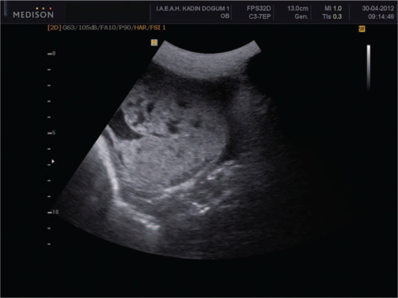
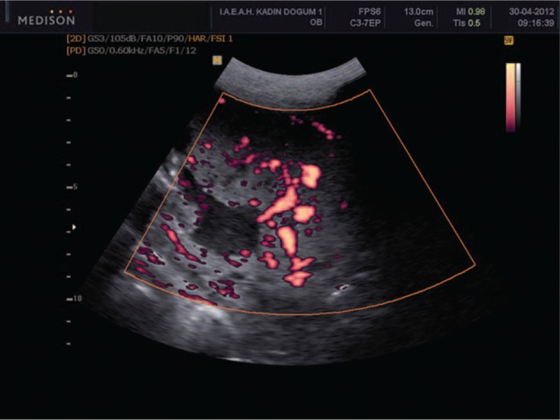
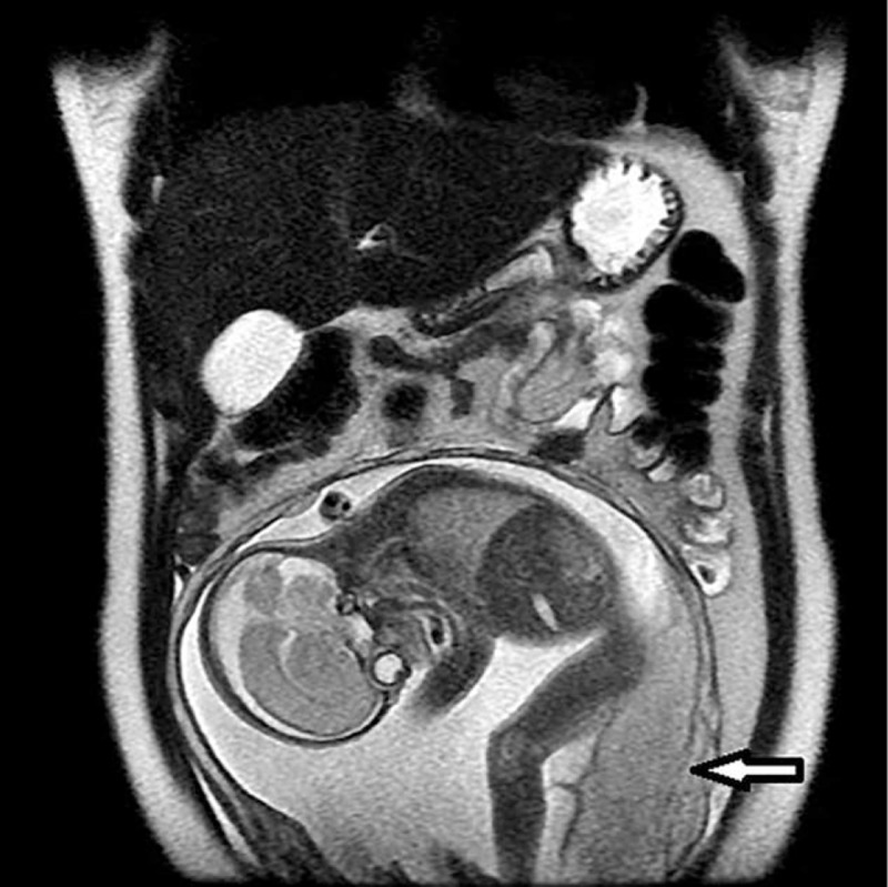
In this 4-year period, among 22,543 deliveries only 16 patients met the diagnostic criteria of placenta accreta (0.71/1000). In our conservative treatment group, the patients’ mean age was 31.5 ± 3.2 years, and their mean body mass index was 28.2 ± 2.1. All cases except 1 were multiparous. One patient had had 3 cesarean sections, 5 patients had 2 cesarean sections, and 4 patients had 1 cesarean section. One patient was primigravid, and she had no uterine curettage history. Two patients had a history of spontaneous abortion, and 4 patients had a history of induced abortion of the pregnancy (Table 1). The placenta was posteriorly located at 4 patients (36%) and anterior localized at 7 patients (64%). Six patients had placenta previa totalis, and in 5 patients placenta was located away from internal cervical os. Vascular lacunae with turbulent flow were noted (Figures 1 and 2). Placental invasion of the myometrium was observed in MRI (Figure 3). Mean gestational age was 35.2 ± 4.1 weeks. The mean operation time was 110 ± 20 minutes. Median 4 (2–7) units of erythrocyte suspensions and median 2 (0–4) units of fresh frozen plasma were transfused intraoperatively and postoperatively (Table 2). In 1 patient, a postoperative wound infection developed. Postoperative febrile reactions developed in 2 patients. One of these was caused by wound infection, and the other was due to atelectasis. The mean hospitalization time was 4.2 ± 0.4 days. β-hCG measurements were negative for all patients in postoperative 6th week and 6th month.
TABLE 1.
Demographic and Obstetric Characteristics of Patients

FIGURE 1.

Lacunar vascular areas in placenta.
FIGURE 2.

Wide lacunar areas by power Doppler sonography.
FIGURE 3.

Invasive placenta in coronal section MRI. MRI = magnetic resonance imaging.
TABLE 2.
Sonographic and Operative Parameters of Patients

DISCUSSION
The incidence of placenta accreta approximates about 1 in 1000 deliveries and, has increased 10-fold in the last 50 years, primarily because of the rise in cesarean section rates.7 In view of the fact that the indications for cesarean delivery seem to be steadily expanding, including cesarean delivery on maternal request, the incidence of placenta accreta is likely to continue to increase as the risk increases with the number of previous cesarean sections.6 Myometrial trauma and scarring resulting from repeated dilatation and curettage or other corrective surgeries also contribute to the risk of developing abnormal placental adherence.8,9
In our study, 10 of the 11 patients had a previous history of cesarean delivery. Also, 5 of them had a history of previous uterine curettage. Only 1 patient was primigravid, and she had no uterine curettage history. The risk of developing placenta accreta increases with the number of previous cesarean deliveries. Up to 88% of the women with placenta accreta have concomitant placenta previa.9–11 Six of our patients had concomitant placenta previa (55%). In 7 patients (64%), placenta was located in anterior uterus, on the previous uterine scar (P = 0.02). In our cohort of patients, we had no placenta percreta cases. Placenta accreta should be suspected in women who have both a placenta previa, particularly anterior, and a history of cesarean or other uterine surgery. Patients at risk of abnormal placentation should be assessed antenatally by ultrasonography, with or without adjunct MRI if indicated.12 Second and third trimester gray-scale sonographic characteristics include loss of continuity of the uterine wall, multiple vascular lacunae within the placenta, lack of hypoechoic border between the placenta and the myometrium, bulging of placental site into the bladder, and increased vasculature evident on color Doppler sonography in placenta.9,13 Multiple vascular lacunae and turbulent flow were observed in Doppler sonography, but in our cases we did not observe any invasion of bladder. If ultrasound findings are not considered definitive or the placenta is located on the posterior wall, MRI can be performed using gadolinium contrast agent intravenously.9,13
The most important factor affecting the outcome is prenatal diagnosis. It gives the opportunity to make a delivery plan that properly anticipates the expected blood loss and other potential complications of delivery. In addition, it gives the opportunity for electively timing the procedure, because the prevention of complications ideally requires the presence of a multidisciplinary surgical team, which is associated with improved outcomes.14 A number of reports have shown that outcomes are worse and morbidity is higher in women who deliver in an emergency or in an unplanned fashion.15 There is a great benefit of planned as opposed to emergency peripartum hysterectomy. In mothers with placenta previa and a suspected placenta accreta who required peripartum hysterectomy, a scheduled delivery has been associated with shorter operative times and lower frequency of transfusions, complications, and intensive care unit admissions.16 When the condition is not diagnosed antenatally, the most imminent and evident hint at diagnosis is profuse postpartum hemorrhage and placental retention after the second stage of labor is completed. Accordingly, the timing of delivery may have a crucial impact on maternal and perinatal outcome. O’Brien et al17 reported that after 35 weeks, 93% of patients with placenta accreta experience hemorrhage necessitating delivery. We planned delivery at the 35th to 36th weeks of pregnancy for our elective patients. We provided betamethasone treatment at about the 31st to 32nd week to encourage fetal pulmonary maturity. In these patients, we did not observe any neonatal morbidity. None of the patients required admission to neonatal intensive care unit.
Conservative, uterine-preserving approaches for the management of placenta accreta have been described to both reduce the morbidity of peripartum hysterectomy as well as allow for future fertility.18,19 A number of different approaches including uterine artery embolization, methotrexate therapy, hemostatic sutures, pelvic devascularization, and balloon tamponade have been described, with varying rates of success.20 One large series study reported a success rate of 78%, with a rate of maternal morbidity of 6%.19 In that study, which included 167 women from 25 hospitals in France, 78% retained their uterus, 11% required a hysterectomy within 24 hours of delivery because of hemorrhage, and 11% underwent hysterectomy within 3 months of delivery because of complications; 6% experienced severe morbidity, including sepsis, vesicouterine fistula, and/or uterine necrosis.19,21 Role of adjuvant methotrexate in cases of conservative management is uncertain. No large-scale studies have compared methotrexate use against no use of methotrexate in the treatment of placenta accreta, and at the present time, there are no convincing data either way.22 In selective cases, the placement of a balloon catheter was performed concurrently with conservative management, with the intent of avoiding hysterectomy and thereby preserving fertility.19
Postoperative complications reported with the established conservative approach include severe postpartum hemorrhage, postoperative disseminated intravascular coagulopathy, and infection resistant to antimicrobial therapy that may require laparotomy and hysterectomy.23 Thus far, the results of using preoperative prophylactic internal iliac artery catheterization as an adjuvant treatment to hysterectomy or in cases of conservative management are equivocal, and are largely limited by the small sample size of the studies. So, there is as yet no effective conservative treatment of placenta accreta, and hysterectomy is the preferred solution for these patients. Therefore, a new conservative treatment approach is required for women who have strong desire for preserving their future fertility.
In this study, we have reported a series of 11 patients who we have treated conservatively with our surgical procedure that we believe has not been reported in literature. There was no need of hysterectomy at intraoperative or postoperative stage. We have dissected placenta from the uterine wall by hydrodissection. Postoperatively, we did not observe hemorrhage, infection, or require a secondary operation. We did not encounter any serious complications. As we described the approach here, hypoperfusion of uterus by ligation of both the utero-ovarian anastomosis and hypogastric arteries may create time for a coagulation cascade in uterine vessels. Incision of the uterus was performed away from the placenta according to placental mapping. We have not made an incision into the placenta, and with this technique we avoided initiation of gross hemorrhage. Infusion of hot saline combined with oxytocin may produce a vasoconstriction of vessels in the placental bed, and can help in dissecting the placenta from the uterus. Hydrodissection technique is recently being used successfully in ophthalmic, neural, and laparoscopic surgery. We think that the role of pressurized saline is mechanical, and it generates a mass effect for dissection of placenta from the uterine wall. As a result of all these actions, perfusion of the uterus decreased, and with vasoconstriction in the placental bed, we could more easily dissect the placenta from the placental bed. Although we needed massive transfusions (≥4 units erythrocyte suspension) for 6 patients, we could control bleeding and did not need hysterectomy.
CONCLUSION
Placenta accreta is becoming a more common complication of pregnancy. Prenatal diagnosis is important in optimizing the counseling, treatment, and outcome of women with placenta accreta. Surgical treatment for placenta accreta is commonly performed as hysterectomy. However, conservative management should be the preferred approach especially for pregnant women who want to retain their future fertility. In this study, we have presented a comprehensive approach to conservative treatment for placenta accreta cases. Our surgical procedure is effective and useful in the conservative management of patients with placenta accreta, and it can be used as an alternative conservative treatment protocol if it can be supported by larger randomized controlled trials.
Footnotes
Abbreviation: MRI = magnetic resonance imaging.
Key message: Because hysterectomy is the most common result in the treatment of placenta accreta, a new approach is required. We described a combined surgical procedure in conservative management of placenta accreta, and it was successful in our cohort of patients.
The authors have no funding and conflicts of interest to disclose.
Supplemental digital content is available for this article. Direct URL citations appear in the printed text and are provided in the HTML and PDF versions of this article on the journal's Website (www.md-journal.com).
REFERENCES
- 1.Bodner LJ, Nosher JL, Gribbin C, et al. Balloon-assisted occlusion of the internal iliac arteries in patients with placenta accreta/percreta. Cardiovas Inter Radiol 2006; 29:354–361. [DOI] [PubMed] [Google Scholar]
- 2.Sinha P, Oniya O, Bewley S. Coping with placenta praevia and accreta in a DGH setting and words of caution. J Obstet Gynaecol 2005; 5:334–338. [DOI] [PubMed] [Google Scholar]
- 3.Faranesh R, Shabtai R, Eliezer S, et al. Suggested approach for management of placenta percreta invading the urinary bladder. Obstet Gynecol 2007; 110:512–515. [DOI] [PubMed] [Google Scholar]
- 4.Wu S, Kocherginsky M, Hibbard JU. Abnormal placentation: twenty-year analysis. Am J Obstet Gynecol 2005; 192:1458–1461. [DOI] [PubMed] [Google Scholar]
- 5.Eller AG, Porter TF, Soisson P, et al. Optimal management strategies for placenta accreta. BJOG 2009; 116:648–654. [DOI] [PubMed] [Google Scholar]
- 6.Warshak CR, Ramos GA, Eskander R, et al. Effect of predelivery diagnosis in 99 consecutive cases of placenta accrete. Obstet Gynecol 2010; 115:65–69. [DOI] [PubMed] [Google Scholar]
- 7.Chou MM, Hwang JI, Tseng JJ, et al. Internal iliac artery embolization before hysterectomy for placenta accreta. J Vasc Int Radiol 2003; 14:1195–1199. [DOI] [PubMed] [Google Scholar]
- 8.Cheung CS-y, Chan BC-p. The sonographic appearance and obstetric management of placenta accreta. Int J of Womens Health 2012; 4:587–594. [DOI] [PMC free article] [PubMed] [Google Scholar]
- 9.Garmi G, Salim R. Epidemiology, etiology, diagnosis, and management of placenta accreta. Obstet Gynecol Int 2012; 2012: Article ID 873929. [DOI] [PMC free article] [PubMed] [Google Scholar]
- 10.Washecka R, Behling A. Urologic complications of placenta percreta invading the urinary bladder: a case report and review of the literature. Hawaii Med J 2002; 61:66–69. [PubMed] [Google Scholar]
- 11.Wu S, Kocherginsky M, Hibbard J. Abnormal placentation: twenty-year analysis. Am J Obstet Gynecol 2005; 192:1458–1461. [DOI] [PubMed] [Google Scholar]
- 12.Oyelese Y, Smulian JC. Placenta previa, placenta accrete and vasa previa. Obstet Gynecol 2006; 107:927–941. [DOI] [PubMed] [Google Scholar]
- 13.Levine D, Hulka CA, Ludmir J, et al. Placenta accreta: evaluation with color Doppler US, power Doppler US, and MR imaging. Radiology 1997; 205:773–776. [DOI] [PubMed] [Google Scholar]
- 14.Eller AG, Bennett MA, Sharshiner M, et al. Maternal morbidity in cases of placenta accreta managed by a multidisciplinary care team compared with standard obstetric care. Obstet Gynecol 2011; 117:331–337. [DOI] [PubMed] [Google Scholar]
- 15.Wright JD, Pri-Paz S, Herzog TJ, et al. Predictors of massive blood loss in women with placenta accreta. Am J Obstet Gynecol 2011; 205:38e1–38e6. [DOI] [PubMed] [Google Scholar]
- 16.Robinson BK, Grobman WA. Effectiveness of timing strategies for delivery of individuals with placenta previa and accrete. Obstet Gynecol 2010; 116:835–842. [DOI] [PubMed] [Google Scholar]
- 17.O’Brien JM, Barton JR, Donaldson ES. The management of placenta percreta: conservative and operative strategies. Am J Obstet Gynecol 1996; 175:1632–1638. [DOI] [PubMed] [Google Scholar]
- 18.Alanis M, Hurst BS, Marshburn PB, et al. Conservative management of placenta increta with selective arterial embolization preserves future fertility and results in a favorable outcome in subsequent pregnancies. Fertil Steril 2006; 86:1514.e3–1514.e7. [DOI] [PubMed] [Google Scholar]
- 19.Perez-Delboy A, Wright JD. Surgical management of placenta accreta: to leave or remove the placenta? BJOG 2014; 121:163–169. [DOI] [PubMed] [Google Scholar]
- 20.Doumouchtsis SK, Papageorghiou AT, Arulkumaran S. Systematic review of conservative management of postpartum hemorrhage: what to do when medical treatment fails. Obstet Gynecol Surv 2007; 62:540–547. [DOI] [PubMed] [Google Scholar]
- 21.Sentilhes L, Ambroselli C, Kayem G, et al. Maternal outcome after conservative treatment of placenta accreta. Obstet Gynecol 2010; 115:526–534. [DOI] [PubMed] [Google Scholar]
- 22.Timmermans S, Van Hof AC, Duvekot JJ. Conservative management of abnormally invasive placentation. Obstet Gynecol Surv 2007; 62:529–539. [DOI] [PubMed] [Google Scholar]
- 23.Paull JD, Smith J, Williams L, et al. Balloon occlusion of the abdominal aorta during caesarean hysterectomy for placenta percreta. Anaesth Intensive Care 1995; 23:731–734. [DOI] [PubMed] [Google Scholar]


