

Abstract
The present investigation reports the occurrence of filarial worm, Setaria digitata, recovered during the surgery of a cow suffering from intussusception. The worms were thread like, milky white, long with tapering ends especially towards the hind end with an average size of 62.8 ± 9.89 mm in length. On examination of anterior end, the cuticular rings surrounding the mouth region with dorsal, ventral and lateral prominences were observed. The posterior end had few spines along with a pair of appendages near the tip of the tail. On the basis of morphological characteristics the worms were identified as female S. digitata.
Keywords: Cow, Jammu, Occurrence, Setariadigitata
Introduction
Setarial parasites are commonly found in the peritoneal cavity of ungulates. The adults in peritoneal cavity are virtually nonpathogenic but may be associated with mild fibrinous peritonitis. On rare occasions, the adult worms migrate erratically to eyes and other abdominal locations (Soulsby 1982; Urquhart et al. 2003). The larval stages of various species found in cattle (Setaria digitata, S. marshalli, and S. labiatopapillosa) have been reported to cause eosinophilic granulomatous lesions in central nervous system resulting into enzootic cerebrospinal nematodosis or “Kumari”. The current study reports the occurrence of S. digitata in a cow from R.S. Pura, Jammu.
Materials and methods
A non-descript cow aged 2 years suffering from intussusception was presented at clinics, Faculty of Veterinary Sciences and Animal Husbandry, Sher-e-Kashmir University of Agricultural Sciences and Technology, R.S. Pura, Jammu. During surgery, four thread-like worms were recovered from the peritoneal cavity. The worms were collected in normal saline, brought to the laboratory, transferred in a petri dish containing glycerol alcohol for clearing and were identified according to the morphological descriptions given elsewhere (Singh 2003; Bhatia et al. 2010).
Results and discussion
The collected worms were milky white, more tapered towards posterior end with an average length of 62.8 ± 9.89 mm (ranging from 40 to 80 mm). On the anterior end, the mouth was surrounded by cuticular rings that have dorsal, ventral and lateral prominences (Fig. 1). The lateral crescentic shaped prominences were not so prominent. The long spirally coiled posterior end ended into a knob which showed few spines (Fig. 2). A pair of lateral appendages was also seen near the tip of the tail. On the basis of morphological features the worms were identified as female S. digitata.
Fig. 1.
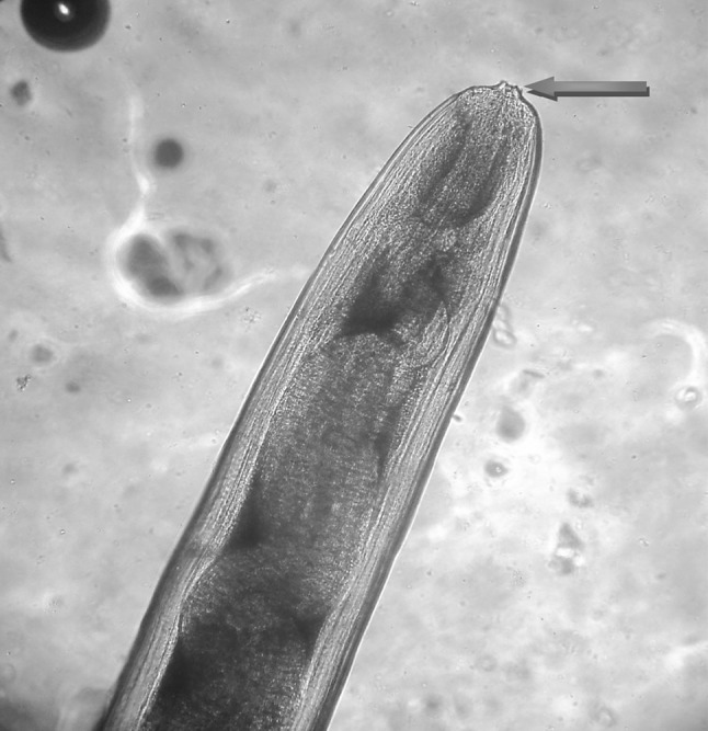
Anterior end of female Setaria digitata showing cuticular rings (arrow)
Fig. 2.

Posterior end of female Setaria digitata showing terminal spines
Setaria digitata is assumed to be the natural parasite of bovidae and the adult parasite in peritoneal cavity is considered nonpathogenic. However, juveniles when enter the abnormal hosts like sheep, goat, horse, etc. show aberrant migration in central nervous system, causing lumbar paralysis or “Kumari” which may even terminates into death (Soulsby 1982; Singh 2003). They also have tendency of invading other visceral organs like liver, kidney and intestine. Earlier various workers have reported the prevalence of Setaria spp. in bovines from different parts of the country (Mohan 1975; Chauhan and Pande 1980; Bhopale et al. 1982; Patnaik 1989; Thirumurthy et al. 1995; Siddiqui et al. 1996; Sundar and Ravindran 2009). Recently, Singh et al. (2013) have documented the occurrence of Setaria labiatopapillosa in a cow from Ludhiana, Punjab.
References
- Bhatia BB, Pathak KML, Juyal PD. Textbook of veterinary parasitology. 2. Ludhiana: Kalyani Publishers; 2010. pp. 249–250. [Google Scholar]
- Bhopale KK, Joshi SC, Shah HL. Occurrence of Setaria labiatopapillosa in buffaloes in MP. Liv Adv. 1982;7:26–27. [Google Scholar]
- Chauhan PPS, Pande BP. On the occurrence of Setaria labiatopapillosa in the intestinal lining of buffalo calf. Indian J Parasitol. 1980;4:89–91. [Google Scholar]
- Mohan RN. A note on Setaria digitata in cattle and buffaloes and cells of the peritoneal exudate. Indian J Anim Sci. 1975;45:914–915. [Google Scholar]
- Patnaik MM. On filarial nematodes in domestic animals in Orissa. Indian Vet J. 1989;66:573–574. [Google Scholar]
- Siddiqui AA, Sharma SP, Kumar M. Prevalence of Setaria infection in buffaloes and horses. Indian J Anim Sci. 1996;66:243–245. [Google Scholar]
- Singh KRS. Veterinary helminthology. 1. New Delhi: Indian Council of Agricultural Research; 2003. pp. 515–520. [Google Scholar]
- Singh H, Singh NK, Singh ND, Jyoti, Rath SS (2013) Occurrence of Setaria labiatopapillosa in peritoneal cavity of crossbred cattle. J Parasit Dis. doi: 10.1007/s12639-013-0308-3 [DOI] [PMC free article] [PubMed]
- Soulsby EJL. Helminths, arthropods and protozoa of domesticated animals. 7. London: ELBS and Bailliere Tindal; 1982. pp. 316–319. [Google Scholar]
- Sundar STB, Ravindran R. Intensity of setarial worm infection among bovines in and around Bangalore. Tamil Nadu J Vet Anim Sci. 2009;5(6):272–274. [Google Scholar]
- Thirumurthy CC, Senthilvel K, Madhavan Pillai K. Setaria digitata in bullock’s urine. J Vet Anim Sci. 1995;26:72. [Google Scholar]
- Urquhart GM, Armour J, Duncan JL, Dunn AM, Jennings FW. Veterinary parasitology. UK: Blackwell Science LTD; 2003. [Google Scholar]