

Abstract
Background:
Arthroscopic scapulothoracic bursectomy with partial superomedial angle scapulectomy has been described as a treatment for persistent scapulothoracic bursitis with varying results.
Purpose:
To evaluate patients after arthroscopic scapulothoracic bursectomy utilizing validated functional outcome instruments.
Study Design:
Case series.
Methods:
Fifteen patients underwent arthroscopic scapulothoracic bursectomy and partial superomedial angle scapulectomy. Thirteen patients were available for review at a mean 27-month follow-up (range, 13-65 months). Patients were evaluated preoperatively with a history, physical examination evaluating medial scapula border tenderness and crepitus, pain visual analog scale (VAS) score, and the simple shoulder test (SST). Postoperatively, patients were evaluated with all preoperative questionnaires as well as a satisfaction survey and the American Shoulder and Elbow Surgeons (ASES) score.
Results:
SST scores improved significantly from a mean 7.7 ± 3.1 preoperatively to 10.3 ± 2.3 postoperatively (P = .03). VAS pain scores reduced significantly from 6.5 ± 2.2 preoperatively to 2.3 ± 2.4 postoperatively (P < .001). Ninety-two percent (12/13) of patients were satisfied, and 92% (12/13) stated they would have the surgical procedure performed again. The mean ASES postoperative score was 80.1 (range, 38-100). The 2 clinical failures (ASES scores <50) had either a workers’ compensation claim with persistent medial border tenderness or ongoing rotator cuff disease. Despite lower ASES scores, these patients were still satisfied with the procedure and would undergo it again.
Conclusion:
Arthroscopic scapulothoracic bursectomy with partial superomedial angle scapulectomy provides significant improvements in pain and functional outcomes. Even in patients at risk for poorer clinical outcomes, patient satisfaction and willingness to undergo the surgical procedure again was still high.
Keywords: scapulothoracic bursitis, arthroscopic, scapulectomy
Scapulothoracic bursitis, or a “snapping” scapula, was first described over 100 years ago as a condition with audible and palpable grating localized to the superomedial angle of the scapula associated with pain.1,7 The etiology is likely secondary to anomalous tissue between the scapula and chest wall, including scarred and inflamed bursal tissue, a hooked superior medial scapula angle, a Luschka tubercle, a malunited rib, scapula fracture, or an osteochondroma.4 Surgical treatment including open partial scapulectomy was first described by Milch.6 Recently, arthroscopic methods, including bursectomy and partial scapulectomy, have been described for the treatment of scapulothoracic bursitis with varying results.3,5,8,10,11
Limited clinical data have been published describing the results of open or arthroscopic treatment of scapulothoracic bursitis. Nicholson and Duckworth9 reported improvements in pain and function after open scapulothoracic bursectomy with superomedial angle resection. Limitations of an open approach include increased pain, larger incisions, a requirement for rhomboid muscle detachment, and the need for postoperative immobilization. Arthroscopic treatment, including scapulothoracic bursectomy with or without superomedial angle resection, has been reported with reasonable results, although failure rates of 13% to 31% have been reported.8,11 Most studies did not include preoperative outcome data, and validated shoulder outcome tools were rarely utilized.3,5,10,11
The purpose of this study was to evaluate the outcomes of arthroscopic scapulothoracic bursectomy with partial superomedial angle scapulectomy for scapulothoracic bursitis.
Methods
Between January 1, 2007, and December 31, 2011, 15 patients underwent arthroscopic scapulothoracic debridement and superomedial angle scapulectomy by a single surgeon. Patient charts were retrospectively reviewed, and patients were recruited to return for a questionnaire evaluation and physical examination at a minimum 1 year postoperative. Institutional review board approval was obtained prior to initiating the study. Inclusion criteria included any patient having the procedure performed at the University of Utah by the primary surgeon (R.Z.T.) during the time period. Thirteen patients (87% follow-up) returned for examination at a mean 27 months (range, 13-65 months) postoperative. There were 5 male and 8 female patients, and the dominant shoulder was affected in 46% (6 of 13). The mean patient age at the time of surgery was 42 years (range, 21-62 years). Patients had a mean preoperative duration of symptoms of 59 months (range, 8-303 months). Rotator cuff disease and cervical spine pain were treated previously in 4 and 3 patients, respectively. Six patients had only isolated scapulothoracic symptoms.
The indications for surgery were failure of nonoperative treatment of scapulothoracic bursitis, including anti-inflammatory medications, physical therapy, and a scapulothoracic cortisone injection. Scapulothoracic bursitis was defined as localized pain to the superior medial angle and/or medial border of the scapula. All patients had tenderness to palpation along the superomedial angle and/or medial border of the scapula. Scapulothoracic crepitus was common but was not required to define the diagnosis. All patients underwent at least 1 cortisone injection performed in the office by the primary surgeon, with all patients confirming a temporary reduction in symptoms. All injections were performed in the prone position with the arm in a “chicken wing” position guiding the needle directly under the scapula along the medial border into the scapulothoracic space. Patients underwent a mean 2.5 injections (range, 1-10 injections) prior to surgery, some of which were not performed while under the primary surgeon’s care. Some patients had a concomitant diagnosis of either rotator cuff disease or cervical spine pain. At the time of the scapulothoracic bursectomy, the scapulothoracic pain was self-reported as most significant in all patients.
All surgical procedures were performed prone with the arm placed in the “chicken wing” position (Figure 1). A 2-portal technique was utilized, with the first portal (inferior portal) created approximately 3 to 4 cm medial to the medial border of the scapula halfway between the scapula spine and the inferior angle in the superior/inferior direction (Figure 2). A 30° scope was utilized in all cases, starting in the inferior portal. A superior portal was created under spinal-needle localization at the level of the scapula spine 3 to 4 cm medial to the medial scapula border (Figure 2). A shaver and cautery were utilized in the superior portal to perform the scapulothoracic debridement, including removal of inflamed bursal tissue and release of adhesions. The superomedial angle was then outlined utilizing several spinal needles, and the underlying serratus anterior was released from this region of the scapula (Figure 3). Utilizing a 4.0-mm bur, a 2 cm × 2 cm × 3 cm triangle of the superomedial scapula corner was completely removed (Figure 4). The scope was then placed in the superior portal, and a shaver and cautery device were used to complete the bursectomy down to the inferior scapula angle. Postoperatively, patients were in a sling for comfort only and were allowed to use the shoulder as tolerated, with a lifting limitation of 10 pounds for 6 weeks. Formalized physical therapy was prescribed between postoperative weeks 2 and 6, including shoulder stretching and rotator cuff, deltoid, and scapula stabilizer strengthening exercises. At 6 weeks postoperative, patients were allowed to return to activities as tolerated.
Figure 1.

Prone positioning with the arm in the “chicken wing” position.
Figure 2.
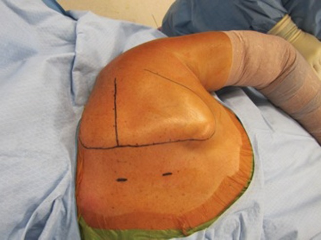
Two-portal technique: superior portal at the level of the scapula spine 3 to 4 cm medial to the medial border of the scapula, and inferior portal halfway between the scapula spine and the inferior scapula angle 3 to 4 cm medial to the medial border of the scapula.
Figure 3.

Undersurface of the superomedial angle of the scapula exposed after the serratus anterior is released utilizing an arthroscopic cautery.
Figure 4.

Undersurface of the scapula after partial superomedial angle scapulectomy removing a 2 cm × 2 cm × 3 cm triangle of superomedial scapular bone.
All patients underwent physical examination preoperatively, including an evaluation of medial scapula border tenderness and the presence of crepitus. All patients completed a preoperative questionnaire including the Simple Shoulder Test (SST) and a visual analog scale (VAS) pain score. Postoperatively, patients underwent physical examination evaluating medial scapula border tenderness as well as scapulothoracic crepitus. Postoperative questionnaire evaluations included the SST, the American Shoulder and Elbow Surgeons (ASES) score, VAS pain, a yes/no question asking whether patients would undergo the surgical procedure again, and a yes/no question asking patients if they were satisfied with the surgical procedure.
Student t tests (2-tailed) were performed comparing preoperative and postoperative SST and VAS pain scores. P values <.05 were considered statistically significant.
Results
SST scores improved significantly from a mean 7.7 ± 3.1 preoperatively to 10.3 ± 2.3 postoperatively (P = .03). VAS pain scores reduced significantly from 6.5 ± 2.2 preoperatively to 2.3 ± 2.4 postoperatively (P < .001). The mean postoperative ASES score was 80.1 (range, 38-100). Twelve patients stated that they were satisfied, and 12 patients stated that they would undergo surgery again. All patients (n = 13) had medial scapula border tenderness preoperatively, while 4 patients still had medial scapula border tenderness postoperatively. Eight patients had painful preoperative scapulothoracic crepitus, while 2 patients had painful postoperative crepitus. Nonpainful crepitus was seen in 3 patients postoperatively. Of those with preoperative crepitus (n = 8), 3 had resolution of crepitus, 3 had persistence of nonpainful crepitus, and 2 had persistence of painful crepitus.
Two patients were defined as failures by having final ASES scores less than 50. One of these patients had an ongoing workers' compensation claim (ASES score, 47); the other patient had ongoing symptoms of rotator cuff disease at final follow-up (ASES score, 38). The patient with a workers' compensation claim had persistent medial scapula border tenderness, while the other failure did not. Despite both failures having poor postoperative ASES scores, both stated they were satisfied and would undergo the procedure again. Both had reductions in pain, with a change in VAS pain score from 10 to 6.8 for the workers' compensation patient and 9 to 7.1 in the patient with residual rotator cuff symptoms.
The single unsatisfied patient had an ASES score of 85 and a reduction in pain from 3 to 1.1. This patient had painful preoperative medial border tenderness as well as crepitus, of which both persisted postoperatively. The patient who reported they would not undergo the surgery again stated that her current social situation would not allow her to undergo surgery at the time of follow-up but was otherwise satisfied with the procedure.
Discussion
Arthroscopic scapulothoracic bursectomy with superomedial angle scapulectomy reliably improves pain and shoulder function in patients with scapulothoracic bursitis. More than 90% of patients treated reported that they were satisfied and that they would undergo the procedure again. Most patients with preoperative crepitus had some residual crepitus postoperatively (62.5%); therefore, warning patients of residual crepitus postoperatively is prudent. Workers' compensation status and residual rotator cuff pathology likely have a negative effect on final outcomes. Nevertheless, these factors should not be considered contraindications to the procedure, as patient satisfaction and willingness to undergo the procedure again is still likely in these patient groups despite lower final outcome scores.
Few studies have evaluated the outcomes of arthroscopic scapulothoracic bursectomy and partial scapulectomy. Harper et al3 originally reported the arthroscopic technique using a 2-portal method, similar to our study. These authors reported on 6 patients successfully undergoing the procedure, although only VAS pain scores were utilized to record outcomes in 4 of 6 patients. Nevertheless, VAS pain scores reduced from a mean 8.5 to 3.5. Pearse et al11 reported on 13 patients undergoing an arthroscopic scapulothoracic bursectomy also using a 2-portal technique. Only 3 patients underwent an associated scapulectomy at the time of bursectomy.11 Sixty-six percent of patients reported an improvement in symptoms, with the median Constant score of those improved being 87 while the median Constant score of those who did not improve was 55. All patients had persistent mechanical symptoms, although it was associated with discomfort only 46% of the time; therefore, continued snapping is predictable but not always associated with pain. It also appears that the results of bursectomy alone are variable, and empiric resection may improve the results. Finally, patients with mild preoperative scapular winging or scoliosis did poorly, which may be risk factors for a worse result.
Chan et al2 reported on a new technique of scapulothoracic arthroscopy using a third superior portal. Pavlik et al10 reported the results of scapulothoracic bursectomy and partial scapulectomy utilizing the 3-portal technique in 10 patients. They reported no neurologic complications due to the third superior portal. Crepitus resolved in only 20% of patients despite a reduction in pain in all cases, consistent with the results of Pearse et al.10,11 Ninety percent of patients returned to their preoperative work level; 90% of patients had good or excellent results based on UCLA scores.10 The one fair result occurred in a workers' compensation patient.
Recently, Millett et al8 reported on the largest series of patients after scapulothoracic bursectomy and partial scapulectomy. Twenty-one shoulders were evaluated at a mean 2.5 years postoperative. Nineteen patients had a bursectomy and scapulectomy while 2 had a bursectomy alone. ASES scores improved from 53 to 73 points, and VAS pain scores decreased from 9 to 5. Patient satisfaction was higher in females than males. Younger patients had smaller improvements in ASES scores and were less satisfied than older patients. Patients who had a bursectomy alone were less satisfied than those undergoing bursectomy and scapulectomy. Thirteen percent of patients required a revision procedure. The authors concluded that while pain and function may be improved after bursectomy and scapuloplasty, the final outcome scores remain lower than expected.
Comparing our results with the published data, our patients had reductions in pain levels similar to Harper et al.3 Similar to the results of Pearse et al11 and Chan et al,2 many patients in our study still had residual mechanical symptoms despite a very limited number of those having pain associated with the symptoms. Chan et al2 had reliable outcomes with high levels of satisfaction, with the only fair outcome in a workers’ compensation patient. While the workers’ compensation patient in our study was also a clinical failure based on the ASES score, the patient was still satisfied and would have undergone the procedure again. Our mean postoperative ASES scores were very similar to those reported by Millett et al8 (80.1 vs 73, respectively). Despite similar values, we determined that those patients with low ASES scores still reported satisfaction with the procedure and that they would undergo the procedure again. Finally, we noted that residual shoulder-related symptoms are likely to reduce outcomes despite maintaining patient satisfaction. This finding has not been reported in prior studies.2,3,8,10,11 Assimilating all information from prior studies as well as the current study, risk factors for inferior clinical outcomes include failure to perform a scapuloplasty, workers’ compensation status, scapular winging, scoliosis, young male patients, and patients with residual shoulder-related pathology.2,3,8,10,11
Limitations of the current study include a small sample size, limiting the ability to perform a strong statistical analysis of factors influencing outcomes. In general, scapulothoracic bursitis resistant to nonoperative measures is an uncommon problem; therefore, the number of surgically treated patients is small. Second, we looked at patients with a minimum 1-year follow-up after the surgical procedure. While the 2-year outcome is often recommended to evaluate the functional outcomes after surgical reconstruction, patients are typically released to full activity without restrictions at 6 weeks postoperative. Twelve months is a reasonable length of time for the improvement in outcomes after this type of debridement procedure to stabilize. Third, more than half of these patients had concomitant pathologies, which may be confounders that influence the outcomes. Finally, data were reviewed retrospectively. Despite the retrospective nature of the study, all outcome data, including the SST and VAS pain scores, were collected prospectively.
Conclusion
Scapulothoracic bursectomy with partial scapulectomy is a reliable treatment for scapulothoracic bursitis with predictably high rates of patient satisfaction. Improvement in functional outcomes and pain relief are reliably achieved after surgical treatment, although poorer outcomes can be seen in workers' compensation patients as well as those with residual symptomatic shoulder pathology. Despite these poorer outcomes, patient satisfaction and willingness to undergo the procedure again remains high. Therefore, these should not be considered strict contraindications.
Footnotes
The authors declared that they have no conflicts of interest in the authorship and publication of this contribution.
References
- 1. Boinet W. Fait clinique. Bull Mem Sec Chir Paris. 1867;8:458. [Google Scholar]
- 2. Chan BK, Chakrabarti AJ, Bell SN. An alternative portal for scapulothoracic arthroscopy. J Shoulder Elbow Surg. 2002;11:235–238. [DOI] [PubMed] [Google Scholar]
- 3. Harper GD, McIlroy S, Bayley JI, Calvert PT. Arthroscopic partial resection of the scapula for snapping scapula: a new technique. J Shoulder Elbow Surg. 1999;8:53–57. [DOI] [PubMed] [Google Scholar]
- 4. Kuhne M, Boniquit N, Ghodadra N, Romeo AA, Provencher MT. The snapping scapula: diagnosis and treatment. Arthroscopy. 2009;25:1298–1311. [DOI] [PubMed] [Google Scholar]
- 5. Lehtinen JT, Macy JC, Cassinelli E, Warner JJ. The painful scapulothoracic articulation: surgical management. Clin Orthop Relat Res. 2004;(423):99–105. [DOI] [PubMed] [Google Scholar]
- 6. Milch H. Partial scapulectomy for snapping of the scapula. J Bone Joint Surg Am. 1950;32:561–566. [PubMed] [Google Scholar]
- 7. Milch H, Burman MS. Snapping scapula and humerus varus: report of six cases. Arch Surg. 1933;26:570–588. [Google Scholar]
- 8. Millett PJ, Gaskill TR, Horan MP, van der Meijden OA. Technique and outcomes of arthroscopic scapulothoracic bursectomy and partial scapulectomy. Arthroscopy. 2012;28:1776–1783. [DOI] [PubMed] [Google Scholar]
- 9. Nicholson GP, Duckworth MA. Scapulothoracic bursectomy for snapping scapula syndrome. J Shoulder Elbow Surg. 2002;11:80–85. [DOI] [PubMed] [Google Scholar]
- 10. Pavlik A, Ang K, Coghlan J, Bell S. Arthroscopic treatment of painful snapping of the scapula by using a new superior portal. Arthroscopy. 2003;19:608–612. [DOI] [PubMed] [Google Scholar]
- 11. Pearse EO, Bruguera J, Massoud SN, Sforza G, Copeland SA, Levy O. Arthroscopic management of the painful snapping scapula. Arthroscopy. 2006;22:755–761. [DOI] [PubMed] [Google Scholar]