

Abstract
Background:
There is a paucity of information regarding the effect of lesion location on surgical outcomes in the treatment of osteochondritis dissecans (OCD) of the humeral capitellum.
Purpose:
To survey the literature for conclusions that can be drawn regarding the effect of lesion location on treatment of capitellar OCD lesion. The hypothesis was that lesion severity and the need for more aggressive surgical interventions are increased for lesions that are located laterally on the capitellum.
Study Design:
Systematic review; Level of evidence, 4.
Methods:
All studies from the past 20 years were determined using a literature search of PubMed, Scopus, and Cochrane databases. Included studies were clinical studies that specifically commented on the location of the OCD defect on the capitellum. Excluded studies were case reports, review articles, and those that did not include information regarding the location of the OCD lesion on the capitellum.
Results:
Six studies met the inclusion criteria. Autograft reconstruction was found to yield reliable outcomes regardless of lesion location, as 87% (26/30) of lateral lesions had excellent or good outcomes using the Timmerman and Andrews score, while 91% (21/23) of central lesions had excellent or good outcomes. There was a trend toward improved outcomes with more aggressive surgical management of lateral lesions, specifically those involving the lateral cartilage margin. The failure rate for nonreconstructive operative management for lateral lesions was noted to be significant, as failure rates for peg fixation of lateral lesions was seen to be as high as 44% (4/9) in one of the studies.
Conclusion:
Studies regarding capitellar OCD lesion location, as it relates to symptom severity and surgical outcome, are limited. The literature suggests that lesions located on the lateral capitellum—particularly those involving the lateral cartilage margin—require more aggressive surgical management than those located medially. A refinement of the Takahara classification is proposed, which includes lesion location as a factor influencing surgical decision making.
Keywords: osteochondritis, pediatric, elbow, capitellum, location
Osteochondritis dissecans (OCD) of the elbow is increasing in prevalence in American pediatric populations. Seen primarily in young overhead athletes such as baseball players, the condition is thought to be secondary to repetitive compressive and shearing forces that are exerted by the radial head on the humeral capitellum during the throwing motion.9,11 These forces have been shown to cause microtrauma of the articular cartilage, leading to avascularity, fracturing, and ultimately, overt detachment from subchondral bone.4 Traumatic cartilage changes invariably lead to the insidious onset of pain and functional limitation, which can be accompanied by mechanical symptoms and significant loss of motion in advanced lesions.
It should be noted that OCD of the elbow is different from Panner disease, an osteochondrosis of the humeral capitellum. With greater similarities to Legg-Calve-Perthes disease than to OCD, Panner disease is thought to be caused by relative avascularity of the growing epiphysis, causing resorption and eventual replacement of subchondral bone. Although presentation may be similar to that of OCD of the elbow, the disease process differs with respect to the inciting event (not thought to be caused by repetitive use and microtrauma), age at presentation (typically 5-12 years old), and prognosis (excellent outcomes with symptomatic management).
Classification
Treatment options for capitellar OCD lesions are numerous, with great surgical innovations occurring within the past 10 years. The options progress along a spectrum of aggressiveness and include simple rest,17 arthroscopic debridement and drilling,19 fragment fixation,8 and autograft or allograft transfer. All management options can be successfully employed when used in the appropriate clinical scenario. Since its publication in 2007, the Takahara classification for pediatric OCD lesions has proven to be a valuable tool used to guide management of OCD lesions (Table 1).16 Based on their experience with 106 adolescent patients, the authors established 2 groups of patients: those with stable lesions, which can heal completely with rest, and those with unstable lesions, which require surgery to obtain satisfactory results. Stable lesions are defined as those that occur in a capitellum with an open physis, display low-grade radiographic changes, and have maintained elbow range of motion. Conversely, unstable lesions are defined as those found in a capitellum with a closed physis, display higher grade radiographic changes, and have restriction of elbow motion greater than 20°.
TABLE 1.
Takahara Classification, 2007
| Capitellar Physis | Radiographic Grade | Range of Motion | Treatment | |
| Stable | Open | I | Normal | Rest |
| Unstable | Closed | II or III | Restricted >20° | Fixation or reconstruction |
It has generally been accepted that stable lesions can effectively and predictably be treated in a nonsurgical manner. What has not been fully elucidated, however, is the surgical decision making regarding unstable lesions. Although the Takahara classification system has proven to be very useful in guiding the decision to pursue nonoperative versus operative management, it does not include discrete guidelines to direct surgical management based on lesion characteristics. One key missing element in this classification scheme is the location of the lesion on the capitellum, as it is becoming increasingly apparent that lesion location on the capitellum has significant implications in surgical management.
Biomechanical Considerations
Mihata et al6 recently published a cadaveric study assessing the effect of valgus torque on contact pressures in the radiocapitellar joint. Using 8 matched pairs of upper extremity cadaver limbs, the authors found that capitellar valgus laxity and contact pressures increase in the presence of OCD lesions. These changes in contact pressures are in accordance with those found in prior cadaveric biomechanical studies.10 Perhaps more important, Mihata et al6 found that contact pressures are greater within lateral defects than in central defects when a valgus torque is applied. This finding caused the authors to speculate that lateral lesions are of greater severity than medial lesions. Similarly, based on their experience treating 27 patients with advanced OCD lesions, Mihara et al5 proposed as early as 2010 that poor prognosis is associated with lesions in which the lateral cartilage margin of the capitellum is not restored. These findings have led to subsequent research looking more closely at the location of OCD lesions on the capitellum as it relates to treatment and outcomes.
Given the growing evidence that the lateral margin of the capitellum is essential to determining OCD severity, prognosis, and treatment, the aim of this review is to evaluate clinical studies that specifically deal with OCD lesions involving the lateral margin of the capitellum. All studies from the past 20 years were determined using a literature search of PubMed, Scopus, and Cochrane databases. Included studies were clinical studies that specifically commented on the location of the OCD defect on the capitellum. Excluded studies were case reports, review articles, and those that did not include information regarding the location of the OCD lesion on the capitellum.
Literature Review
This review of the literature yielded 6 studies that referenced lesion location specifically (Table 2). A variety of surgical techniques were employed, including debridement, fixation, and autograft. All the studies involved management of adolescent patients with advanced OCD lesions.
TABLE 2.
Summary of Articles Meeting Inclusion Criteriaa
| Authors (Year) | No. of Patients | Lesion Location | Surgical Management | Outcome | Comments |
|---|---|---|---|---|---|
| Iwasaki et al1 (2006) | 8 | 5 central 3 lateral | Mosaicplasty (knee) | Increased T&A score: 141.6 to 193.3 in lateral, 130 to 177 in central 7 of 8 patients pain free at follow-up | 2 of 8 did not return to baseball (both central) |
| Iwasaki et al2 (2009) | 19 | 10 central 9 lateral | Mosaicplasty (knee) | Improved extension: −13.3 to 0 in lateral, −12.5 to −8 in central Increased T&A score: 131 to 191 | 2 nonexcellent results (both central, >300 mm2) |
| Takeba et al18 (2010) | 4 | 4 central | Absorbable pin fixation | Improved extension: –20 to –3.75 | |
| Shimada et al14 (2012) | 26 | 8 central 18 lateral | Costal autograft | Improved extension: −17.5 to −0.3 in lateral, −14.5 to −1.25 in central Improved T&A score: 112.5 to 187.5 in lateral, 121.9 to 194.4 in central | 5 repeat surgeries: 4 for lateral lesions, 2 of which had debridement of protruding cartilage |
| Shi et al13 (2012) | 43 | 29 central (“contained”) 14 lateral (“uncontained”) | Debridement (32 drilling, 6 pin fixation) | Preoperative extension: −14.3 contained vs −24.8 uncontained Preoperative effusion: 24% contained vs 64% uncontained Postoperative extension: −3.3 contained vs −13.4 uncontained | Uncontained lesions larger and shallower |
| Kosaka et al3 (2013) | 32 | 9 central 4 local lateral 19 wide lateral | Peg fixation or autograft | Improved extension: –6.4 to –4.7 Improved T&A score: 133 to 177 | 4 of 9 wide lateral lesions treated with peg fixation required reoperation |
aT&A score, Timmerman and Andrews score.
The earliest study to comment on lesion location was that of Iwasaki et al1 in 2006. This study of 8 adolescent baseball players with advanced OCD lesions included 3 anterolateral lesions and 5 anterocentral lesions. Each of the lesions was treated aggressively with autologous osteochondral mosaicplasty. Results were excellent across the board, with 7 of 8 patients exhibiting pain-free motion at a mean follow-up of 24 months. In addition, Timmerman and Andrews scores increased drastically for both groups, increasing from 141.6 to 193.3 in the anterolateral group and from 130 to 177 in the anterocentral group. Six of 8 players returned to baseball, including all 3 of those with anterolateral lesions.
A 2010 study by Takeba et al18 reported on 4 adolescent baseball players with “anterior” lesions. Stabilization with a poly-l-lactide absorbable pin following local debridement and loose body removal yielded encouraging short-term results (as little as 3 months). Three of 4 patients demonstrated computed tomography evidence of union, and the remaining patient displayed computed tomography evidence of gradual improvement at 3-month follow-up. However, it should be noted that though this article referenced lesion location (and thus fit our inclusion criteria), the authors did not specify whether lesions were located medially or laterally on the capitellum. Therefore, conclusions regarding the effect of lesion location on outcome cannot be drawn from this study.
Later, Iwasaki et al2 published their results of mosaicplasty for the treatment of 19 adolescent males with advanced OCD lesions. Ten lesions were located centrally, while 9 were located laterally. The series displayed excellent results regardless of lesion location. At an average of 4 years postoperative, 18 of 19 patients were pain free, and a statistically significant increase in range of motion was seen (112°-128°). Two nonexcellent results were seen using the Timmerman and Andrews scoring system. Both of these outcomes occurred in patients with central lesions that were greater than 300 mm2 in size.
Shimada et al14 published their results using costal allograft for OCD lesion reconstruction in 2012. Their series included 26 patients with advanced capitellar OCD. Using allograft primarily from the sixth rib, the authors treated 8 central lesions and 18 lesions that involved the lateral margin of the capitellum. The authors found results that were comparable with prior studies using osteochondral mosaicplasty. Overall, range of motion and Timmerman and Andrews score increased similarly for lesions that were located centrally and those located laterally. Near-full postoperative extension was seen in both groups. The method was shown to be an effective manner of reconstructing the lateral aspect of the capitellum to obtain a stable margin.
In 2012, Shi et al13 published their results of 43 elbows in 42 patients who underwent surgery for OCD lesions. Various surgeries were performed, including loose body removal (n = 22), osteochondral drilling (n = 32), and internal fixation (n = 6). Location of the lesions was defined as “contained” (29 elbows) or “uncontained” (14 elbows). Uncontained lesions were defined as those involving and extending beyond the lateral margin of the capitellar cartilage. Uncontained lesions were found to have increased preoperative flexion contracture (24.8° vs 14.3°, not statistically significant) and were more likely to present with effusion. Postoperatively, the lateral, uncontained lesions were found to have a statistically significant limitation to extension (13.4° vs 3.3°). The authors hypothesized that the increased severity of uncontained lesions was because of the fact that the most lateral capitellar cartilage experiences both shear and compressive forces because of a lack of support at the lateral edge. This hypothesis is supported by the biomechanical findings of Mihara et al5 referenced earlier.
Finally, Kosaka et al3 published the most recent study in 2013, highlighting their experience with 32 male patients with advanced capitellar OCD who were treated surgically. All patients underwent either osteochondral peg fixation or osteochondral autograft. Within the series, 9 lesions were central on the capitellum, 4 were localized exclusively to the lateral margin, and 19 were widespread and involved the lateral margin. Outcomes were very encouraging, with 26 of 32 patients returning to sports. Timmerman and Andrews scores increased in a statistically significant manner, with improvements from 133 preoperatively to 177 postoperatively. However, 4 patients were noted to have lesion instability and developed free bodies, all requiring a second surgery. In each of these 4 poor outcomes, the initial surgery involved peg fixation of a widespread lateral lesion. The authors opined that worse outcomes are seen in large, lateral lesions because of their inherent instability. They advocated the use of a more aggressive approach to obtain a satisfactory outcome in lesions involving the lateral capitellar margin.
An Updated Classification
A few discrete trends emerge from this review of the literature. First, as would be anticipated because of the uncommon nature of this problem, research regarding the location of OCD lesions on the capitellum is limited. Few studies have directly addressed location, and the studies that do lack standardized outcome measures (Timmerman and Andrews scores, return to sport, range of motion, etc), which would allow for direct comparison. Second, it appears that lateral lesions tend to be more severe with regard to symptomatology and loss of function. This is demonstrated most notably in the studies by Shi et al,13 Shimada et al,14 and Iwasaki et al,1,2 in which preoperative extension was more limited, effusion was seen, and preoperative Timmerman and Andrews scores were lower for lateral lesions. This more severe symptomatology coincides with the findings of greater contact pressures in recent cadaveric biomechanical studies.6 Third, the use of autograft provides reliable results regardless of the location of the lesion for which it is used. This includes the use of costal or femoral sources as a donor site. When osteochondral autograft alone is evaluated in these studies, outcomes are excellent regardless of lesion location on the capitellum. Comparison of the 2 studies by Iwasaki et al1,2 and Shimada et al14 demonstrates 87% (26/30) excellent or good outcomes using the Timmerman and Andrews score for lateral lesions; central lesions had 91% (21/23) excellent or good outcomes. Finally, much better outcomes are seen when lateral lesions are treated aggressively, with autograft reconstruction instead of simple debridement or fixation. The failure rate for nonreconstructive operative management for lateral lesions is striking, specifically in the study by Kosaka et al,3 in which 4 of 9 wide lateral lesions treated with peg fixation required reoperation. Additionally, reconstruction of the lateral margin proved to be a reliable intervention, as 10 of 10 patients with reconstruction of wide lateral lesions had satisfactory results.3
Given the recent literature findings, we propose a refinement of the Takahara classification for OCD lesions of the elbow (Table 3). This is based on the growing evidence in the literature that large, unstable lesions located laterally on the capitellum tend to have a poorer prognosis than do those located more medially, a suspicion that other authors have voiced recently.13,15 This modification is based on the location of lesion on the capitellum in relation to radial head on 45° flexed, supinated anteroposterior view (Figure 1). A radial head center line is created by bisecting the radial head and continuing the line superiorly through the center of the capitellum, thus dividing the capitellum into 2 halves. Radiographic grade is assessed using the Minami classification.7
TABLE 3.
Proposed Update to the Takahara Classificationa
| Capitellar Physis | Radiographic Grade | Range of Motion | Location | Management | |
|---|---|---|---|---|---|
| Type I | Open | I | Normal | N/A | Rest |
| Type II | Closed | II/III | Restricted >20° | Medial to the radial head center line | Debridement |
| Type IIIa | Closed | II/III | Restricted >20° | Extending lateral to the radial head center line | Repair or reconstruction |
| Type IIIb | Closed | II/III | Restricted >20° | Extending lateral to the radial head center line, including the lateral margin | Reconstruction |
aN/A, not applicable.
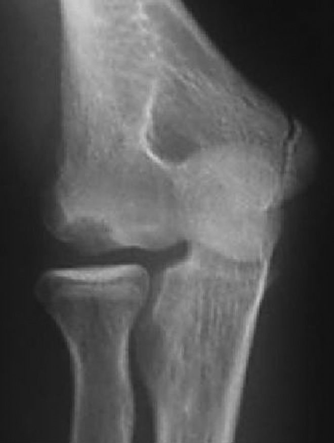
Figure 1.

Radiograph of 45° flexed, supinated anteroposterior view with lesion locations. II, type II lesions (medial); III, type III lesions (lateral).
In the proposed refined classification, type I lesions (“stable lesions”) have an open capitellar physis, a grade I radiographic lesion, and nearly full range of motion (ROM) at the time of diagnosis. Most of these lesions, if treated with thorough rest, will heal completely. Type II lesions (“unstable lesions”) are those with a closed capitellar physis, a grade II/III lesion radiographically or present with restricted elbow ROM, and have a lesion that lies medial to the radial head center line (Figure 2). These lesions tend to respond well to simple debridement or repair. Type IIIa lesions (“unstable lesions”) are those with a closed capitellar physis, a grade II/III lesion radiographically or present with restricted elbow ROM, and have a lesion that lies laterally to the radial head center line (Figure 3). These lesions tend to do better with more aggressive therapies (eg, repair or reconstruction). Type IIIb lesions (“unstable lesions”) are those with a closed capitellar physis, a grade II/III lesion radiographically or present with restricted elbow ROM, and have a lesion that lies laterally to the radial head center line, including the lateral cartilage margin (Figure 4). These lesions necessitate reconstruction.
Figure 2.

Type II osteochondritis dissecans lesion.
Figure 3.
Type IIIa osteochondritis dissecans lesion.
Figure 4.

Type IIIb osteochondritis dissecans lesion.
Discussion
A few important caveats should be discussed in more depth. Type IIIa lesions are located on the lateral half of the capitellum and do not involve the lateral cartilage margin. These would be akin to the “contained” lesions described by Shi et al.13 These lesions may be treated with a variety of methods, such as fragment fixation. Likewise, autograft mosaicplasty has demonstrated to be a reliable treatment method in this group as well. Care should be taken, however, to recognize that the severity of these lesions seems to increase as they become more laterally located, and what is abundantly clear is that involvement of the lateral margin of the capitellum necessitates reconstruction, especially with large lesions. This is undertaken to enhance stability and reduce shear forces that the lateral capitellum experiences, and attempts at fragment fixation of large lateral lesions yield poor results, as was demonstrated in the study by Kosaka et al.3
The effect of lesion size on outcome and proper surgical treatment has not yet been fully elucidated, and thus, is not directly included in this classification scheme. Though it is intuitive that larger defects would be more severe and require more aggressive surgical techniques, the literature does not fully support that suspicion to this point. In their landmark article, Takahara et al16 found that better results were seen for lesions that were <50% of the capitellar articular width than for large lesions that were >50% of the width. More recent studies, however, have been less conclusive. Evaluating microfracture with follow-up magnetic resonance imaging in 10 adolescent patients, Wulf et al20 found no correlation between lesion size (range, 50-180 mm2) and Timmerman and Andrews outcome scores. Likewise, in their study of 13 adolescent patients undergoing arthroscopic debridement, Schoch and Wolf12 found no correlation between Disabilities of the Arm, Shoulder and Hand (DASH) score and lesion size on magnetic resonance imaging (range, 49-240 mm2). It should be noted that this update to the classification scheme includes lesion size indirectly, as the size of a lesion affects its location. For example, once a type II medial lesion becomes greater than 50% of the capitellar width, it becomes a type IIIa lesion, indicating that it would require a more aggressive intervention.
Patients who are skeletally mature (closed physis), have nonadvanced lesions (type I), and do not have great limitation to function (maintained ROM) pose an interesting dilemma. It is the fate of the lesions in this group that has not yet been fully established. Indeed, patients with more advanced lesions (type II or III in this classification scheme) undoubtedly progressed through a relatively benign period such as this as their disease progressed. However, it has not yet been determined if the OCD disease process is doomed to progress in these skeletally mature individuals, regardless of the severity of their lesion. Thus, further work is necessary to determine the character of the lesions in this patient group, determining if these lesions are certain to progress to more advanced defects despite surgical or nonsurgical interventions.
Inherent limitations of this analysis are the limited number of studies available, the small number of patients within these studies, and a lack of standardized outcome measures obtained throughout the studies. As additional discussions regarding capitellar OCD evolve within the literature, the ability to draw definitive conclusions will be enhanced by greater collaboration between researchers and/or research centers and consistency with regard to outcome measures. Additionally, further studies evaluating surgical outcomes as they pertain to lesion size and depth will be important in this discussion. Likewise, evaluations of various techniques that reconstruct the lateral cartilage margin in an efficient manner will also be beneficial. It is our hypothesis that, with time, results will demonstrate that larger, thicker, lateral lesions are more severe and, thus, require a more aggressive surgical intervention to obtain satisfactory outcomes.
Conclusion
After a thorough review of the literature, we have refined the Takahara classification scheme to include the important factor of lesion location. This adaptation better reflects the clinical scenario that physicians are encountering and the current questions that are being addressed in the literature. In an effort to improve the way that capitellar OCD lesions are characterized and systematically treated, we propose this update to the present Takahara classification scheme with the hope that it will more effectively guide surgical management and further research efforts.
Footnotes
The authors declared that they have no conflicts of interest in the authorship and publication of this contribution.
References
- 1. Iwasaki N, Kato H, Ishikawa J, Saitoh S, Minami A. Autologous osteochondral mosaiclasty for capitellar osteochondritis dissecans in teenaged patients. Am J Sports Med. 2006;34:1233–1239. [DOI] [PubMed] [Google Scholar]
- 2. Iwasaki N, Kato H, Ishikawa J, Masuko T, Funakoshi T, Minami A. Autologous osteochondral mosaicplasty for osteochondritis dissecans of the elbow in teenage athletes. J Bone Joint Surg Am. 2009;91:2359–2366. [DOI] [PubMed] [Google Scholar]
- 3. Kosaka M, Nakase J, Takahashi R, et al. Outcomes and failure factors in surgical treatment for osteochondritis dissecans of the capitellum. J Pediatr Orthop. 2013;33:719–724. [DOI] [PubMed] [Google Scholar]
- 4. Kusumi T, Ishibashi Y, Tsuda E, et al. Osteochondritis dissecans of the elbow: histopathological assessment of the articular cartilage and subchondral bone with emphasis on their damage and repair. Pathol Int. 2006;56:604–612. [DOI] [PubMed] [Google Scholar]
- 5. Mihara K, Suzuki K, Makiuchi D, Nishinaka N, Yamaguchi K, Tsutsui H. Surgical treatment for osteochondritis dissecans of the humeral capitellum. J Shoulder Elbow Surg. 2010;19:31–37. [DOI] [PubMed] [Google Scholar]
- 6. Mihata T, Quigley R, Robicheaux G, McGarry MH, Neo M, Lee TQ. Biomechanical characteristics of osteochondral defects of the humeral capitellum. Am J Sports Med. 2013;41:1909–1914. [DOI] [PubMed] [Google Scholar]
- 7. Minami M, Nakashita K, Ishii S, Usui M, Muramatsu I. Twenty-five cases of osteochondritis dissecans of the elbow. Rinsho Seikei Geka. 1979;14:805–810. [Google Scholar]
- 8. Nobuta S, Ogawa K, Sato K, Nakagawa T, Hatori M, Itoi E. Clinical outcome of fragment fixation for osteochondritis dissecans of the elbow. Ups J Med Sci. 2008;113:201–208. [DOI] [PubMed] [Google Scholar]
- 9. Ruchelsman DE, Hall MP, Youm T. Osteochondritis dissecans of the capitellum: current concepts. J Am Acad Orthop Surg. 2010;18:557–567. [DOI] [PubMed] [Google Scholar]
- 10. Sabo MT, McDonald CP, Ferreira LM, Johnson JA, King GA. Osteochondral lesions of the capitellum do not affect elbow kinematics and stability with intact collateral ligaments: an in vitro biomechanical study. J Hand Surg Am. 2011;36:74–80. [DOI] [PubMed] [Google Scholar]
- 11. Schenck RC, Jr, Goodnight JM. Osteochondritis dissecans. J Bone Joint Surg Am. 1996;78:439–456. [PubMed] [Google Scholar]
- 12. Schoch B, Wolf BR. Osteochondritis dissecans of the capitellum: minimum 1-year follow-up after arthroscopic debridement. Arthroscopy. 2010;26:1469–1473. [DOI] [PubMed] [Google Scholar]
- 13. Shi LL, Bae DS, Kocher MS, Micheli LJ, Waters PM. Contained versus uncontained lesions in juvenile elbow osteochondritis dissecans. J Pediatr Orthop. 2012;32:221–225. [DOI] [PubMed] [Google Scholar]
- 14. Shimada K, Tanaka H, Matsumoto T, et al. Cylindrical costal osteochondral autograft for reconstruction of large defects of the capitellum due to osteochondritis dissecans. J Bone Joint Surg Am. 2012;94:992–1002. [DOI] [PMC free article] [PubMed] [Google Scholar]
- 15. Smith MV, Bedi A, Chen NC. Surgical treatment for osteochondritis dissecans of the capitellum. Sports Health. 2012;4:425–432. [DOI] [PMC free article] [PubMed] [Google Scholar]
- 16. Takahara M, Mura N, Sasaki J, Harada M, Ogino T. Classification, treatment, and outcome of osteochondritis dissecans of the humeral capitellum. J Bone Joint Surg Am. 2007;89:1205–1214. [DOI] [PubMed] [Google Scholar]
- 17. Takahara M, Ogino T, Fukushima S, Tsuchida H, Kaneda K. Nonoperative treatment of osteochondritis dissecans of the humeral capitellum. Am J Sports Med. 1999;27:728–732. [DOI] [PubMed] [Google Scholar]
- 18. Takeba J, Takahashi T, Hino K, Watanabe S, Imai H, Yamamoto H. Arthroscopic technique for fragment fixation using absorbable pins for osteochondritis dissecans of the humeral capitellum: a report of 4 cases. Knee Surg Sports Traumatol Arthrosc. 2010;18:831–835. [DOI] [PubMed] [Google Scholar]
- 19. Tis JE, Edmonds EW, Bastrom T, Chambers HG. Short-term results of arthroscopic treatment of osteochondritis dissecans in skeletally immature patients. J Pediatr Orthop. 2012;32:226–231. [DOI] [PubMed] [Google Scholar]
- 20. Wulf CA, Stone RM, Giveans MR, Lervick GN. Magnetic resonance imaging after arthroscopic mircofracture of capitellar osteochondritis dissecans. Am J Sports Med. 2012;40;2549–2556. [DOI] [PubMed] [Google Scholar]