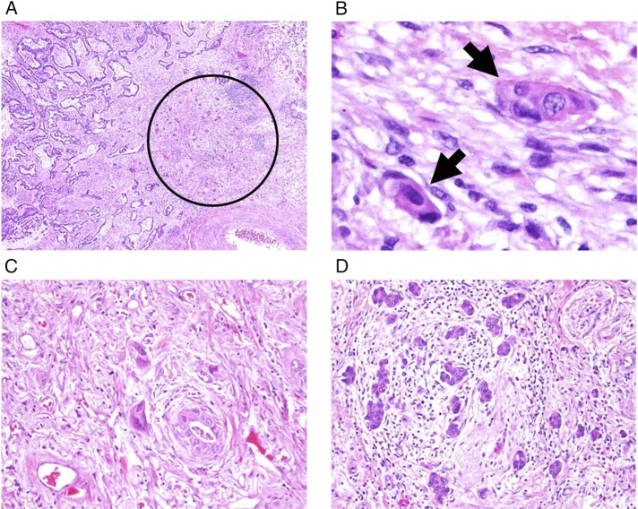
Figure 2 –
A-D, Histologic findings of tumor budding (original magnifications: A, ×40; B, ×400; C, D, ×200). A, Tumor budding identified in stroma of the invasive tumor edge (circle). B, Tumor budding defined as isolated small tumor nests composed of fewer than five tumor cells (arrows). C, Low-grade tumor budding. D, High-grade tumor budding.

