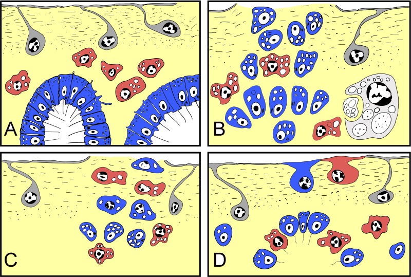
Figure 10. Schematic representation of Halisarca dujardini regeneration and the origin of new exopinacocytes and choanocytes.
(A) Intact sponge. (B) I stage of regeneration: formation of “regenerative plug”. (C) II stage of regeneration: wound healing and formation of a “blastema”. (D) III stage of regeneration: restoration of ectosome and choanosome. Grey—exopinacocytes, blue—choanocytes, red—archaeocytes.

