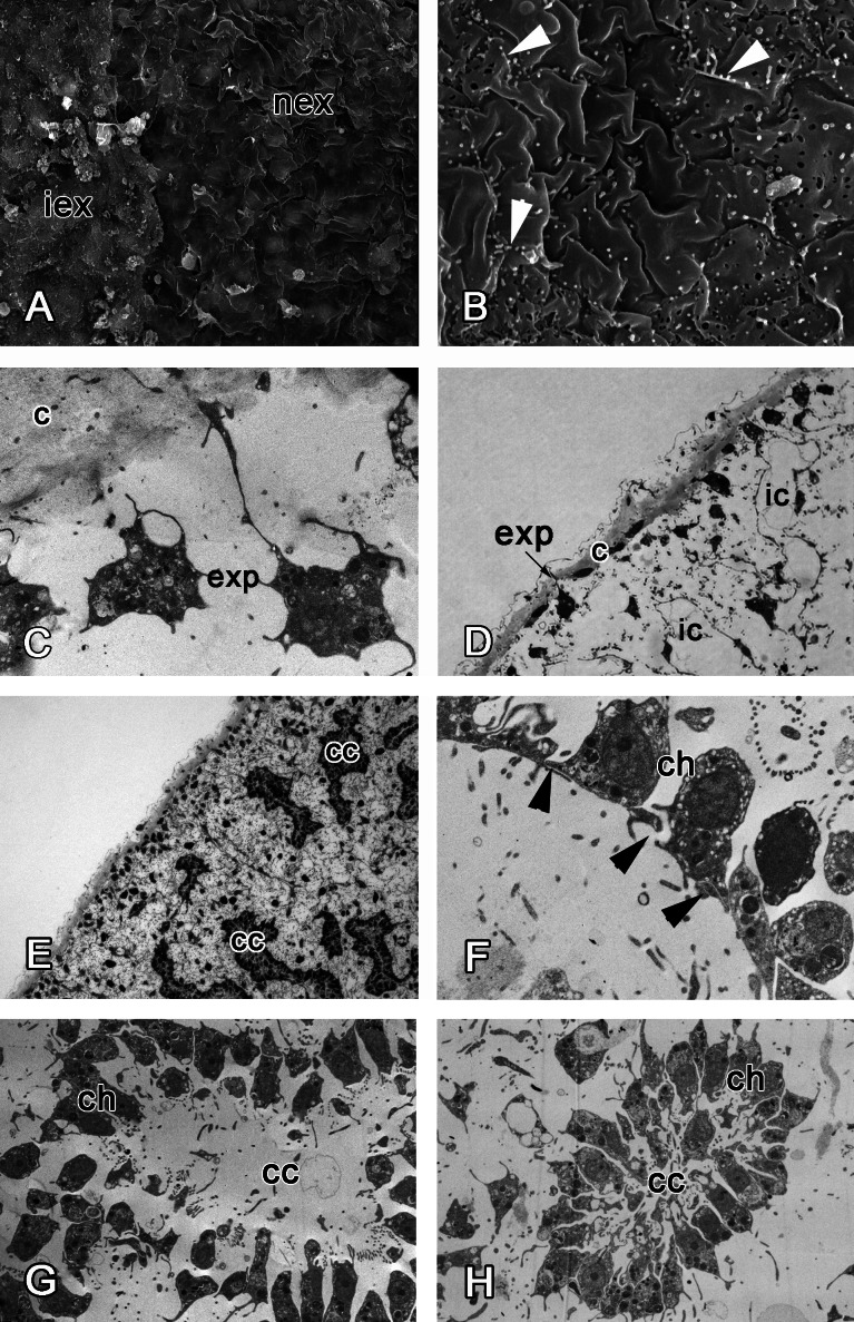
Figure 7. 48 h after injury.
(A) SEM of the intact and new exopinacoderm. (B) SEM of details of new exopinacoderm, showed numerous small cytoplasm microvilli form at the upper (apical) surface (arrowheads). (C) TEM of cell body of new differentiated exopinacocytes. (D) Semi-thin section of regenerated ectosome with cortical layer and inhalant canals. (E) Semi-thin section of regenerated ectosome and choanosome. (F) Connections of choanocytes of new-formed chamber by interdigitations in their basal parts (arrowheads); (G) TEM of newly-formed choanocyte chamber. (H) TEM of intact choanocyte chamber of the same sponge. Scale bars: A—10 µm; B, C—5 µm; D—20 µm; E—100 µm; F—5 µm; G, H—10 µm. c, cortical layer; cc, choanocyte chamber; ch, choanocyte; exp, exopinacocytes; ic, inhalant canal; iex, intact exopinacocytes; nex, new exopinacocytes.

