

Abstract
We evaluated the chondroprotective effects of wogonin by investigating its effects on the gene expression and production of matrix metalloproteinase-3 (MMP-3) in primary cultured rabbit articular chondrocytes, as well as on production of MMP-3 in the rat knee. Rabbit articular chondrocytes were cultured in a monolayer, and RT-PCR was used to measure interleukin-1β (IL-1β)-induced expression of MMP-3, MMP-1, MMP-13, a disintegrin and metalloproteinase with thrombospondin motifs-4 (ADAMTS-4), and type II collagen. In rabbit articular chondrocytes, the effects of wogonin on IL-1β-induced production and proteolytic activity of MMP-3 were investigated using western blot analysis and casein zymography, respectively. The effect of wogonin on MMP-3 protein production was also examined in vivo. In rabbit articular chondrocytes, wogonin inhibited the expression of MMP-3, MMP-1, MMP-13, and ADAMTS-4, but increased expression of type II collagen. Furthermore, wogonin inhibited the production and proteolytic activity of MMP-3 in vitro, and inhibited production of MMP-3 protein in vivo. These results suggest that wogonin can regulate the gene expression and production of MMP-3, by directly acting on articular chondrocytes.
Keywords: Osteoarthritis, Wogonin, Chondrocyte, Metalloproteinase
INTRODUCTION
It has been reported that osteoarthritis is the most common degenerative articular disease, and affects millions of people, especially in the elderly. The major pathophysiologic features of osteoarthritis are synovial inflammation, formation of osteophytes, changes in subchondral bone, and degeneration of articular cartilage. The cause of osteoarthritis is unclear, and involves multiple biochemical and mechanical factors. The equilibrium between physiologic synthesis and degradation of articular cartilage is disrupted during the progression of osteoarthritis (Mankin, 1982; Aigner and McKenna, 2002).
The activation of degradative enzymes leads to the loss and degradation of proteoglycans and collagen in articular cartilage, and the matrix metalloproteinases (MMP) play a pivotal role in the destruction of articular cartilage in osteoarthritis patients (Dean et al., 1989; Kullich et al., 2007). The types of matrix metalloproteinases include collagenases (MMP-1, -8 and -13), gelatinases (MMP-2 and -9), and stromelysins (MMP-3, -7, -10 and -11) (Birkedal-Hansen et al., 1993; Burrage et al., 2006). Among them, MMP-3 degrades proteoglycans and activates procollagenase in articular cartilage (Garnero et al., 2000; Lin et al., 2004). Therefore, exploration of the mechanisms through which the proteolytic activity, production, and expression of MMP-3 are inhibited by plant-derived natural products used as arthritis remedies will support the effective treatment of osteoarthritis, and may lead to new therapeutic strategies.
According to a number of reports, wogonin derived from Scutellariae Radix, a medicinal plant used for controlling the inflammatory diseases, showed the diverse biological activities including anti-inflammatory and anticancer effects (Lin et al., 2006; Kimura and Sumiyoshi, 2013; Kwak et al., 2014; Lee et al., 2015). Lee and his colleagues reported that wogonin inhibited TNF-alpha-induced MMP-9 expression in human aortic smooth muscle cells (Lee et al., 2006). Also, Lim and his colleagues reported that wogonin inhibited MMP-13 induction in IL-1β-treated SW1353 cells, a chondrosarcoma cell line (Lim et al., 2011).
However, to the best of our knowledge, there are no reports about the effects of wogonin on gene expression and production of MMP-3, an articular cartilage-degradative enzyme that decomposes proteoglycans, in primary cultured rabbit articular chondrocytes, or on production of MMP-3 in the rat knee. Therefore, to evaluate the chondroprotective potential of wogonin, we examined its effects on IL-1β-induced gene expression and production of MMP-3 in vitro, and on production of MMP-3 in vivo.
MATERIALS AND METHODS
Materials
All chemicals and reagents used in this experiment, including wogonin (purity: 98.0%), were purchased from Sigma-Aldrich (St. Louis, MO, USA) unless otherwise specified. Dulbecco’s Modified Eagle’s Medium (DMEM) was purchased from Gibco-BRL (Grand Island, NY, USA) and recombinant human IL-1β was purchased from R&D Systems (Minneapolis, MN, USA).
Primary cultures of chondrocytes from rabbit articular cartilage
Male New Zealand White rabbits were obtained from Daehan Biolink (Seoul, South Korea) at 2 weeks of age. Animals were housed 1 animal per cage, provided with distilled water and food ad libitum, and kept under a 12 h light/dark cycle (lights on from 08:00–20:00) at constant temperature (22.5°C) and humidity (55%). Animals were cared for in accordance with the Guide for the Care and Use of Laboratory Animals, and care was regulated by Chungnam National University (the approval number of animal experiment: CNU-00555) (Daejeon, Korea). Rabbit articular chondrocytes were isolated from the tibial plateau and femoral condyle in cartilage of the knee joint. Cartilage was washed in phosphate-buffered saline (PBS) and minced into pieces measuring 2 mm3, approximately. Cartilage tissue was digested for 4 h with 0.2% type II collagenase at 37°C. After collection of individual cells by brief centrifugation, the cells were transferred to 100-mm culture dishes (seeding density: 105 cells/cm2) in 12-mL DMEM supplemented with 10% fetal bovine serum (FBS), in the presence of penicillin (100 units/mL) and streptomycin (100 μg/mL). Cells were cultured at 37°C in a humidified, 5% CO2/95% air, water-jacketed incubator, and medium was replaced every other day.
Treatment of cells with wogonin
Chondrocytes were seeded on 6-well culture plates at a density of 105 cells/cm2. After 2 days in monolayer culture, the cells were incubated for 2 h in growth medium with 1, 10, 50, or 100 μM of wogonin, followed by incubation in the presence or absence of IL-1β (10 ng/mL) for 24 h. Wogonin was dissolved in dimethylsulfoxide, diluted in PBS, and administered in culture medium (final concentrations of dimethylsulfoxide were 0.5%). The final pH values of these solutions were between 7.0 and 7.4. Culture medium and 0.5% dimethylsulfoxide in medium did not affect gene expression, production, or proteolytic activity of MMP-3 in primary cultured chondrocytes. The supernatant was collected and centrifuged, and cell and supernatant fractions were stored at −80°C until use.
Cytotoxicity assay
Chondrocytes were seeded at a density of 2×10⁵/mL (0.1 mL/well) in a 96-well microtiter plate, and allowed to attach for 24 h to keep the log phase growth at the time of drug treatment. Wogonin was dissolved in DMSO, and administered in DMEM supplemented with 10% FBS (final concentrations of DMSO were under 0.5%). 0.5% DMSO alone did not affect the proliferation of chondrocytes. After incubation with the indicated drug concentrations for 72 h, cell proliferation was determined using the sulforhodamine B (SRB) assay (Skehan et al., 1990).
Isolation of total RNA and RT-PCR
Total RNA was isolated from chondrocytes using the Easy-BLUE Extraction Kit (INTRON Biotechnology, Inc. Kyung-kido, South Korea), and reverse transcribed using AccuPower RT Premix (BIONEER Corporation, Daejeon, South Korea) according to the manufacturer’s instructions. About 2 μg of total RNA was primed with 1 μg of oligo (dT) in a final reaction volume of 30 μL. 2 μL of RT reaction product was amplified in 20 μL using Thermoprime Plus DNA Polymerase (ABgene, Rochester, NY, USA). PCR was performed with the following primers: MMP-3 (5′ATG GAC CTT CTT CAG CAA 3′, 5′TCA TTA TGT CAG CCT CTC 3′), MMP-13 (5′AGG AGC ATG GCG ACT TCT AC 3′, 5′TAA AAA CAG CTC CGC ATC AA 3′), MMP-1 (5′TCA GTT CGT CCT CAC TCC AG 3′, 5′TTG GTC CAC CTG TCA TCT TC 3′), ADAMTS-4 (5′CAA GGT CCC ATG TGC AAC GT 3′, 5′CAT CTG CCA CCA CCA GTG TCT 3′), and type II collagen (5′AAC ACT GCC AAC GTC CAG AT 3′, 5′CTG ACG CAC GGT ATA GGT GA 3′). GAPDH (5′ACT GGC GTC TTC ACC ACC AT 3′; 5′AAG GCC ATG CCA GTG AGC TT 3′) was used as a quantitative control. The PCR products increased as the concentration of RNA increased. The amplified fragment sizes were 350 base pairs (bp) for MMP-3, 458 bp for MMP-13, 300 bp for MMP-1, 90 bp for ADAMTS-4, 220 bp for type II collagen, and 400 bp for GAPDH. After PCR, 15 μL of PCR products were subjected to 2% agarose gel electrophoresis and visualized with ethidium bromide under a transilluminator (Moon et al., 2011).
Western blot analysis for measuring production of MMP-3 in culture supernatant
The Bradford assay was used to measure protein concentrations in culture supernatants to ensure consistent weight of protein samples subjected to electrophoresis. Culture supernatant samples containing MMP-3 protein (50 μg each) were subjected to 10% sodium dodecyl sulfate-polyacrylamide gel electrophoresis (SDS-PAGE), and then transferred onto a polyvinylidene difluoride (PVDF) membrane. Blots were blocked using 5% skim milk in Tris-buffered saline/Tween 20 (TBS-T), and probed overnight with MMP-3 antibody in blocking buffer at 4°C. Antibody against MMP-3 was purchased from Santa Cruz Biotechnology (Santa Cruz, CA, USA). Membranes were washed with TBS-T and probed for 1 h with a secondary antibody conjugated with horseradish peroxidase (Calbiochem, CA, USA). After 4 washes with TBS-T, immunoreactive bands were detected using an enhanced chemiluminescence kit (Pierce ECL western blotting substrate, Thermo Scientific, Waltham, MA, USA).
Casein zymography to measure the proteolytic activity of produced MMP-3
A modified casein-substrate zymography was carried out using culture supernatants from chondrocytes pretreated for 2 h with wogonin and stimulated for 24 h with IL-1β in DMEM containing 0.5% FBS. The Bradford assay was used to measure protein concentrations in culture media to ensure consistency across samples. Samples were electrophoresed at 4°C in a 10% SDS gel containing 0.1% casein. After electrophoresis, gels were washed with 10 mM Tris-HCl (pH 8.0) containing 2.5% Triton X-100. Next, gels were incubated at 37°C for 48 h in 50 mM Tris-HCl (pH 8.0) containing 1% Triton X-100, 0.2 M NaCl, and 5 mM CaCl2. Finally, gels were stained with 1% Coomassie Brilliant Blue, destained, and photographs were taken (Moon et al., 2011).
In vivo experiments
Male Sprague-Dawley rats (Daehan Biolink, Seoul, South Korea) weighing 200–210 g were used to investigate the effect of wogonin on production of MMP-3 in articular cartilage in vivo. Animals were housed 5 per cage, provided with distilled water and food ad libitum, and kept under a 12 h light/dark cycle (lights on from 08:00–20:00) at constant temperature (22.5°C) and humidity (55%). Animals were cared for in accordance with the Guide for the Care and Use of Laboratory Animals, and care was regulated by Chungnam National University (the approval number of animal experiment: CNU-00555) (Daejeon, South Korea). Rats were randomly divided into 4 groups: control, IL-1β only, 50 μM wogonin plus IL-1β, or 100 μM wogonin plus IL-1β. Rats were anesthetized with vaporized diethyl ether, and those from the 50 μM wogonin plus IL-1β and 100 μM wogonin plus IL-1β treatment groups received a 30-μL injection of 50 μM or 100 μM wogonin, respectively, into the right knee joint. After 3 h, rats from the IL-1β only group, the 50 μM wogonin plus IL-1β group, and the 100 μM wogonin plus IL-1β group received a 30-μL injection of 20-ng IL-1β in sterile PBS into the right knee joint. Rats from the control group were injected with 30 μL of sterile PBS (Hu et al., 2011). Rats were euthanized via CO2 asphyxiation 72 h after injections. Articular cartilage (tibial plateau and femoral condyle) was isolated from each animal, homogenized, and prepared for measurement of MMP-3 protein by western blot analysis. Tissue lysates from articular cartilage homogenates containing MMP-3 protein (50 μg each) were subjected to 10% SDS-PAGE, and transferred onto a PVDF membrane. Blots were blocked with 5% skim milk in TBS-T, and probed with MMP-3 antibody (Santa Cruz Biotechnology, Santa Cruz, CA, USA) in blocking buffer overnight at 4°C. Membranes were washed with TBS-T, and probed for 1 h with a secondary antibody conjugated with horseradish peroxidase (Calbiochem, CA, USA). After 4 washes with TBS-T, immunoreactive bands were detected using an enhanced chemiluminescence kit (Pierce ECL western blotting substrate, Thermo Scientific, Waltham, MA, USA).
Statistics
Means of individual group were converted to percent control and expressed as mean ± S.E.M. The difference between groups was assessed using one-way ANOVA and Holm-Sidak test as a post-hoc test. p<0.05 was considered as significantly different.
RESULTS
Effect of wogonin on MMP-3 gene expression in rabbit chondrocytes
To examine the potential activity of wogonin (Fig. 1) on gene expression of MMP-3, the key matrix metalloproteinase involved in destruction of articular cartilage, MMP-3 gene expression was measured after pretreatment with wogonin. As shown in Fig. 2, wogonin inhibited IL-1β-induced MMP-3 gene expression.
Fig. 1.
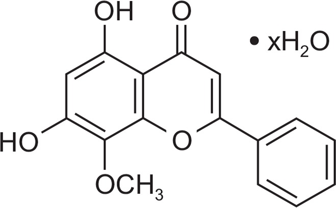
Chemical structure of wogonin.
Fig. 2.

Effect of wogonin on MMP-3 gene expression in rabbit chondrocytes. Primary cultured rabbit articular chondrocytes were pretreated with varying concentrations (1, 10, 50, and 100 μM) of wogonin for 2 h and then stimulated with IL-1β (10 ng/mL) for 24 h. MMP-3 gene expression level was measured by RT-PCR. Three independent experiments were performed and the representative data were shown (cont: control, concentration unit is μM). *Significantly different from control (p<0.05). †Significantly different from IL-1β alone (p<0.05).
Effect of wogonin on proliferation of rabbit chondrocytes (cytotoxicity assay)
To check the potential cytotoxicity of wogonin to cultured rabbit chondrocytes, effect of wogonin on proliferation of rabbit chondrocytes using SRB assay. As can be seen in Fig. 3, wogonin showed no significant cytotoxicity at the concentrations of 1, 10, 50, and 100 μM. The numbers of cells in wogonin-treated cultures were 100 ± 15%, 102 ± 13%, 92 ± 16%, 91 ± 14%, and 85 ± 11% for control, 1, 10, 50, and 100 μM wogonin, respectively.
Fig. 3.

Effect of wogonin on proliferation of rabbit chondrocytes. Chondrocytes were incubated for 72 h in the presence of varying concentrations of wogonin. Cell viability was determined using SRB assay as described in Materials and Methods. Each bar represents a mean ± S.E.M. of three independent experiments in comparison with that of the control set at 100%.
Effect of wogonin on gene expression of MMP-1, MMP-13, ADAMTS-4, and type II collagen in rabbit chondrocytes
If wogonin can affect the gene expression of MMP-3, the key matrix metalloproteinase involved in destruction of articular cartilage, it should be investigated whether wogonin affect the gene expression of MMP-1, MMP-13, ADAMTS-4, the other degradative enzymes related to destruction of articular cartilage, and type II collagen, in rabbit chondrocytes. As shown in Fig. 4, wogonin also inhibited IL-1β-induced gene expression of MMP-1, MMP-13, and ADAMTS-4, the other degradative enzymes related to destruction of articular cartilage, in rabbit chondrocytes. Furthermore, wogonin showed an additional chondroprotective effect by recovering the compromised gene expression of type II collagen by IL-1β, in rabbit chondrocytes.
Fig. 4.

Effect of wogonin on gene expression of MMP-1, MMP-13, or collagen type II in rabbit chondrocytes. Primary cultured rabbit articular chondrocytes were pretreated with varying concentrations (1, 10, 50, and 100 μM) of wogonin for 2 h and then stimulated with IL-1β (10 ng/mL) for 24 h. The gene expression level of MMP-1, MMP-13, ADAMTS-4, or collagen type II was measured by RTPCR. Three independent experiments were performed and the representative data were shown (cont: control, concentration unit is μM). *Significantly different from control (p<0.05). †Significantly different from IL-1β alone (p<0.05).
Effect of wogonin on IL-1β-induced production of MMP-3 from rabbit articular chondrocytes
If wogonin can affect the MMP-3 gene expression at the transcriptional level, it should be investigated whether wogonin affects IL-1β-induced production of MMP-3 proteins from rabbit articular chondrocytes. As shown in Fig. 5, stimulation with IL-1β (10 ng/mL) increased production of MMP-3 from chondrocytes. However, wogonin inhibited the effects of IL-1β on MMP-3 production. This result means that wogonin can control the steps of protein synthesis (production) of MMP-3.
Fig. 5.

Effect of wogonin on IL-1β-induced production of MMP-3 from rabbit articular chondrocytes. Primary cultured rabbit articular chondrocytes were pretreated with varying concentrations (1, 10, 50, and 100 μM) of wogonin for 2 h and then stimulated with IL-1β (10 ng/mL) for 24 h. Culture supernatants were collected for measurement of the levels of produced MMP-3 by western blot analysis. Three independent experiments were performed and the representative data were shown (cont: control, concentration unit is μM). *Significantly different from control (p<0.05). †Significantly different from IL-1β alone (p<0.05).
Effect of wogonin on caseinolytic activity of produced MMP-3 in rabbit articular chondrocytes
To investigate the effect of wogonin on the proteolytic activity of produced MMP-3, which is known to degrade proteoglycans, 1 of the 2 major matrix components of cartilage, culture supernatants from rabbit articular chondrocytes were analyzed for caseinolytic activity by casein zymography, after treatment with IL-1β for 24 h. As can be seen in Fig. 6, IL-1β increased the production and thus caseinolytic activity of MMP-3 in rabbit articular chondrocytes, and this effect was inhibited by pretreatment with wogonin.
Fig. 6.

Effect of wogonin on caseinolytic activity of produced MMP-3 in rabbit articular chondrocytes. Primary cultured rabbit articular chondrocytes were pretreated with varying concentrations (1, 10, 50, and 100 μM) of wogonin for 2 h and then stimulated with IL-1β (10 ng/mL) for 24 h. Culture supernatants were collected for measurement of the proteolytic activity of produced MMP-3 by casein zymography. Three independent experiments were performed and the representative data were shown (cont: control, concentration unit is μM). *Significantly different from control (p<0.05). †Significantly different from IL-1β alone (p<0.05).
Effect of wogonin on MMP-3 production in vivo
To examine whether wogonin shows the potential effect in vivo, we checked the effect of intraarticular injection of wogonin into the knee joint of rats on IL-1β-stimulated production of MMP-3 from articular cartilage tissues. As shown in Fig. 7, treatment with IL-1β (20 ng/30 μL) increased MMP-3 production in articular cartilage tissues. However, wogonin inhibited IL-1β-induced MMP-3 production, in vivo.
Fig. 7.

Effect of wogonin on production of MMP-3 in vivo. The knee joint of rats were pretreated with 50 or 100 μM of wogonin for 3 h and then stimulated with IL-1β (20 ng/30 μL) for 72 h, by intraarticular injection. Tissue lysates from articular cartilage homogenates containing MMP-3 proteins were collected for measurement of the level of produced MMP-3 in vivo, by western blot analysis. The representative data were shown. Equal protein loading was evaluated by β-actin levels (cont: control, concentration unit is μM). *Significantly different from control (p<0.05). †Significantly different from IL-1β alone (p<0.05).
DISCUSSION
Restoration of the equilibrium between synthesis and degradation of articular cartilage during the progression of osteoarthritis is a promising approach to the effective control of this condition. Wogonin is a medicinal plant-derived flavonoid that has shown anti-inflammatory, anti-carcinogenic, and anti-matrix metalloproteinase effects (Lin et al., 2006; Lee et al., 2006; Lim et al., 2011; Kimura and Sumiyoshi, 2013; Kwak et al., 2014; Lee et al., 2015). However, to the best of our knowledge, there have been no reports on the effects of wogonin on gene expression and production of MMP-3, which is an articular cartilage-degradative enzyme that decomposes proteoglycans. Therefore, we examined the effects of wogonin on MMP-3 gene expression and production in primary cultured rabbit articular chondrocytes, as well as on MMP-3 production in the rat knee.
Although osteoarthritis can be defined as a non-inflammatory disease, its development and progression have been attributed to low-grade inflammation in intraarticular sites, as well as to various inflammatory cytokines in articular tissues and fluids that are produced by chondrocytes and/or interact with chondrocytes (Bonnet and Walsh, 2005; Kobayashi et al., 2005; Loeser, 2006; Goldring et al., 2008). IL-1β is an inflammatory cytokine that is produced by cells in articular tissues, including chondrocytes, and which can increase expression of MMPs and stimulate the progression of osteoarthritis. IL-1β plays a pivotal role in the initiation and progression of destruction of articular cartilage by inhibiting synthesis of collagen and stimulating MMP expression (Aida et al., 2005; Kobayashi et al., 2005; Pantsulaia et al., 2010). MMP-3, in particular, has been reported to play an important pathophysiological role in osteoarthritis by degrading components of the extracellular matrix, such as proteoglycans. MMP-3 levels were increased more than MMP-1 levels in patients suffering from osteoarthritis in knee joints compared to the control group (Garnero et al., 2000; Lijnen, 2002).
As shown in Results, we found that wogonin inhibited IL-1β-induced MMP-3 gene expression (Fig. 2). This result suggests that wogonin suppresses the gene expression of MMP-3 at the transcriptional level.
In addition to MMP-3, MMP-1 and MMP-13 have been reported to play important roles in the destruction of cartilage in osteoarthritis. MMP-1 is a commonly detected metalloproteinase in synovial fluid from patients suffering from osteoarthritis (Freemont et al., 1997; Goupille et al., 1998; Kanyama et al., 2000; Yoshihara et al., 2000; Neuhold et al., 2001; Jo et al., 2003; Little et al., 2009). Another degradative enzyme, ADAMTS-4, is a major aggrecanase in mouse cartilage, and involved in cartilage matrix destruction during osteoarthritis (Stanton et al., 2005; Echtermeyer et al., 2009).
Therefore, we examined the effect of wogonin on expression of MMP-1, MMP-13, and ADAMTS-4. As can be seen in Fig. 4, wogonin inhibited IL-1β-induced gene expression of MMP-1, MMP-13, and ADAMTS-4, in rabbit chondrocytes. At the same time, wogonin restored the gene expression of type II collagen that had been inhibited by IL-1β. Thus, the chondroprotective effect of wogonin are supported by its regulation of the gene expression of diverse proteases involved in the destruction of articular cartilage in osteoarthritis, as well as by its promotion of the gene expression of type II collagen at the transcriptional level.
Next, if wogonin can affect the MMP-3 gene expression at the transcriptional level, it should be investigated whether wogonin affects IL-1β-induced production of MMP-3 proteins from rabbit articular chondrocytes. As can be seen in Fig. 5, stimulation with IL-1β increased production of MMP-3 from chondrocytes, and this effect was inhibited by wogonin. This result shows that wogonin can regulate the step of protein synthesis of MMP-3.
To investigate the effect of wogonin on the proteolytic activity of produced MMP-3, culture supernatants from rabbit articular chondrocytes stimulated with IL-1β for 24 h were analyzed for caseinolytic activity by casein zymography. As shown in Fig. 6, the proteolytic activity of MMP-3 was increased when rabbit articular chondrocytes were stimulated with IL-1β, and this effect was inhibited by pretreatment with wogonin. This result suggests that wogonin can also affect the proteolytic activity of produced MMP-3 in osteoarthritic articular cartilage tissues.
Finally, we examined the effect of intraarticular injection of wogonin into the knee joint of rats on IL-1β-stimulated production of MMP-3 in articular cartilage tissue. As shown in Fig. 7, wogonin inhibited IL-1β-stimulated production of MMP-3 in articular cartilage tissue. This result shows that, in addition to its in vitro effects, wogonin exerts chondroprotective effects in vivo when administered via intraarticular injection.
Taken together, these results show that the chondroprotective effects of wogonin are produced by its regulation of the gene expression and production of MMP-3 in articular chondrocytes. Future studies will develop wogonin as a novel agent for the control of cartilage damage in osteoarthritis via intraarticular administration.
Acknowledgments
This work was supported by biomedical research institute fund (GNUHBIF-2015-0004) from the Gyeongsang National Univeristy Hospital.
Footnotes
CONFLICTS OF INTEREST
The authors have no conflicts of interest to declare.
REFERENCES
- Aida Y, Maeno M, Suzuki N, Shiratsuchi H, Motohashi M, Matsumura H. The effect of IL-1beta on the expression of matrix metalloproteinases and tissue inhibitors of matrix metalloproteinases in human chondrocytes. Life Sci. 2005;77:3210–3221. doi: 10.1016/j.lfs.2005.05.052. [DOI] [PubMed] [Google Scholar]
- Aigner T, McKenna L. Molecular pathology and pathobiology of osteoarthritic cartilage. Cell Mol Life Sci. 2002;59:5–18. doi: 10.1007/s00018-002-8400-3. [DOI] [PMC free article] [PubMed] [Google Scholar]
- Birkedal-Hansen H, Moore WG, Bodden MK. Matrix metalloproteinases: a review. Crit Rev Oral Biol Med. 1993;4:197–250. doi: 10.1177/10454411930040020401. [DOI] [PubMed] [Google Scholar]
- Bonnet CS, Walsh DA. Osteoarthritis, angiogenesis and inflammation. Rheumatology. 2005;44:7–16. doi: 10.1093/rheumatology/keh344. [DOI] [PubMed] [Google Scholar]
- Burrage PS, Mix KS, Brinckerhoff CE. Matrix metalloproteinases: role in arthritis. Front Biosci. 2006;11:529–543. doi: 10.2741/1817. [DOI] [PubMed] [Google Scholar]
- Dean DD, Martel-Pelletier J, Pelletier JP, Howell DS, Woessner JF., Jr Evidence for metalloproteinase and metalloproteinase inhibitor imbalance in human osteoarthritic cartilage. J Clin Invest. 1989;84:678–685. doi: 10.1172/JCI114215. [DOI] [PMC free article] [PubMed] [Google Scholar]
- Echtermeyer F, Bertrand J, Dreier R. Syndecan-4 regulates ADAMTS-5 activation and cartilage breakdown in osteoarthritis. Nat Med. 2009;15:1072–1076. doi: 10.1038/nm.1998. [DOI] [PubMed] [Google Scholar]
- Freemont AJ, Hampson V, Tilman R, Goupille P, Taiwo Y, Hoyland JA. Gene expression of matrix metalloproteinases 1, 3, and 9 by chondrocytes in osteoarthritic human knee articular cartilage is zone and grade specific. Ann Rheum Dis. 1997;56:542–549. doi: 10.1136/ard.56.9.542. [DOI] [PMC free article] [PubMed] [Google Scholar]
- Garnero P, Rousseau JC, Delmas PD. Molecular basis and clinical use of biochemical markers of bone, cartilage, and synovium in joint diseases. Arthritis Rheum. 2000;43:953–968. doi: 10.1002/1529-0131(200005)43:5<953::AID-ANR1>3.0.CO;2-Q. [DOI] [PubMed] [Google Scholar]
- Goldring MB, Otero M, Tsuchimochi K, Ijiri K, Li Y. Defining the roles of inflammatory and anabolic cytokines in cartilage metabolism. Ann Rheum Dis. 2008;67(Suppl 3):iii75–iii82. doi: 10.1136/ard.2008.098764. [DOI] [PMC free article] [PubMed] [Google Scholar]
- Goupille P, Jayson MI, Valat JP, Freemont AJ. Matrix metalloproteinases: the clue to intervertebral disc degeneration. Spine (Phila Pa 1976) 1998;23:1612–1626. doi: 10.1097/00007632-199807150-00021. [DOI] [PubMed] [Google Scholar]
- Hu PF, Chen WP, Tang JL, Bao JP, Wu LD. Protective effects of berberine in an experimental rat osteoarthritis model. Phytother Res. 2011;25:878–885. doi: 10.1002/ptr.3359. [DOI] [PubMed] [Google Scholar]
- Jo H, Park JS, Kim EM. The in vitro effects of dehydroepiandrosterone on human osteoarthritic chondrocytes. Osteoarthritis Cartilage. 2003;11:585–594. doi: 10.1016/S1063-4584(03)00094-3. [DOI] [PubMed] [Google Scholar]
- Kanyama M, Kuboki T, Kojima S. Matrix metalloproteinases and tissue inhibitors of metalloproteinases in synovial fluids of patients with temporomandibular joint osteoarthritis. J. Orofac. Pain. 2000;14:20–30. [PubMed] [Google Scholar]
- Kimura Y, Sumiyoshi M. Anti-tumor and anti-metastatic actions of wogonin isolated from Scutellaria baicalensis roots through anti-lymphangiogenesis. Phytomedicine. 2013;20:328–336. doi: 10.1016/j.phymed.2012.10.016. [DOI] [PubMed] [Google Scholar]
- Kobayashi M, Squires GR, Mousa A. Role of interleukin-1 and tumor necrosis factor alpha in matrix degradation of human osteoarthritic cartilage. Arthritis Rheum. 2005;52:128–135. doi: 10.1002/art.20776. [DOI] [PubMed] [Google Scholar]
- Kullich W, Fagerer N, Schwann H. Effect of the NSAID nimesulide on the radical scavenger glutathione S-transferase in patients with osteoarthritis of the knee. Curr Med Res Opin. 2007;23:1981–1986. doi: 10.1185/030079907X223486. [DOI] [PubMed] [Google Scholar]
- Kwak S, Ku SK, Han MS, Bae JS. Vascular barrier protective effects of baicalin, baicalein and wogonin in vitro and in vivo. Toxicol Appl Pharmacol. 2014;281:30–38. doi: 10.1016/j.taap.2014.09.003. [DOI] [PubMed] [Google Scholar]
- Lee SO, Jeong YJ, Yu MH, Lee JW, Hwangbo MH, Kim CH, Lee IS. Wogonin suppresses TNF-alpha-induced MMP-9 expression by blocking the NF-kappaB activation via MAPK signaling pathways in human aortic smooth muscle cells. Biochem Biophys Res Commun. 2006;351:118–125. doi: 10.1016/j.bbrc.2006.10.006. [DOI] [PubMed] [Google Scholar]
- Lee W, Ku SK, Bae JS. Anti-inflammatory effects of Baicalin, Baicalein, and Wogonin in vitro and in vivo. Inflammation. 2015;38:110–125. doi: 10.1007/s10753-014-0013-0. [DOI] [PubMed] [Google Scholar]
- Lijnen HR. Matrix metalloproteinases and cellular fibrinolytic proteolytic activity. Biochemistry. 2002;67:92–98. doi: 10.1023/a:1013908332232. [DOI] [PubMed] [Google Scholar]
- Lim H, Park H, Kim HP. Effects of flavonoids on matrix metalloproteinase-13 expression of interleukin-1β-treated articular chondrocytes and their cellular mechanisms: inhibition of c-Fos/AP-1 and JAK/STAT signaling pathways. J Pharmacol Sci. 2011;116:221–231. doi: 10.1254/jphs.11014FP. [DOI] [PubMed] [Google Scholar]
- Lin CM, Chang H, Chen YH, Li SY, Wu IH, Chiu JH. Protective role of wogonin against lipopolysaccharide-induced angiogenesis via VEGFR-2, not VEGFR-1. Int. Immunopharmacol. 2006;6:1690–1698. doi: 10.1016/j.intimp.2006.07.003. [DOI] [PubMed] [Google Scholar]
- Lin PM, Chen CT, Torzilli PA. Increased stromelysin-1 (MMP-3), proteoglycan degradation (3B3- and 7D4) and collagen damage in cyclically load-injured articular cartilage. Osteoarthritis Cartilage. 2004;12:485–496. doi: 10.1016/j.joca.2004.02.012. [DOI] [PubMed] [Google Scholar]
- Little CB, Barai A, Burkhardt D. Matrix metalloproteinase 13-deficient mice are resistant to osteoarthritic cartilage erosion but not chondrocyte hypertrophy or osteophyte development. Arthritis Rheum. 2009;60:3723–3733. doi: 10.1002/art.25002. [DOI] [PMC free article] [PubMed] [Google Scholar]
- Loeser RF. Molecular mechanisms of cartilage destruction: mechanics, inflammatory mediators and aging collide. Arthritis Rheum. 2006;54:1357–1360. doi: 10.1002/art.21813. [DOI] [PMC free article] [PubMed] [Google Scholar]
- Mankin HJ. The response for articular cartilage to mechanical injury. J Bone Joint Surg Am. 1982;64:460–466. [PubMed] [Google Scholar]
- Moon PD, Jeong HS, Chun CS, Kim HM. Baekjeolyusin-tang and its active component berberine block the release of collagen and proteoglycan from IL-1β-stimulated rabbit cartilage and down-regulate matrix metalloproteinases in rabbit chondrocytes. Phytother Res. 2011;25:844–850. doi: 10.1002/ptr.3353. [DOI] [PubMed] [Google Scholar]
- Neuhold LA, Killar L, Zhao W. Postnatal expression in hyaline cartilage of constitutively active human collagenase-3 (MMP-13) induces osteoarthritis in mice. J Clin Invest. 2001;107:35–44. doi: 10.1172/JCI10564. [DOI] [PMC free article] [PubMed] [Google Scholar]
- Pantsulaia I, Kalichman L, Kobyliansky E. Association between radiographic hand osteoarthritis and RANKL, OPG and inflammatory markers. Osteoarthritis Cartilage. 2010;18:1448–1453. doi: 10.1016/j.joca.2010.06.009. [DOI] [PubMed] [Google Scholar]
- Skehan P, Storeng R, Scudiero D, Monks A, McMahon J, Vistica D, Warren JT, Bokesch H, Kenney S, Boyd MR. New colorimetric cytotoxicity assay for anticancer-drug screening. J Natl Cancer Inst. 1990;82:1107–1112. doi: 10.1093/jnci/82.13.1107. [DOI] [PubMed] [Google Scholar]
- Stanton H, Rogerson FM, East CJ. ADAMTS-5 is the major aggrecanase in mouse cartilage in vivo and in vitro. Nature. 2005;434:648–652. doi: 10.1038/nature03417. [DOI] [PubMed] [Google Scholar]
- Yoshihara Y, Nakamura H, Obata K. Matrix metalloproteinases and tissue inhibitors of metalloproteinases in synovial fluids from patients with rheumatoid arthritis or osteoarthritis. Ann Rheum Dis. 2000;59:455–461. doi: 10.1136/ard.59.6.455. [DOI] [PMC free article] [PubMed] [Google Scholar]