Abstract
Context:
Supernumerary teeth or hyperdontia is an additional tooth, teeth or tooth like structures that either have erupted or remain unerupted in addition to the 20 deciduous and 32 permanent teeth. Supernumerary teeth may occur in isolation or as part of a syndrome or developmental abnormality.
Aims:
A retrospective study was conducted to analyze the prevalence of supernumerary teeth in a group of South Indian nonsyndromic population.
Settings and Design:
A total of 2400 radiographs were examined for the presence of supernumerary teeth.
Subjects and Methods:
All the radiographs were examined for the presence of supernumerary teeth, their location, morphology, and number.
Statistical Analysis Used:
Cross-tabulation using statistical analysis software (SPSS version 16).
Results:
The study results showed the prevalence to be 1.2% with 44.83% of them having single supernumerary teeth. Their prevalence was more in males and the maxillary posterior region was the most common location.
Conclusions:
Knowledge about the supernumerary teeth is important for dental clinicians as they are relatively common but are detected as an incidental finding in a radiograph. A routine screening panoramic radiograph is mandatory for every patient to prevent the possible complications associated with it.
Keywords: Hyperdontia, nonsyndromic, panoramic radiograph, supernumerary teeth
INTRODUCTION
Dental practitioners routinely encounter with the anomalies affecting the teeth such as those affecting the size, shape or number of teeth. Supernumerary teeth or hyperdontia is an additional tooth, teeth or tooth-like structures that either have erupted or remain unerupted in addition to the 20 deciduous and 32 permanent teeth. Supernumerary teeth can be single or multiple, unilateral or bilateral, and seen in one or both the jaws. They can develop in any region of the dental arch. They may erupt normally, stay impacted, appear inverted, or take an abnormal route of the eruption.[1]
Majority cases of hyperdontia show only one additional tooth in a normal series. Supernumerary teeth may occur in isolation or as part of a syndrome or developmental abnormalities, such as cleidocranial dysplasia, Gardner's syndrome or cleft lip and palate.[2] The presence of one or two supernumerary teeth is quite common when compared with the hyperdontia involving multiple teeth. The discovery of impacted supernumerary teeth is commonly an incidental finding on a routine panoramic radiograph.
Supernumerary teeth can be classified on the basis of their location or morphology. According to the location, they can be classified as mesiodens, paramolars, distomolars, and parapremolars. Various morphological forms of supernumerary teeth are conical, tuberculate, supplemental, and odontome.[2] Supernumerary teeth, whether impacted or erupted may remain in position for years together, without causing any disturbances and clinical manifestations. However, in some cases, they may create various clinical problems such as crowding, delayed eruption, diastema, rotations, cystic lesions, and resorption of adjacent teeth.[3] It is important to diagnose correctly such conditions and institute suitable treatment to these patients at the appropriate time.[2]
SUBJECTS AND METHODS
After obtaining consent from the authorities concerned, this retrospective study was conducted to examine the records of nonsyndromic patients who attended the Department of Oral Medicine and Radiology, A. J. Institute of Dental Sciences, Mangalore between June 2008 and June 2014. Panoramic radiographs and clinical records of patients above the age of 18 years and without any syndromic features were selected for the study. These radiographs were taken with a digital panoramic system (Kodak 8000C Digital Panoramic and Cephalometric System made in France by Trophy for Carestream Health, Inc., Toronto, Canada) under standard exposure factors, as recommended by the manufacturer. A total 2400 radiographs were considered for the study. All the radiographs were examined for the presence of supernumerary teeth, their location, morphology, and number. Morphologically, teeth were classified as conical, tuberculate, supplemental, and odontoma.[4] Supplemental forms were further classified as those resembling incisor, canine, premolar, and molar. The collected data were entered into a spreadsheet (Microsoft Excel 2007) and were analyzed using statistical analysis software SPSS (SPSS Inc. 1989-2007, Version 16, Team EQX, Chicago).
RESULTS
Among the 2400 radiographs selected only 29 radiographs showed the presence of supernumerary teeth with a prevalence rate of 1.2%. Number of supernumerary teeth varied from 1 to 11 with a total of 64 supernumerary teeth. 44.83% of patients showed single supernumerary teeth whereas 24% showed two supernumerary teeth. One patient had eleven impacted supernumerary teeth [Figure 1].
Figure 1.
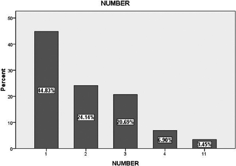
Distribution of supernumerary teeth, according to number
Prevalence of supernumerary teeth was more in males compared to females with a ratio of 3:1. Among the 64 teeth, 92.19% were impacted, and 7.81% were erupted. Maxillary posterior region (53.12%) was the most common location, followed by mandibular posterior region (32.81%) [Figure 2].
Figure 2.
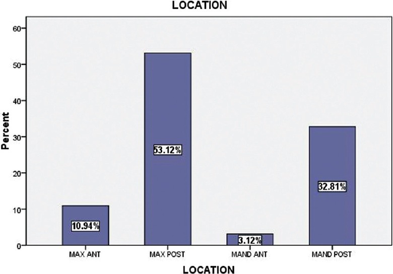
Distribution of supernumerary teeth, according to location
Maxillary right quadrant (39.06%) had a higher incidence of supernumerary teeth followed by maxillary left quadrant (25%). 39% of them were fully developed, whereas 23.44% of them had an evidence incomplete crown formation and 23.44% showed incomplete root formation [Figure 3].
Figure 3.
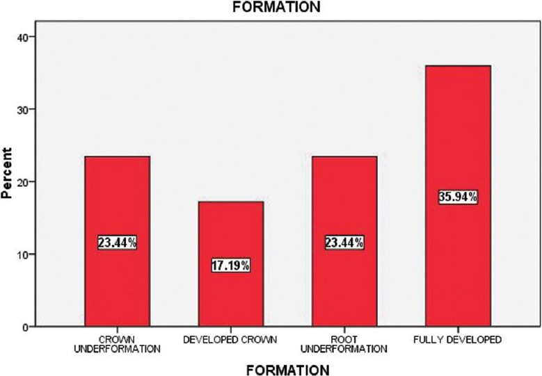
Distribution of supernumerary teeth, according to formation
Based on morphology, supplemental form (59.38%) was the most common type followed by 32.81% showing tuberculate form and 7.81% odontomas [Figure 4]. 73.68% of supplemental form were resembling premolars.
Figure 4.
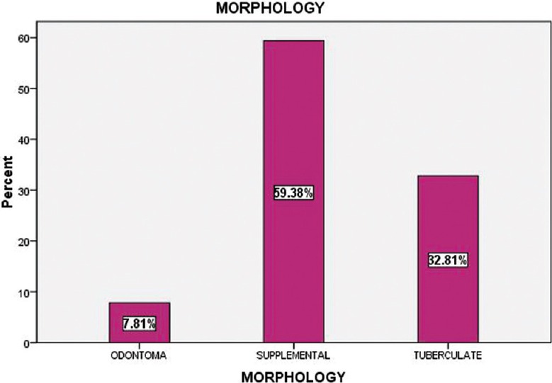
Distribution of supernumerary teeth, according to morphology
DISCUSSION
Nonsyndromic hyperdontia is a relatively common dental anomaly encountered in dental practice. The reported prevalence of supernumerary teeth in various populations ranges from 0.1% to 3.8%.[5] Minimal studies are available about the prevalence of supernumerary teeth in Indian population. Our study showed a prevalence rate of 1.2%, which is similar to the prevalence rate in a previous study conducted on the Indian population.[6]
The etiology for supernumerary teeth remains unclear. Both genetic and environmental factors are considered as the etiology of supernumerary teeth. Accordingly, various concepts have been suggested for their occurrence, such as, heredity, atavistic tendency, dichotomy of the tooth bud, genetic syndromes, “postpermanent” dentition and dental lamina hyperactivity. Localized and independent hyperactivity of the dental lamina is the most accepted theory for the development of supernumerary teeth.[2]
Various studies have shown the prevalence of single supernumerary teeth in 76–86%, double supernumeraries in 12–23% and multiple supernumeraries in <1% of cases.[2] The same trend was observed in our study with more subjects having single supernumerary teeth than two or more. In our study, male to female ratio is 3:1 similar to the male predominance seen in other studies.[4] Although some studies have mentioned that there are no differences in the number of teeth, according to gender,[7] our study clearly showed the incidence as well as the number to be more in males when compared to females [Figure 5].
Figure 5.
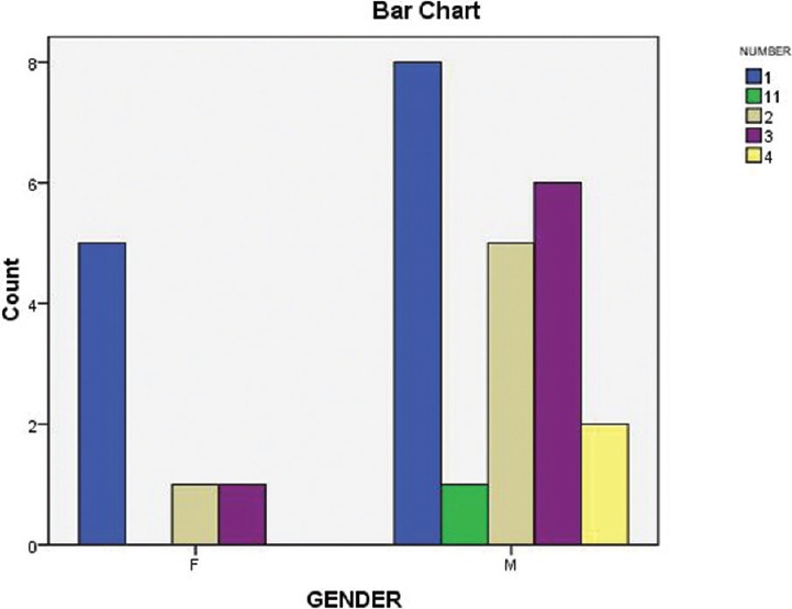
Distribution of supernumerary teeth, according to gender
In most of the cases, supernumerary teeth are incidental findings on panoramic examination. Similarly, in our study, among the 64 supernumerary teeth, 59 were impacted, and 5 were erupted. Location-wise, most of the previous studies have mentioned anterior maxilla as the most common location for the presence of supernumerary teeth. However in our study, maxillary posterior was the most predominant position, followed by mandibular posterior. Maxillary right quadrant had higher incidence compared to maxillary left quadrant. In contradiction to previous studies, supplemental was the most common morphological type observed in our study, predominantly, those resembling premolar. Most other studies have mentioned conical form as the most prevalent morphological form.[8]
Supernumerary teeth, whether impacted or erupted may remain in position for years together, without causing any disturbances and clinical manifestations. However, in some cases, they may cause complications like impaction of permanent teeth, delayed or ectopic eruption of adjacent teeth, malocclusions like midline diastema, or crowding and formation of cysts with bone destruction and root resorption of adjacent teeth.[3] In our patients crowding was the only complication noted due to supernumerary teeth.
It is important to diagnose correctly such conditions and institute suitable treatment to these patients at the appropriate time. Treatment of the supernumerary teeth can be achieved with either of the following options: (1) Removal of the supernumerary teeth, if complications are found or anticipated. (2) Leaving the supernumerary teeth in situ with periodic follow-up, if they are asymptomatic and without any associated pathology.[2]
CONCLUSION
Knowledge about the supernumerary teeth is important for dental clinicians as they are relatively common but are detected as an incidental finding in a radiograph. A routine screening panoramic radiograph is mandatory for every patient to unveil this condition so as to enable the dentist in early diagnosis, intervention and prevent the possible complications associated with it.
Financial support and sponsorship
Nil.
Conflicts of interest
There are no conflicts of interest.
REFERENCES
- 1.Ezirganlı S, İsa Kara M, Hüseyin Köşger H, Özer K, Kırtay M. Non-syndromic multiple permanent impacted supernumerary teeth: A retrospective study. Clin Dent Res. 2011;35:59–63. [Google Scholar]
- 2.Shah AK, Joshi MU. Nonsyndromic multiple supernumerary premolars: Report of three cases. Health Agenda. 2013;1:85–89. [Google Scholar]
- 3.Anegundi RT, Tegginmani VS, Battepati P, Tavargeri A, Patil S, Trasad V, et al. Prevalence and characteristics of supernumerary teeth in a non-syndromic South Indian pediatric population. J Indian Soc Pedod Prev Dent. 2014;32:9–12. doi: 10.4103/0970-4388.127041. [DOI] [PubMed] [Google Scholar]
- 4.Rajab LD, Hamdan MA. Supernumerary teeth: Review of the literature and a survey of 152 cases. Int J Paediatr Dent. 2002;12:244–54. doi: 10.1046/j.1365-263x.2002.00366.x. [DOI] [PubMed] [Google Scholar]
- 5.Karayilmaz H, Kirzioğlu Z, Saritekin A. Characteristics of nonsyndromic supernumerary teeth in children and adolescents. Turk J Med Sci. 2013;43:1013–8. [Google Scholar]
- 6.Mahabob MN, Anbuselvan GJ, Kumar BS, Raja S, Kothari S. Prevalence rate of supernumerary teeth among non-syndromic South Indian population: An analysis. J Pharm Bioallied Sci. 2012;4:S373–5. doi: 10.4103/0975-7406.100279. [DOI] [PMC free article] [PubMed] [Google Scholar]
- 7.Alvira-gonzález J, Gay-Escoda C. Non-syndromic multiple supernumerary teeth: Meta-analysis. J Oral Pathol Med. 2012;41:361–6. doi: 10.1111/j.1600-0714.2011.01111.x. [DOI] [PubMed] [Google Scholar]
- 8.Sharma A, Singh VP. Supernumerary teeth in Indian children: A survey of 300 cases. Int J Dent 2012. 2012 doi: 10.1155/2012/745265. 745265. [DOI] [PMC free article] [PubMed] [Google Scholar]


