Abstract
We have designed and constructed a small-animal adaptive SPECT imaging system as a prototype for quantifying the potential benefit of adaptive SPECT imaging over the traditional fixed geometry approach. The optical design of the system is based on filling the detector with the object for each viewing angle, maximizing the sensitivity, and optimizing the resolution in the projection images. Additional feedback rules for determining the optimal geometry of the system can be easily added to the existing control software. Preliminary data have been taken of a phantom with a small, hot, offset lesion in a flat background in both adaptive and fixed geometry modes. Comparison of the predicted system behavior with the actual system behavior is presented along with recommendations for system improvements.
Keywords: SPECT, Imaging, Instrumentation, Adaptive
1. INTRODUCTION AND SYSTEM OVERVIEW
Traditional SPECT imaging systems have fixed geometries meant to accomodate objects of various dimensions and characteristics as well as a variety of different imaging tasks. This means that the system is generally not optimized for any given patient and task that needs to be performed. This can mean things like decreased sensitivity and resolution, which translate into higher required doses for the subject as well as images that result in decreased observer performance (i.e. less accurate diagnoses). In this work, we have developed a prototype imager as a test-bed for adaptive SPECT imaging. Adaptive SPECT imaging puts the geometry of the system under continuously variable real-time computer control.1 With this capability, the system can optimize itself for every given subject and task to be performed. The basic approach to determining the optimal system geometry is to take an initial set of coarsely sampled data from which properties of the object can be calculated. This information is used in conjunction with a set of feedback rules to compute the optimal system geometry. The final data are then taken on a more finely sampled grid in the optimal configuration. Various different feedback rules can be applied and ideally they would depend on the task being performed. However, for design purposes, a more general feedback approach was implemented and is described in the following sections. The benefits of this type of optimization have the potential to decrease subject dose and improve image quality.
In order to make the prototype design tractable, we have started out with a set of assumptions about the system design. The first is that the system will be a one-detector system with object viewing angles sampled by rotating a vertically-oriented object. In addition, the system will have one on-axis pinhole with discreet diameter control. In this framework the adjustable parameters of the system are the object-to-pinhole distance, the pinhole-to-detector distance, and the pinhole diameter. Complications such as multiple pinhole functionality are left to a future design or an upgrade of the current design. Future iterations of this prototype design could be used as one module in a multi-detector, horizontally-oriented system by taking advantage of the type of gantry system developed for the FastSPECT II instrument.2
2. THE DETECTOR
Our prototype instrument has drawn on much of the existing technology developed in the Center for Gamma-Ray Imaging (CGRI) group at the University of Arizona, particularly in terms of the gamma-ray detection process. The detector module and control electronics are virtually identical to those used in the pre-existing FastSPECT II system. A description of that system can be found in Ref. 2 and Ref. 3 and will be briefly summarized here.
The modular gamma-ray detector system consists of three main components, the detector itself, the control electronics, and the control software. The detector is made up of a solid 114.3 mm × 114.3 mm × 5 mm NaI(Tl) scintillation crystal attached to a 10 mm thick quartz light guide and a 3 × 3 array of 1.5 inch diameter endon Hamamatsu photomultiplier tubes (PMTs). A single gamma-ray hit on the detector face will produce an output current pulse from each of the 9 PMTs. These current pulses are then sent to the control electronics for processing.
The control electronics consist of a front-end board that processes the current pulses and a back-end board that interfaces to the computer. The control software, written in Labview and C, communicates with the back-end board to save the processed PMT outputs for each qualified gamma-ray event. Data acquired using this system are saved in list-mode format. That is, each qualified gamma-ray event that hits the detector face produces 9 values that represent the number of secondary photons hitting each PMT. These 9 values are saved as one line in an output list. To recover a traditional binned-mode image (i.e. number of gamma-ray hits as a function of position on the detector face), the 9 PMT values for each qualified gamma-ray event are converted into a position estimate via a maximum likelihood algorithm.3
Using that procedure, we also calculate an error in our ability to determine the correct position by looking at our position estimates for a series of gamma-rays with known incident locations. To do this we simply collect about 6000 events hitting the detector at a known location and perform the position estimation procedure. An error in the estimated position is then determined as simply the average difference between the known and estimated positions. The mean radial position error in the center of the detector is about 2.3 mm and increases with distance from the center.
3. OPTICAL DESIGN
Our basic system assumptions (described in Section 1) as well as the existing detector design already give us a good general idea of how the system will look and behave. In this section, we go through the details of the optical design to determine the exact behavior of the system for a given object. This means that, given a particular object, our optical design will determine what system geometry (object-to-pinhole distance, pinhole-to-detector distance, and pinhole diameter) will give the “optimal” system performance as well as calculate the predicted performance of the system.
In order to do this, we must first define what we mean by “optimal” performance. Ideally, the “optimal” performance of the system is that which maximizes the observer's performance for a specific task.4–6 For example, if the task is tumor volume estimation, then the “optimal” performance is that which allows the observer to perform the most accurate tumor volume estimation. This would imply that the “optimal” performance of the system is dependent not only on the object, but also on the task being performed. In this case, a new optical design is needed for each task that one would like to perform with the instrument. Since this prototype instrument is being designed for use with many possible tasks, we have chosen to focus on a more general optimization to define the possible geometry ranges of the system. The initial system control software will use this optical design to control the system geometry. However, in the future, more complex, task-dependent geometry control codes (e.g. Ref. 1) can be written for use with the same instrument.
The system performance parameters that we have chosen to consider in the design are the field-of-view (FOV), sensitivity, and resolution of the system. Therefore, in the context of this prototype system, the geometry is considered “optimal” when the image of the region of interest (ROI) approximately fills the detector face, the sensitivity of the system is “as high as possible”, and the resolution in object space of a projection image is “as small as possible”. Note that the ROI could include the entire animal or a smaller, localized region of the animal (e.g. a lesion). The remainder of this section will be devoted to calculating how the FOV, sensitivity, and resolution depend on the system geometry as well as determining how to control the system geometry to get the desired values for FOV, sensitivity, and resolution. For Sections 3.1-3.3 we consider the ROI as the entire object, and in Section 3.4 we examine the case of a small ROI contained within the entire object.
3.1. Calculating system performance as a function of system geometry
To calculate expressions for how the image size, sensitivity, and resolution depend on the relevant system parameters, we used simple ray-tracing and radiometric arguments. By ray-tracing the extreme rays of the system, we derive an expression for the image size as a function of system parameters as
| (1) |
where d is the diameter of the pinhole, do is the object-to-pinhole distance, di is the pinhole-to-detector distance, Ho is the height of the object, and Hi is the height of the image.
The sensitivity of the system can then be determined by looking at how the solid angle subtended by the pinhole compares to 4π sr, because the object is emitting in all directions, but only light that passes through the pinhole can reach the detector. This gives a sensitivity of
| (2) |
as long as the image is entirely contained within the detector face.
Finally, the resolution of the system is calculated by looking at three contributions; blur due to the finite pinhole diameter, blur due to our ability to estimate the position on the detector face of a perpendicular incoming gamma-ray, and blur due to a gamma-ray hitting the scintillation crystal at an angle. The overall resolution of the system can be calculated by adding all of its components in quadrature, assuming that the components are all independent and gaussian. The blur due to the pinhole diameter can be calculated by looking at how a single point in object space maps to image space. Since we have a finite pinhole diameter this will map to a disk in image space whose size we can calculate by drawing rays from one point in object space through the top and bottom of the pinhole for a point in object space offset in the z direction. For the blur due to our ability to estimate the position on the detector face of a perpendicular incoming gamma-ray, we simply take a value of 2.5 mm, which is based on the analysis done in Section 2. The blur due to a gamma-ray hitting the scintillation crystal at an angle is caused by the fact that the interaction depth of the photon in the crystal corresponds to slightly different detected positions in image space. This calculation took into account the exponential absorption profile of the NaI scintillation crystal and defined the blur as corresponding to the spread induced when 75% of the incoming photons are absorbed.
Since we are interested in how small of a thing we can resolve in the object we project these calculations from image space back into object space. This gives a resolution that represents the smallest thing we can resolve in the object in a single projection image. After performing this projection and combining all three of these resolution contributors we get an estimated overall system resolution of
| (3) |
where , and μ is the attenuation coeffcient for NaI at 140 keV.
3.2. Controlling geometry to get “optimal” values for system performance
From equations derived in the previous section, we can easily calculate the system FOV, sensitivity, and resolution for a specific system geometry. The next step is understanding how to vary the system geometry in order to get the “optimal” values for FOV, sensitivity, and resolution. There are, presumably, many different approaches to dealing with this problem. The approach that we have taken here is one possibility.
3.2.1. Choosing the object-to-pinhole distance
We have started off by examining the equation for sensitivity and noticing that, for a given source strength and pinhole diameter, the sensitivity is completely determined by the object-to-pinhole distance. This means that if we minimize the object-to-pinhole distance, we maximize the sensitivity. As a result, we begin by making the object-to-pinhole distance as small as possible given a series of constraints. The constraints that we consider are mechanical, object size, obliquity, and vignetting constraints.
The mechanical and object size constraints are practical considerations that limit how close we can get the pinhole plate to the object. The obliquity constraint has to do with the fact that the scintillation crystal has a finite thickness, so that photons entering the crystal at an angle will have a blur in their position because the interaction depth of the photon in the crystal corresponds to slightly different positions in image space. The smaller the object-to-pinhole distance is, the larger this blur is. Therefore, a lower limit is imposed on do in order to keep the resolution in object space approximately equal to the blur due to our ability to estimate the position of a perpendicular incoming gamma ray.
As a result of the pinhole not being ideal (i.e. having a thickness), rays from the object can be vignetted if their angles through the pinhole plate are too steep. A smaller object-to-pinhole distance means steeper rays and more vignetting. In this context, the vignetting constraint is then set as a minimum possible do in order to eliminate vignetting.
3.2.2. Choosing the pinhole-to-detector distance
Once the object-to-pinhole distance is fixed, the pinhole-to-detector distance can be used to control the magnification of the object on the detector. The objective is to keep the size of the image equal to a fraction, f, of the detector face, where f is chosen to nearly fill the detector and the image is taken to be the image of the ROI. The main limitation on the pinhole-to-detector distance is that there is a maximum overall length of the system, Dmax, where Dmax is the distance from the center of the object to the front of the detector face. This restriction is simply to enforce a reasonable system size. For this system, Dmax was chosen to be 283 mm (or 11.14 inches), a value based partially on the commercially available linear motorized stages. In this scenario, the pinhole-to-detector distance can be calculated by setting Equation 1 equal to f (114.3 mm) and solving for the pinhole to detector size, where 114.3 mm × 114.3 mm is the size of the detector face. If this equation gives a di that results in a total system length greater than Dmax then the total system length is fixed to Dmax and do is changed to enforce filling the fraction f of the detector. In this case do is no longer determined by the constraints previously mentioned.
3.2.3. Choosing pinhole diameter
Once the system parameters are defined by the above calculations, the pinhole diameter can be varied using a resolution-throughput tradeoff. To do this, the resolution blur due to the pinhole diameter is compared with the other resolution contributions. If the resolution contribution from the pinhole diameter is smaller than the other contributions, and increasing the pinhole diameter to the next available size would not make the pinhole contribution larger than the other contributions, then the pinhole diameter is increased to the next available diameter. In this case, the sensitivity of the system can be increased without significantly affecting the resolution of the system. If, however, the pinhole contribution is larger than the other contributions and enough photons are hitting the detector, then the pinhole diameter is decreased in order to improve the resolution. Enough photons are considered to be hitting the detector when more than 400 counts/s are detected. This is a somewhat arbitrary threshold and in future implementation of the algorithm, this threshold should linearly depend on the source strength (see discussion in Section 7).
3.3. System Performance versus Object Size
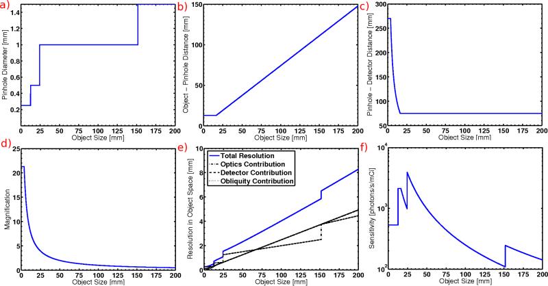
From the equations derived in Sections 3.1 and 3.2 we can calculate the system behavior and performance as a function of object size. Figures 1(a)-(f) show results of these calculations. For reference, a typical length for a rat is about 186 mm, while a mouse is typically 83 mm long. The width of a mouse is generally about 25 mm. While the system is designed to have the capability to image an entire animal, imaging a smaller ROI inside of the animal (i.e. ROIs on the order of ~25 mm or smaller) is the main focus of this study.
Figure 1.
These figures show the following parameters as a function of object size: a) pinhole diameter, (b) object-to-pinhole distance, (c) pinhole-to-detector distance, (d) magnification, (e) resolution in object space of a projection image (overall system resolution is shown as well as the three individual components, two of the individual components are overlapping by construction), (f) sensitivity [counts/s/mCi]
Figures 1(a)-(d) show how the system mechanically behaves as a function of object size. The discritized nature of the pinhole plate is seen in the discontinuous nature of Figure 1(a) and the large range of magnifications achievable with the system is displayed in Figured 1(d).
Figure 1(e) shows the system resolution in object space of a single projection image as a function of object size. The overall system resolution is shown as a solid line, whereas the three individual components as discussed in section 3.1 are shown in broken lines. The contribution from rays hitting the detector face at an angle is overlapping the contribution from our ability to estimate where a gamma-ray hits the detector face. This happens simply because we chose that to be the case when we set our constraints on the object-to-pinhole distance (see Section 3.2.1). Since the obliquity is the dominating factor in those constraints for most of the object sizes, these two lines match most of the time. The contribution due to the pinhole diameter is discontinuous because of the fact that there are only four discrete pinhole diameters to choose from. If the system only had a single 1 mm pinhole that line would remain flat, following its slope near the middle of the object size range. In this case, the resolution at very small object sizes would be significantly worse, however the system is able to adjust itself to get the best resolution possible from the available geometries. The achievable overall resolution for a size about equal to the width of a mouse is approximately 1 mm, which is good for a research small-animal system. For smaller object sizes, the resolutions improve significantly. For example, the predicted resolution for a 5 mm object is only 0.31 mm.
Figure 1(f) shows the sensitivity of the system as a function of object size. For comparison, one camera in the FastSPECTII system would look like a flat line with a value of about 600 counts/s/mCi.7 For small object sizes we generally do better than this value. In order to improve the sensitivity further we can simply modify the comparison value used to perform the resolution/throughput tradeoff when determining the pinhole diameter. Sensitivities at larger object sizes are less than the equivalent FastSPECTII value, but only because the object is being demagnified so that it can fit onto the detector face.
3.4. An Example Object: Small Lesion Within Larger Object
In order to demonstrate the type of performance that this system can achieve, here we examine more carefully the type of situation that might occur in a real imaging scenario. The specific example that we consider here is of a mouse-sized object (a cylinder with a diameter of 25.4 mm and a height of 83 mm) with an interior spherical tumor. The tumor has a diameter of 5 mm and is offset 5 mm from the center of axis of rotation of the object. It is assumed that all of the activity in the object is localized in the spherical tumor. A typical task that might be performed in this situation is tumor volume or activity estimation for determining the effcacy of a particular treatment.
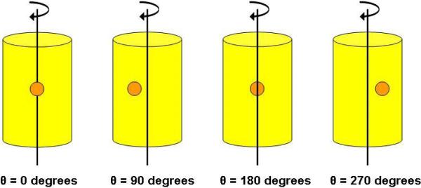
In the previous calculations we assumed that the object of interest had a fixed extent, independent of viewing angle. Here, the extent of the object of interest, namely the tumor, changes with viewing angle of the object (see Figure 2). Although the tumor has a fixed size, its offset from the center of the overall object means that its apparent distance from the center of the overall object changes with viewing angle. Since the detector of the system is not steerable, this implies that the field of view must be adjusted for each angle in order to ensure that the tumor is utilizing the maximum amount of detector space at all times. As a result, the optimal system geometry will change for each viewing angle.
Figure 2.
Diagram showing the object geometry for our example case study. The object is a mouse-sized cylinder with an interior 5 mm diameter spherical tumor offset 5 mm from the center of rotation of the object. The orientation angles correspond to the angles in Figures 3(a)-(f). The reader is viewing the images as if she/he were the detector (i.e. the tumor is closest to the detector at θ=0 degrees).
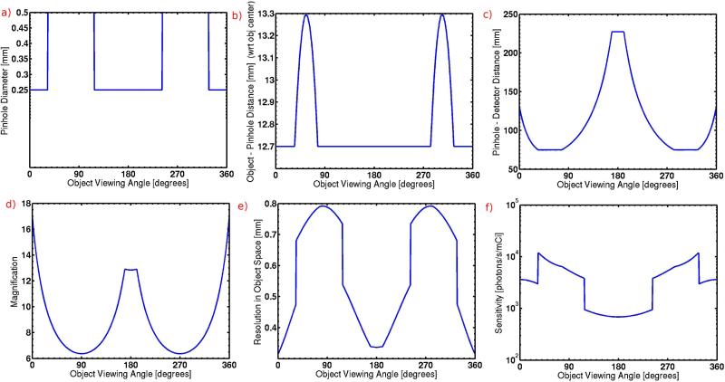
In the beginning of this section we calculated the predicted system behavior as a function of object size. Here we do the same calculations, but this time as a function of object viewing angle for this specific example case. Figure 2 shows the angle orientation as if the reader is the detector and Figures 3(a)-(f) show the results of these calculations. In Figures 3(a)-(d) we see how the geometry of the system changes over the imaging sequence and Figures 3(e)-(f) show the range of resolution and sensitivity values that would be acquired for an adaptive data set. For this particular task, the expected resolution values for a projection image are all sub-mm, ranging between 0.3 and 0.8 mm. The sensitivity values range from 700 - 12000 counts/s/mCi, all higher than the equivalent FastSPECTII system. These simulation results clearly show that adaptive imaging has the potential to significantly improve image quality over standard, fixed geometry systems.
Figure 3.
These figures all show the following parameters as a function of example object viewing angle: a) pinhole diameter, b) object-to-pinhole distance, c) pinhole-to-detector distance, d) magnification, e) overall resolution in object space of a projection image, f) sensitivity
4. MECHANICAL DESIGN
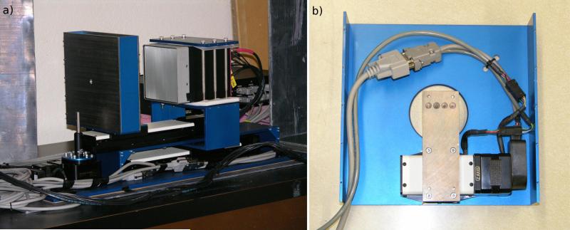
The basic design of the adaptive system has a vertically-oriented animal, one on-axis pinhole and one detector. Viewing angles are achieved by rotating the object. To allow real-time adaptability of the system geometry, the camera position, pinhole position, pinhole diameter, and object viewing angle are all mounted on a series of four motorized stages manufactured by Newmark Systems, Inc. All four stages are controlled via a single Galil Motion Control, Inc. controller so that one command can produce simultaneous motion of all four stages. Figure 4(a) shows a picture of the assembled system.
Figure 4.
a) Closeup of the adaptive imaging system itself. The object stand can be seen sitting on top of the rotation stage. To the right of that is the pinhole assembly and slightly further to the right the detector is shown. b) Picture showing detail of the pinhole assembly.
The entire system sits on a single aluminum plate that can be bolted to a breadboard via a series of 1/4-20 holes for stability. The animal is mounted on a rotation stage that sits at one end of the board. A linear stage with an 8 inch travel range bolts to the baseplate. Above this is a small aluminum stand on which bolts another linear stage with a 6 inch travel range. An extension piece bolts to the slide of the 8 inch linear stage and wraps around the upper stage. The camera is attached to this extension piece via a set of six 1/4-20 bolts that clamp an aluminum plate down on the top of the camera. Nylon sheets are used between the camera and the aluminum clamp to eliminate electrical contact. The pinhole assembly (see Figure 4(b)) bolts to the slide on the linear stage with a 6 inch travel range. The assembly consists of two tungsten plates with oversized holes for the gamma-ray beam to pass without vignetting. Behind the first tungsten plate is a smaller tungsten sandwich that has four small pinholes with diameters of approximately 0.25 mm, 0.5 mm, 1.0 mm, and 1.5 mm, where the two larger diameters are drilled directly into the tungsten and the two smaller diameters are drilled through a small piece of gold foil placed between the two tungsten plates. This entire tungsten sandwich is bolted to a small linear stage with a 1 inch travel range that then bolts on the large back tungsten plate. This allows the small pinhole plate to slide against the large front tungsten plate to pick off the desired pinhole diameter.
The majority of the shielding for the system is provided by the tungsten in the large and small pinhole plates. The large pinhole plates are 1/8 inch thick, while the small pinhole plate has an overall thickness of 1/4 inch. Additional shielding for the system is provided via a large 1/8 inch thick lead-lined box that fits snugly over the entire assembly. The entire system also sits on an additional sheet of 1/8 inch lead and small sheets of loose lead have also been placed near the bottom of the object rotation stage.
5. SOFTWARE CONTROL
The adaptive system has a total of 6 different mechanical components that must be electronically controlled. These include the camera, its power supply, and the four motorized stages. Computer interface software for the camera, written in Labview and C, has already been developed for several similar detectors in the CGRI group and is used with little modification here. Additional LabView and C code has been written to handle control of the camera power supply, the four motorized stages, and the adaptive algorithms. The camera power supply is a National Instruments device and therefore can be controlled easily via LabView with pre-existing software provided by the manufacturer. The four motorized stages are controlled via an RS-232 interface with a single controller. The manufacterer has a series of programs written in C and LabView that can be interfaced through LabView for relatively straightforward user control.
The user interface for the adaptive system brings all of these components together into a single, simple control panel. Upon startup, the software automatically initializes the system and allows the user to re-orient the object to any desired starting rotation angle. The user must then take a set of fixed-geometry images before any adaptive images can be acquired. To do this, the user simply specifies the total exposure time, the number of imaging angles, and the geometry of the system (pinhole diameter, object-to-pinhole distance, and pinhole-to-detector distance). The software proceeds to take and save the data and motor positions to a series of output files. The motor positions are saved for every image, so that the exact geometry is known and can be used in subsequent reconstructions. The total exposure time is distributed evenly over each of the viewing angles. After acquiring the data, the software automatically performs an estimation of the object size for each imaging angle and saves this information for later use. The details of the object size estimation algorithm are described in the following subsection. Once an initial set of fixed-geometry images have been acquired, the user can choose to take either fixed or adaptive images in any combination they choose. The system will automatically re-calculate and update estimates of the object size for each imaging iteration.
In order to take adaptive images, the user enters the total exposure time and number of imaging angles. Information about the object size determined in the last set of images are used here to calculate the optimal geometry of the system. If more angles are desired for the current imaging sequence than the previous imaging sequence, object sizes are linearly interpolated onto the finer angle grid. For the adaptive images, the total exposure time is divided among the different viewing angles in order to keep the number of counts in each projection image constant. This calculation relies on the comparisons between the throughput for the chosen geometry of each viewing angle and ignores leakage.
Note that the current software is implemented using the estimated object size to determine the optimal geometry based on the optical design described in Section 3. However, the software has been written such that alternative feedback rules could be easily added to the current software.
5.1. Object Size Estimation
The estimation of the size of the ROI is perhaps one of the most diffcult tasks of the adaptive system and can have a significant effect on performance of the system. The ideal algorithm would be able to determine the size of the ROI with little or no user input. However, this is diffcult to achieve since the region of interest is object and task dependent. As a result, some user interaction may be required for effective implementation in real imaging systems. One might envision an algorithm that requires the user to pick from a pre-determined set of tasks, independently calculates the size of the ROI, then requests user verification.
The current algorithm uses the fact that the size of the lesion is known and simply calculates the position of the lesion in order to determine its distance off-axis. It works by binning the image and selecting the maximum value as the position of the lesion. By shifting the binning template and calculating the largest maximum value of all the possible binned images, a more accurate estimated position is determined. Once the position of the lesion is known, the size of the FOV is calculated by combining that position with the known 8.1 mm size of the lesion and the known magnification of the system to get the full estimated “object size”. There are two main drawbacks to this specific approach. First, the known size of the lesion must be used in the calculation and, second, the magnification of the lesion is not exactly known. The magnification of the lesion is different if it is located in front of or behind the center of object space. Since the position of the lesion is not known during the scout image acquisition, the magnification of the center of the object space is used. This results in the system underestimating the FOV when the lesion is closer to the detector and overestimating the FOV when the lesion is further from the detector.
Although the algorithm does have drawbacks, it does a reasonable job of calculating the estimated object size in this scenario (see Section 6). As a result, it can be used to take adaptive phantom data that gives an indication of the utility of the adaptive nature of the system. However, future work is required to develop an object size determination algorithm more applicable to real-life situations.
6. A PHANTOM STUDY
We have collected phantom data with the adaptive system in fixed geometry and adaptive modes in order to verify that the system performs as it was designed. In the following sections, the phantom is described, projection images are presented, analysis of leakage fraction is discussed, and comparison of the predicted and actual adaptive behavior is performed.
6.1. The Phantom
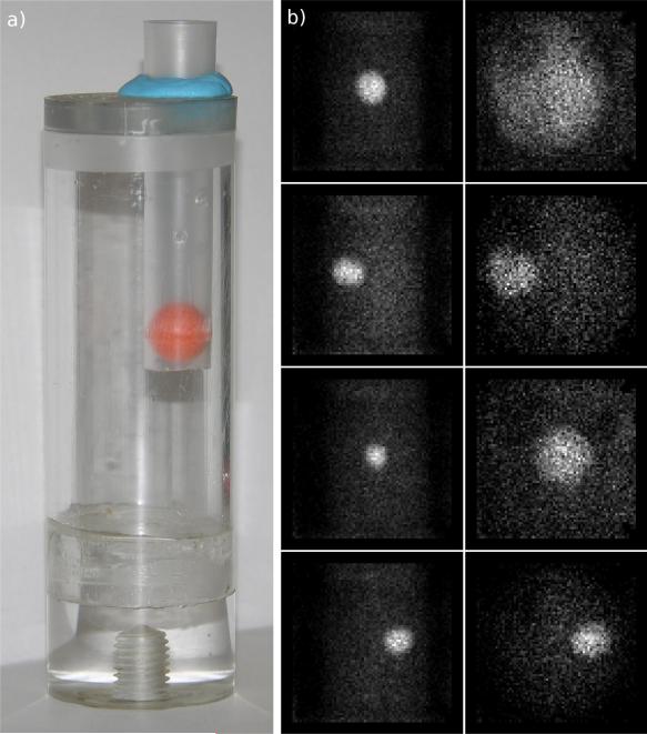
We have developed a phantom to simulate a spherical, offset lesion in a flat background (see Figure 5(a)). The background reservoir is a hollow acrylic tube with an inner diameter of 28.65 mm and an inner height of 66.91 mm. A cap fits over the top of this background reservoir that has two offset holes drilled into it. The first hole has a diameter of just over 12 mm and is 0.3125 inches from the center of the cap. This is the position from which the phantom lesion is suspended. The second hole is also offset from the center, which simply serves as a fill access to the background reservoir when the phantom is fully assembled. The spherical lesion consists of a 8.1 mm inner diameter hollow polypropylene ball with a small puncture that allows the ball to be filled with radioactive source. This spherical ball is secured into a 12 mm outer diameter plastic test tube, which has several punctures in the sides to allow fluid flow. The top of this test tube is attached to the large offset hole in the cap of the background reservoir with a small amount of putty. After the phantom is fully assembled, a syringe is used to fill the lesion with a radioactive source. This is then sealed off with super glue and allowed to dry. The background reservoir is then filled with a cooler radioactive source to produce the flat background.
Figure 5.
a) Phantom used for imaging study. The large cylinder holds the background source while the orange sphere holds the lesion source. The orange sphere is a plastic, hollow ball. b) (left) Projection images from fixed geometry data set (right) Analogous projection images from adaptive data set.
6.2. The Data
Tomographic data were acquired of the phantom in both adaptive and fixed geometry modes with the same instrument. The adaptive data consists of one set of scout images with 12 angles and a final set of adaptive images with 50 angles. The fixed geometry data consists of a single set of images with 50 angles where the exposure time was set to be equal to that of the scout plus adaptive imaging sequence when radioactive decay was taken into account. To make the imaging sequences comparable, both the scout data and the fixed geometry data were taken with FastSPECTII standard geometry (pinhole diameter of 1.0 mm, object-to-pinhole distance of 48.26 mm, and pinhole-to-detector distance of 116.84 mm). Figure 5(b) shows a sequence of four projection images at different angles in the fixed and adaptive geometry configurations.
6.3. Leakage Fraction
In order to estimate the fraction of detected leaked events, data with the pinhole covered and uncovered were taken for the adaptive system in the FastSPECTII configuration. Analysis of these data gives a leakage fraction of approximately 39% for the listmode data (i.e. before MDRF processing with likelihood windowing), 27% after MDRF processing, and 15% after MDRF processing and with the outer 5 rows and columns of pixels set to zero. While this data gives an indication of what the leakage fraction is for this particular geometry, the flexibility of the adaptive system means that the leakage fraction will vary for any given geometry. Therefore, a more complete leakage analysis should be done to fully understand the leakage behavior. This level of leakage is high for a SPECT imaging system and could introduce artifacts in the reconstructed images of the system. The mobility of the system makes shielding diffcult and future modifications of the system should attempt to reduce this effect to get better system performance.
6.4. Actual vs. Predicted System Behavior
Since the positions of each motorized stage are recorded for every image, the actual behavior of the system can be compared with its predicted behavior. Figures 6(a)-(c) show the actual system behavior versus predicted system behavior.
Figure 6.
These figures all show the following parameters as a function of imaging angle: a) Predicted and actual pinhole diameters, b) Predicted and actual object to pinhole distances, c) Predicted and actual pinhole-to-detector distances.
Figure 6(a) shows the predicted and actual pinhole diameters used as a function of viewing angle. The actual pinhole diameter seems to follow the general trend of the predicted pinhole diameter with some mild deviations. The source of these deviations could arise from differences between the modelled and actual phantom, object size estimation errors, errors in machining of the pinhole plates, photon noise, and leakage in the system.
Figure 6(b) plots the predicted and actual object-to-pinhole distance as a function of viewing angle. In this case, the system behaves exactly as expected simply because the pinhole plate is running into a limit the entire time. This limit is not due to mechanical abilities of the system, but rather the extent of the overall object. The distance of 21 mm is the closest that the pinhole plate can get to the phantom edge without hitting it. The system does have the capability to provide smaller object-to-pinhole distances for objects with smaller overall diameters.
Finally, Figure 6(c) shows the predicted and actual pinhole-to-detector distances. Again, the system behavior follows the predicted trend, but does have some systematic deviations. In this case, the deviations are mainly caused by errors in the object size estimation and zero angle positioning errors in conjunction with sparse angular sampling of the scout images. In this case, errors in the object size strongly affect the actual pinhole-to-detector distance used because this distance is what controls the magnification of the system. The strong dependency of the pinhole-to-detector distance on object size can clearly be seen by examining Figure 1(c). For this study, the object size is relatively small, around 8.1 mm, so that the pinhole-to-detector values are chosen from the area of the curve that is steepest on this plot. Between object sizes of 5 and 15 mm the pinhole-to-detector distance ranges from 270 mm to 75 mm. That means that an error in estimating the object size by only 1 mm will cause an error in the pinhole-to-detector distance of roughly 20 mm.
The other main contributing factor to the error in the chosen pinhole-to-detector distance is the sparse angle sampling of the scout images. For these data, the scout images consist of measurements taken over 12 angles. In order to get the object sizes for all 50 angles of the final adaptive data, estimated object sizes from the scout images are linearly interpolated. This means that if the extreme angles are not seen in the scout images, they will not be properly accounted for in the adaptive images. The reason that 12 angles were chosen for the scout images was to eliminate this effect since this results in data being taken for angles of 0, 90, 180, and 270 degrees in addition to others. This means that, provided the object of interest is at an extreme value for angle 0, all the extreme values of the object orientation will be properly sampled. However, if the zero angle orientation of the object is not properly aligned, errors will be incurred during the angle interpolation. In these data, the zero angle orientation was offset by approximately 8 degrees, causing the above mentioned interpolation problems. This can be seen in Figure 6(c) by the fact that the maximum pinhole-to-detector distances are not separated by 180 degrees. With the current software setup, it is diffcult to align the zero angle properly because it requires visual inspection of the actual object by the user. Fortunately, this error could be easily remedied by including a real-time projection image viewer that displays an acquired image of the object to the user after each angle update of the object orientation.
7. DISCUSSION
We have successfully designed and constructed an adaptive small-animal SPECT imaging system. The current system is a one-detector module with a vertically-oriented animal that serves as a testbed for a future, large-scale, SPECT imaging system with a horizontally-oriented animal. We have demonstrated that construction of the adaptive system is not only feasible, but that it also performs as designed. The system has a complete software package that handles user input, controls all of the mechanical adaptive components and saves their encoder-based position information, computes geometry feedback rules, and communicates with the detector to take and save all relevant data for future data processing. A phantom has been designed for system testing that has a hot offset lesion in a flat background. Data have been successfully acquired with this phantom in both adaptive and fixed geometry configurations. For these data, feedback rules that force the lesion to fill the detector were employed, although the system software can be easily modified to handle additional feedback rules.
During the construction and testing of the adaptive SPECT system, many valuable lessons were learned that should contribute to improvement of its performance in future modifications. In terms of hardware, the main issue that needs to be addressed is the large leakage fraction of the system as well as the fact that it varies with the system geometry. In terms of software, improved zero angle animal orientation adjustment as well as a more accurate object size estimation algorithm could both lead to better performance of the adaptive system by allowing more accurate determination of the object properties from the scout imaging sequence data.
The feedback rules employed for this study were based on filling the detector with the object for every viewing angle, maximizing the sensitivity, and minimizing the resolution. While these are useful rules to apply for initial testing purposes, they do not take task-based measures of image quality into account. Appropriate task-based feedback rules are currently in development1 and should be relatively straightforward to implement with the system given the flexible software interface. Addition of these types of feedback rules have the potential to produce significantly improved image quality in comparison to traditional, fixed geometry systems.
ACKNOWLEDGMENTS
Thanks to Jacob Hesterman, Kevin Gross, Bill Hunter, Jean Chen, Don Wilson, Eric Clarkson, Gail Stevenson, Steven Moore, Robert Hunter, Corrie Thies, Nancy Preble, and all of the students for their help and advice. We are also grateful for support from the following grants: NIH/NIBIB R01 EB002146, NIH/NIBIB R37 EB000803, and NIH/NIBIB P41 EB002035.
REFERENCES
- 1.Barrett H, Furenlid L, Freed M, Hesterman J, Kupinski M, Clarkson E, Whitaker M. Adaptive SPECT. Trans. Med. Imag. 2007. submitted. [DOI] [PMC free article] [PubMed]
- 2.Furenlid L, Wilson D, Chen Y.-c., Kim H, Pietraski P, Crawford M, Barrett H. FastSPECTII: A Second-Generation High-Resolution Dynamic SPECT Imager. IEEE. Trans. Nuc. Sci. 2004;51(3):631–635. doi: 10.1109/TNS.2004.830975. [DOI] [PMC free article] [PubMed] [Google Scholar]
- 3.Chen Y, Furenlid L, Wilson D, Barrett H. Calibration of Scintillation Cameras and and Pinhole SPECT Imaging Systems. In: Kupinski M, Barrett H, editors. Small-Animal SPECT Imaging. Springer Publishing; 2005. pp. 195–202. [Google Scholar]
- 4.Barrett H, Myers K. Foundations of Image Science. John Wiley and Sons, Inc.; 2004. [Google Scholar]
- 5.Hesterman J, Kupinski M, Furenlid M, Wilson D. Experimental task-based optimization of a four-camera variable-pinhole small-animal SPECT system. Proc. SPIE. 2005;5749:300–309. doi: 10.1117/12.594386. [DOI] [PMC free article] [PubMed] [Google Scholar]
- 6.Kupinski M, Clarkson E, Gross K, Hoppin J. Optimizing imaging hardware for estimation tasks. Proc. SPIE. 2003;5034:309–313. [Google Scholar]
- 7.Chen Y-C. PhD thesis. University of Arizona; 2006. System Calibration and Image Reconstruction for a New Small-Animal SPECT System. [Google Scholar]


