Abstract
Objective:
To discuss return to play after femur fractures in several professional athletes.
Background:
Femur fractures are rare injuries and can be associated with significant morbidity and mortality. No reports exist, to our knowledge, on return to play after treatment of isolated femur fractures in professional athletes. Return to play is expected in patients with femur fractures, but recovery can take more than 1 year, with an expected decrease in performance.
Treatment:
Four professional athletes sustained isolated femur fractures during regular-season games. Two athletes played hockey, 1 played football, and 1 played baseball. Three players were treated with anterograde intramedullary nails, and 1 was treated with retrograde nailing. All players missed the remainder of the season. At an average of 9.5 months (range, 7–13 months) from the time of injury, all athletes were able to return to play. One player required the removal of painful hardware, which delayed his return to sport. Final radiographs revealed that all fractures were well healed. No athletes had subjective complaints or concerns that performance was affected by the injury at an average final follow-up of 25 months (range, 22–29 months).
Uniqueness:
As the size and speed of players increase, on-field trauma may result in significant injury. All players returned to previous levels of performance or exceeded previous statistical performance levels.
Conclusions:
In professional athletes, return to play from isolated femur fractures treated with either an anterograde or retrograde intramedullary nail is possible within 1 year. Return to the previous level of performance is possible, and it is important to develop management protocols, including rehabilitation guidelines, for such injuries. However, return to play may be delayed by subsequent procedures, including hardware removal.
Key Words: football, sports, lower extremity injuries
Each year, in the United States, approximately 60 000 patients sustain midshaft femoral fractures.1 These injuries often occur from high-velocity trauma and are frequently associated with concomitant injuries to the pelvis and lower extremity.2 Almost one-half of the patients treated for femur fractures at level I trauma centers have some residual disability 12 months after the injury.3 Up to 20% of patients treated surgically for femoral-shaft fractures are unable to return to their original work 3 years after the injury, but most patients can return to work in some capacity.3,4 Functional recovery after long-bone fractures usually depends on concomitant injuries and the general health of the patient.3,5–8 Femur fractures are rarely seen in sports. Isolated fractures are thought to result in good functional recovery, but return to play has not been documented in professional athletes.9
Both anterograde and retrograde intramedullary nailing are considered acceptable treatments of midshaft femur fractures, and both are thought to result in good functional outcomes.7,8 Anterograde and retrograde nailing, however, each present specific complication profiles that should be considered before treatment. Postinjury limitations may include hip-abductor weakness with a resultant Trendelenburg gait pattern, quadriceps femoris muscle weakness, anterior knee pain, and trochanteric bursitis, depending on concomitant injury and surgical technique.1,9–11 Newer implants that allow for more-lateral entry may reduce the risk of abductor weakness after anterograde intramedullary nailing.
To date, we have been unable to find any reports documenting return to play after long bone fractures. In particular, no authors, to our knowledge, have addressed return to play after intramedullary nailing of femur fractures in athletes.
We treated 2 professional athletes who underwent intramedullary nailing of midshaft femur fractures with anterograde or retrograde intramedullary nailing. Their clinical results are summarized in this article (Table 1). We identified and contacted 2 other professional athletes who sustained midshaft femur fractures; they had similar treatment and rehabilitation protocols, and we present their clinical data for analysis. Additionally, we discuss treatment options that must be considered by the surgeon and team physician. We hypothesized that return to play could be expected in patients with femur fractures but that recovery would take more than 1 year and that patients would not return to the same level of performance.
Table 1.
Professional Athletes With Femur Fractures: Injury, Surgery, and Outcomes
| Athlete's Sport |
Mechanism of Injury |
Method of Intramedullary Nailing |
Concomitant Injuries |
Time to Full Weight Bearing, mo |
Time to Return to On-Field Practice, mo |
Time Missed, Games (mo) |
Subjective Complaints |
Objective Outcomes |
| Football | Torsional stress | Retrograde | None | 2 | 8 | 5 (9) | None | Pro Bowl following season |
| Hockey | Axial load because of checking from behind and crashing into boards | Anterograde | Patellar fracture (treated nonoperatively) | 3 | 8 | 80 (9) | None | Career high in points following year; second-place Comeback Player of the Year |
| Hockey | Axial load because of checking from behind and crashing into boards | Anterograde | None | 2 | 6 | 59 (7) | None | Led team in playoff points following season |
| Baseball | Axial load while running bases | Anterograde | None | 2 | 10 | 177 (13) | Leg-length discrepancy | Won Comeback Player of the Year for organization |
Case 1
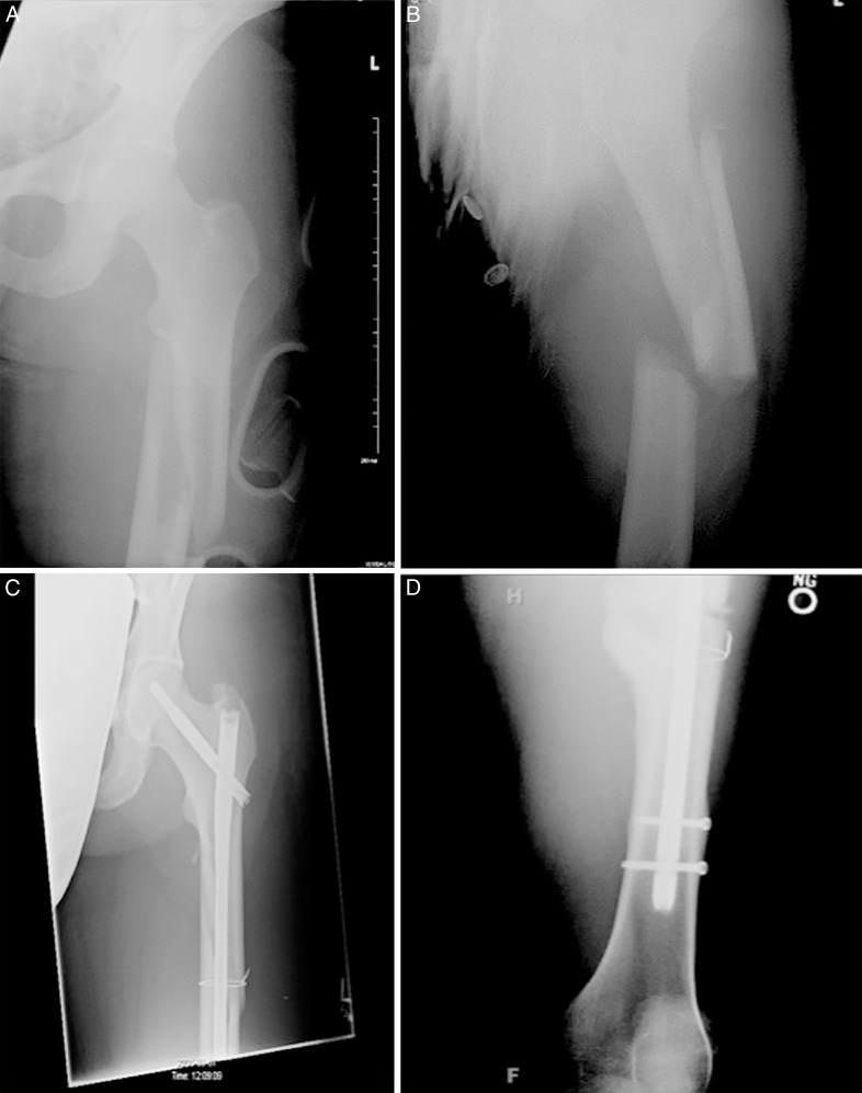
A 26-year-old male professional hockey player was injured during an away game. The player was checked from behind while skating and slid into the boards, sustaining an axial load to his left leg with his foot hitting the boards. The player was unable to bear weight on his left leg and was evaluated by a certified athletic trainer. An obvious deformity was noted: the patient's leg was significantly medially rotated relative to the hip and he localized pain to his thigh. The injury was closed, and he had no loss of consciousness. The patient was splinted on the ice, emergency medical services were activated, and he was transferred to the hospital. En route to the hospital, the patient complained of numbness in his leg and an inability to sense movement. He had palpable dorsalis pedis and popliteal pulses and a normal neurologic examination, but compartment pressures were not assessed. Plain radiographs at the hospital revealed a comminuted fracture of the left femur with a butterfly fragment, as well as a fracture of the superior pole of his patella (Figure 1A and B). Anterograde intramedullary nailing was performed with the patient in the supine position on a fracture table. A standard piriformis entry nail (Synthes Corporation, West Chester, PA) was used. There was difficulty obtaining reduction secondary to extensive comminution. After the proximal fragment was reamed, secondary fracture lines were noted in the distal fragment. Open reduction was then performed, and cerclage wires were used to maintain reduction. A Synthes 13- by 400-mm nail (Synthes, Inc, West Chester, PA) was then inserted after overreaming by 1 mm, and both proximal and distal static interlocking screws were placed. The patient's patellar fracture was treated nonoperatively with a hinged knee brace locked in extension.
Figure 1.
Anterior-posterior radiographs of the patient in case 1 (National Hockey League player). A and B, Comminuted fracture of the left femur with a butterfly fragment. C and D, At 1 year postoperatively, the fracture was healed and the hardware remained intact.
Postoperatively, the patient was placed on aspirin 325 mg daily for 4 weeks for prophylaxis of deep vein thrombosis. He was kept to toe-touch weight bearing for 4 weeks and then advanced to weight bearing as tolerated with crutches for weeks 4 to 8. The patient was transitioned from 2 crutches to 1 during weeks 8 to 12 and then began full weight bearing without assistance. Passive range of motion was begun on the knee at 2 weeks, with active range of motion begun at 4 weeks. The rehabilitation program addressed weight-bearing status, range of motion, quadriceps femoris muscle control, hip-abductor strength, stretching, progressive resistive exercises, balance, proprioception activities, and conditioning. Pool therapy was initiated at 2 weeks after surgery, and he began daily range-of-motion exercises at 4 weeks after surgery (Table 2).
Table 2.
Rehabilitation Protocol for National Hockey League Player
| Phase 1: Wk 1–12 |
Phase 2: Wk 12–24 |
Phase 3: Wk 24–30 |
Phase 4: Wk 30–40 |
| • Protected weight bearing • Prone knee flexion• Heel slides • Seated knee extension (active and passive range of motion) at 4 wk with goal of 90° at 1 mo • Exercise bike• Straight-leg raises• Pool exercises at 2 wk, 2–3×/wk• Standing transfers at 3 wk; weight bearing as tolerated with crutches at 4 wk• Transitioned to 1 crutch at wk 8–12; transition to no crutches at wk 12 | • Weight bearing as tolerated• Walking and gait exercises• Minisquats and squats• Calf raises• Bridges• Lunges• Supine plank hold• Lateral step-outs• Pelvic raises | • Endurance• Jogging in pool (HydroWorx, Middletown, PA)• Endurance biking• Skating• Pool running• Elliptical• 2-Legged lifting exercises• Resistance walking• Balance and proprioception• Jumping rope | • On-ice drills• Conditioning• Cone drills• Explosion and acceleration• Lateral-movement drills• Pursuit drills• Sprint drills• Hurdle and ladder drills• Sled pushes• Power-skating drills• Gradual progression to contact drills• Checking• Return to play |
The patient began skating at 5 months. At 8 months, he continued to have a slightly antalgic gait, which was noticeable after workouts, but during day-to-day activities, his gait appeared normal. We believed that the antalgic gait was due to continued weakness of the hip abductors; the patient did not have any pain on examination and was able to participate in full-contact practice with the team. At 8 months, he had 0.5 cm of thigh atrophy compared with the contralateral side. Dynamometry testing showed the hip abductors had 85% strength and 73% power compared with the contralateral side. In the week before the injury, the athlete lifted a maximum of 150 lb (68 kg) during hip abduction with 10 repetitions and 100 lb (45 kg) during hip flexion with 8 repetitions. At 9 months postoperatively, the patient played in 6 minor-league games before returning for the final 10 games of the regular season at the National Hockey League level. At the time of return to play, the athlete lifted a maximum of 150 lb (68 kg) during hip abduction with 10 repetitions and 100 lb (45 kg) during hip flexion with 8 repetitions. He missed 80 regular-season games (Table 1). The following season, the athlete continued to play in the National Hockey League and had career highs in points (goals and assists). Radiographs at 1 year after surgery showed the patient's fracture was well healed with intact hardware (Figure 1C and D). His weight-lifting performance had returned to preinjury levels. At the 2-year follow-up, the player had no functional complaints, and examination revealed no thigh atrophy compared with the contralateral side.
Case 2
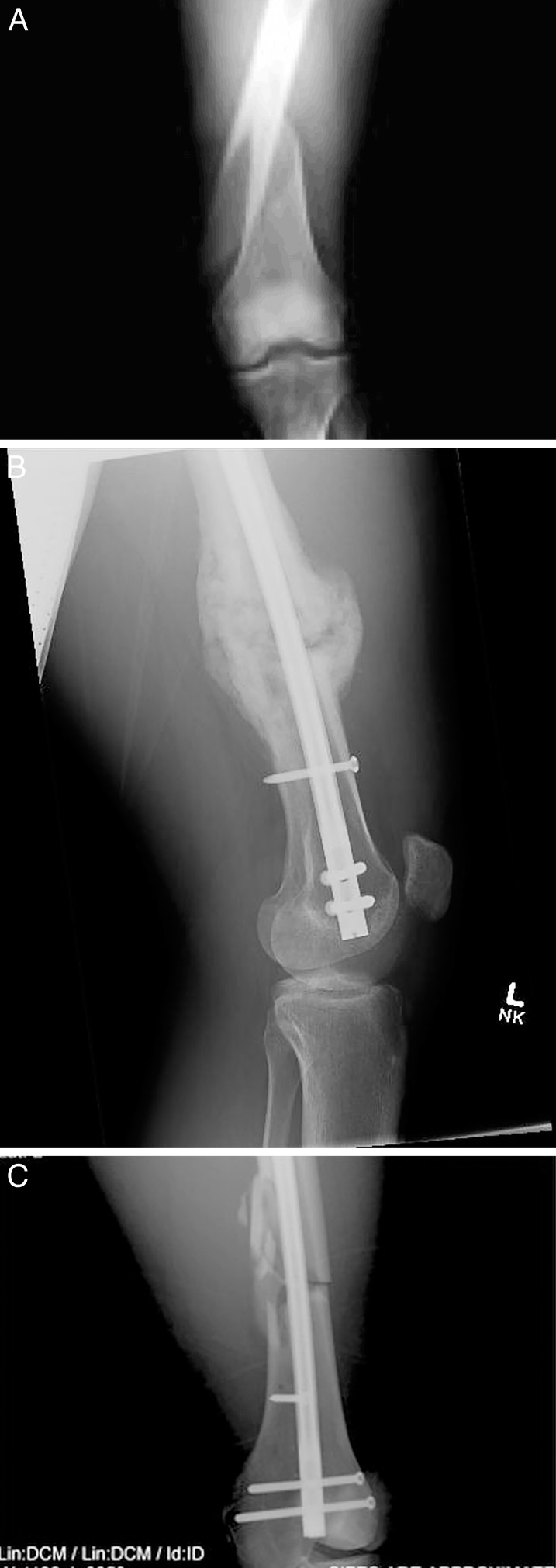
A 27-year-old male professional football player was injured while participating in an away game. The player collided with another player, who struck his left anterior thigh while his left foot was planted in the ground. The patient localized his pain to his left thigh and was unable to bear weight. The skin was intact, but obvious deformity was noted, with the patient's thigh significantly rotated medially relative to the hip. There was no loss of consciousness. Emergency medical services were activated, and the patient was transferred to the hospital. Buck traction was used en route to the hospital. Plain radiographs at the hospital revealed a comminuted left femoral-shaft fracture at the distal metadiaphyseal transition. The medial cortex was significantly comminuted (Figure 2A). No other injuries were noted on radiographs. The patient was positioned supine on a radiolucent table for placement of a retrograde intramedullary nail. A midline anterior incision with a medial parapatellar approach was used, and reduction was confirmed by intraoperative fluoroscopy. Overreaming by 1 mm was performed, and a Synthes 13- by 400-mm, retrograde intramedullary nail was placed. Some residual varus angulation was noted after initial nail placement; an anterior-posterior blocking screw was placed, and the nail was readvanced past the fracture site. The nail was countersunk distally, and distal femoral locking screws were placed. Proximal locking screws were placed with 1 in a static position and 1 in a dynamic position to allow for future dynamization if the fracture healing was delayed. The knee was irrigated, and no articular damage was seen on the patellar surface.
Figure 2.
Anterior-posterior radiographs of the patient in case 2 (National Football League player). A, Comminuted fracture of the left femur with comminution of the medial cortex. B and C, At 9 months postoperatively, the fracture was healed and the hardware remained intact.
The patient flew home on postoperative day 2 and remained partial weight bearing for 6 weeks. Enoxaparin sodium injection (40 mg subcutaneously each day; Lovenox; Sanofi US, Bridgewater, NJ) was given for anticoagulation for the first week, followed by daily aspirin (81 mg) for 4 weeks. Range-of-motion exercises were initiated on day 2, and formal rehabilitation began at 1 week. The detailed rehabilitation protocol used is described in Table 3. The patient was allowed to bear full weight at 8 weeks after surgery and began on-field activities at 3 months. At 6 months, he had no subjective complaints, and his thigh circumference measurements were symmetric bilaterally. Final radiographs at 6 months after surgery showed a well-healed fracture and stable hardware (Figure 2B and C). In the week before the injury, he had lifted 185 lb (84 kg) during the leg extension with 15 repetitions. The athlete was able to lift a maximum of 500 lb (227 kg) during the leg press with 3 sets.
Table 3.
Rehabilitation Protocol for National Football League Player
| Phase 1: Wk 1–8 |
Phase 2: Wk 8–12 |
Phase 3: Wk 12–28 |
Phase 4: Wk 28–40 |
| • Protected weight bearing• Prone knee flexion• Heel slides• Seated knee extension (active and passive range of motion)• Exercise bike• Straight-leg raises• Pool exercises• Standing transfers at 3 wk; weight bearing as tolerated with crutches at 4 wk• Transitioned to 1 crutch at wk 4–6; transitioned to no crutches at wk 6–8 | • Weight bearing as tolerated• Walking and gait exercises• Minisquats and squats• Calf raises• Bridges• Lunges• Supine plank hold• Lateral step-outs• Pelvic raises | • Endurance• Jogging in pool (HydroWorx, Middletown, PA)• Endurance biking• Field running• Pool running• Elliptical• 2-Legged lifting exercises• Resistance walking• Balance and proprioception• Jumping rope | • On-field exercises• Conditioning• Cone and box drills• Explosion and acceleration• Jumping exercises and plyometrics• Hurdle and ladder drills• Sled pushes• Sprints and half-gasser test• Gradual progression to contact drills• Return to play |
The player missed 2 regular-season games and 2 postseason games during the remainder of the season and 1 preseason game the following season. He was able to participate fully as a starter in the first regular-season game, approximately 9 months postinjury (Table 3). At the time of return to play, the athlete was able to lift a maximum of 200 lb (91 kg) during the leg extension with 15 repetitions and 425 lb (193 kg) during the leg press. One year after injury, his maximum lift during the leg press was 500 lb (227 kg), and he had regained his preinjury weight-lifting performance levels.
DISCUSSION
Both anterograde and retrograde intramedullary nails are considered acceptable treatments for midshaft femur fractures.7,8 Both treatments can provide relative stability with predictable fracture healing, and each has a unique risk profile. Harris et al12 reviewed outcomes after intramedullary nailing of midshaft femur fractures at a minimum of 6 months postinjury and noted that 60% of patients had pain at the hip or knee. Significantly worse outcomes occurred in those who had sustained multiple injuries, whereas those with isolated femur fractures had better outcomes. Helmy et al9 reported on 21 patients with isolated femoral-shaft fractures treated with anterograde nailing. At final follow-up at least 1 year after injury, isokinetic muscle testing showed less peak torque generation by the hip abductors (P = .003) and hip extensors (P = .046) compared with the uninjured contralateral side. Ten patients underwent gait analysis and did not show important changes in gait patterns. The authors concluded that anterograde reamed interlocking intramedullary nailing of femoral-shaft fractures using a standard piriformis fossa starting point was associated with a mild hip–abductor muscle-strength deficit; however, gait patterns returned to normal, and functional outcomes were good.
Violation of the hip-abductor musculature and other potential complications are important considerations when discussing return to athletic performance. Anterograde intramedullary nailing may be associated with hip-abductor weakness, but newer lateral-entry implants may reduce the risk of that complication.11,13 Additionally, the latter technique may be easier in larger patients such as athletes, as was true in case 1. Residual peritrochanteric pain was not uncommon after anterograde nailing and may be minimized by less soft tissue dissection during nail placement.14 Also, the end branches of the medial femoral circumflex artery are in close proximity to the insertion site. Dora et al11 noted risk profiles with each insertion location and stated that the benefits of nail insertion must be weighed against the resulting soft tissue damage at the site. The authors recommended the lateral-entry nail, which allows introduction of the nail into the medullar cavity without difficulty.11
Reports of return to work and sports after anterograde intramedullary nailing for femur fractures are limited. Bednar and Ali3 reviewed 47 patients with femoral-shaft fractures treated with intramedullary nails between 1987 and 1990. A total of 80% of patients were able to return to work at their previous jobs, and another 10% were able to return to another full-time job. No patients were athletes, and only 1 patient was unable to return to work. Implant-related pain affected 43% of patients; 85% had relief of pain after implant removal.3 Butcher et al5 reported that only 72% of people treated for lower extremity fractures at level I trauma facilities were able to return to work at 12 months after injury and only 82% were able to return to work at 30 months after injury. After 1 year, the chance of returning to work declined. Benirschke et al15 reviewed 144 patients at a minimum of 12 months postinjury and noted that 39% had some limitation in ability to ambulate or stand, and 9% sought either new employment or job modifications. We are the first, to our knowledge, to report on return to play for professional athletes after femur fractures. Our patients had no subjective complaints of weakness or pain after intramedullary fixation using either anterograde or retrograde nailing. It is difficult to compare our results with those of other authors in the absence of preoperative or postoperative activity scores, such as the Tegner or the University of California, Los Angeles scales.
Retrograde intramedullary nailing may be associated with knee pain.7,8,14,16–18 Although that knee pain may be more associated with concomitant injury to the knee during the trauma, potential alterations in patellofemoral mechanics may lead to weakness and knee pain, and it can be challenging to distinguish the cause of knee pain postoperatively. In a cadaveric study, Morgan et al19 noted no differences in mean contact pressure when nails were placed properly, but 1 mm of prominence significantly increased mean and maximum contact pressures in the patellofemoral joint.16 This nail prominence may slow rehabilitation and make return to play difficult. Although this was not a concern in case 2 (the player did not describe any knee pain after the first 3 weeks of rehabilitation), any potential source of altered patellofemoral mechanics should be considered when developing postoperative rehabilitation protocols. Ostrum et al14 showed no difference in the healing rates between the anterograde and retrograde techniques, but at final follow-up, the anterograde group had a higher rate of thigh pain and the retrograde group had a higher rate of knee pain. More recent studies18 have shown complaints of knee pain in up to 23% of patients after retrograde nailing. Despite good functional outcomes after more than 7 years of follow-up, 17% of patients continued to report anterior knee pain in 1 study.18 Daglar et al20 evaluated knee function in patients with anterograde or retrograde intramedullary nailing of femoral-shaft fractures and noted no difference in isokinetic results at 3.7 years; however, their patients were not athletes.
Both patients in our study were limited to partial weight bearing for 4 weeks after surgery. Protocols for increasing weight bearing were based on the stability of the reduction and the patient's pain tolerance. The patient described in case 1 had significant comminution and was advanced more slowly through partial weight bearing, not being allowed to bear full weight until 12 weeks. The patient in case 2 was allowed full weight bearing at 6 to 8 weeks after the injury. Paterno et al1 described an accelerated physical therapy program with immediate weight bearing as tolerated in a 29-year-old manual laborer who underwent anterograde intramedullary nailing and returned to work in less than 6 months. In the absence of significant comminution, weight bearing may begin sooner in the athlete, and he or she may be allowed an earlier return to sports.
Functional limitations that impaired outcomes after femur fractures included hip-abductor weakness, quadriceps femoris muscle weakness, and anterior knee pain (J. Powell, written communication, 2010).9,10,15 These are potential causes for delayed return to play or inability to return to play. Bain et al10 demonstrated significant hip-abductor weakness of 10% to 20% in patients as long as 49 months after surgery. However, in the absence of significant functional limitations, concomitant injury, or other complications, return to play or work may occur faster than in the patients reported in this study. One report21 documented a mean return-to-work date of 6 months postsurgery (range, 4–20 months) after intramedullary nailing of femoral fractures. The authors, however, did not indicate their patients' occupations or levels of physical ability necessary to return to work. Thus, return to sport or work is theoretically possible even within 6 months.
We have presented a retrospective review of 2 patients we treated. Our review of other players who sustained femur fractures likely fails to include all players with this injury (Table 1). There are probably patient-athletes who could not return to their sport that we were unable to identify. Our retrospective analysis was not meant to show which factors prevented those patients from returning to play but rather to document that return to play is possible in ideal circumstances. We are aware of several collegiate athletes with a history of previous femur fractures, but none participated in professional sports, and we were unable to obtain specific clinical follow-up. When considering return to play by professional athletes, a variety of factors are important, including social and economic factors. Our patients were highly motivated professional athletes with significant financial incentive to return. In many ways, that represents the ideal circumstance for rehabilitation, with daily physician-guided and focused treatment, dedicated certified athletic trainers and physical therapists, and access to numerous resources not always available to the general public. However, any strength deficit compared with the normal contralateral side can be devastating to an athlete and can preclude return to play. The patient in case 1 had a strength deficit 1 month before returning to play, but when he did return to play, he had resumed his preinjury weight-lifting performance and, thus, had no functional strength deficit. He showed decreases in strength, power, and endurance of the hip abductors at 8 months from the time of injury but believed he was symmetric with the other side when he returned to play regular-season hockey 6 weeks later.
In our experience, 2 players with femur fractures were able to return to play within 1 year of injury. We identified only 2 other professional athletes as having isolated femur fractures treated with intramedullary nailing in the past 20 years and both returned to play with good subjective and objective outcomes (Table 1). No other isolated femur fractures have occurred, to our knowledge, in the National Football League during the past 20 years, although several have occurred at the collegiate level, with various results regarding return to play. Our results show that return to play is possible and that athletes must be counseled that it is possible to return within 1 year under ideal circumstances. However, return to play can be delayed by continued pain, weakness, and concomitant injury. If the athlete continues to have pain, despite adequate fracture healing, hardware removal and further evaluation of concomitant soft tissue injury should be considered.
Future researchers should focus on return to play in other sports and sport-specific rehabilitation. Additionally, documenting strength testing after anterograde and retrograde nailing in these patients at the time of return to play may aid rehabilitation efforts. Finally, documenting return to play in patients with similar long-bone injuries, including tibial fractures, is another possible area of study. The risk factors for failure to return to play should also be pursued.
In conclusion, returning to play after intramedullary nailing of femur fractures using an anterograde or retrograde nail can happen within 1 year of injury in professional athletes. Return to previous performance levels is possible, although return may be delayed by the need for subsequent procedures.
REFERENCES
- 1.Paterno MV, Archdeacon MT, Ford KR, Galvin D, Hewett TE. Early rehabilitation following surgical fixation of a femoral shaft fracture. Phys Ther. 2006;86(4):558–572. [PubMed] [Google Scholar]
- 2.Bone LB, Johnson KD, Weigelt J, Scheinberg R. Early versus delayed stabilization of femoral fractures: a prospective randomized study. J Bone Joint Surg Am. 1989;71(3):336–340. [PubMed] [Google Scholar]
- 3.Bednar DA, Ali P. Intramedullary nailing of femoral shaft fractures: reoperation and return to work. Can J Surg. 1993;36(5):464–466. [PubMed] [Google Scholar]
- 4.Jurkovich G, Mock C, MacKenzie E, et al. The Sickness Impact Profile as a tool to evaluate functional outcome in trauma patients. J Trauma. 1995;39(4):625–631. doi: 10.1097/00005373-199510000-00001. [DOI] [PubMed] [Google Scholar]
- 5.Butcher JL, MacKenzie EJ, Cushing B, et al. Long-term outcomes after lower extremity trauma. J Trauma. 1996;41(1):4–9. doi: 10.1097/00005373-199607000-00002. [DOI] [PubMed] [Google Scholar]
- 6.Harris I, Hatfield A, Donald G, Walton J. Outcome after intramedullary nailing of femoral shaft fractures. ANZ J Surg. 2003;73(6):387–389. doi: 10.1046/j.1445-2197.2003.t01-1-02655.x. [DOI] [PubMed] [Google Scholar]
- 7.Ricci EM, Bellabarba C, Evanoff B, Herscovici D, DiPasquale T, Sanders R. Retrograde versus antegrade nailing of femoral shaft fractures. J Orthop Trauma. 2001;15(3):161–169. doi: 10.1097/00005131-200103000-00003. [DOI] [PubMed] [Google Scholar]
- 8.Tornetta P, III, Tiburzi D. Antegrade or retrograde reamed femoral nailing: a prospective randomized trial. J Bone Joint Surg Br. 2000;82(5):652–654. doi: 10.1302/0301-620x.82b5.10038. [DOI] [PubMed] [Google Scholar]
- 9.Helmy N, Jando VT, Lu T, Chan H, O'Brien PJ. Muscle function and functional outcome following standard antegrade reamed intramedullary nailing of isolated femoral shaft fractures. J Orthop Trauma. 2008;22(1):10–15. doi: 10.1097/BOT.0b013e31815f5357. [DOI] [PubMed] [Google Scholar]
- 10.Bain GI, Zacest AC, Paterson DC, Middleton J, Pohl AP. Abduction strength following intramedullary nailing of the femur. J Orthop Trauma. 1997;11(2):93–97. doi: 10.1097/00005131-199702000-00004. [DOI] [PubMed] [Google Scholar]
- 11.Dora C, Leunig M, Beck M, Rothenfluh D, Ganz R. Entry point soft tissue damage in antegrade femoral nailing: a cadaver study. J Orthop Trauma. 2001;15(7):488–493. doi: 10.1097/00005131-200109000-00005. [DOI] [PubMed] [Google Scholar]
- 12.Harris I, Hatfield A, Donald G, Walton J. Outcome after intramedullary nailing of femoral shaft fractures. ANZ J Surg. 2003;73(6):387–389. doi: 10.1046/j.1445-2197.2003.t01-1-02655.x. [DOI] [PubMed] [Google Scholar]
- 13.Ansari Moein C, ten Duis HJ, Oey L, et al. Functional outcome after antegrade femoral nailing: a comparison of trochanteric fossa versus tip of greater trochanter entry point. J Orthop Trauma. 2011;25(4):196–201. doi: 10.1097/BOT.0b013e3181eaa049. [DOI] [PubMed] [Google Scholar]
- 14.Ostrum RF, Agarwal A, Lakatos R, Poka A. Prospective comparison of retrograde versus antegrade femoral intramedullary nailing. J Orthop Trauma. 2000;14(7):496–501. doi: 10.1097/00005131-200009000-00006. [DOI] [PubMed] [Google Scholar]
- 15.Benirschke SK, Melder I, Henley MB, et al. Closed interlocking nailing of femoral shaft fractures: assessment of technical complications and functional outcomes by comparison of a prospective database with retrospective review. J Orthop Trauma. 1993;7(2):118–122. [PubMed] [Google Scholar]
- 16.Moed BR, Watson JT. Retrograde nailing of the femoral shaft. J Am Acad Orthop Surg. 1999;7(4):209–216. doi: 10.5435/00124635-199907000-00001. [DOI] [PubMed] [Google Scholar]
- 17.el Moumni M, Schraven P, ten Duis HJ, Wendt K. Persistent knee complaints after retrograde unreamed nailing of femoral shaft fractures. Acta Orthop Belg. 2010;76(2):219–225. [PubMed] [Google Scholar]
- 18.el Moumni M, Voogd EH, ten Duis HJ, Wendt KW. Long-term functional outcome following intramedullary nailing of femoral shaft fractures. Injury. 2012;43(7):1154–1158. doi: 10.1016/j.injury.2012.03.011. [DOI] [PubMed] [Google Scholar]
- 19.Morgan E, Ostrum RF, DiCiccio J, McElroy J, Poka A. Effects of retrograde femoral intramedullary nailing on the patellofemoral articulation. J Orthop Trauma. 1999;13(1):13–16. doi: 10.1097/00005131-199901000-00004. [DOI] [PubMed] [Google Scholar]
- 20.Daglar B, Gungor E, Deliaoglu OM, et al. Comparison of knee function after antegrade and retrograde intramedullary nailing for diaphyseal femoral fractures: results of isokinetic evaluation. J Orthop Trauma. 2009;23(9):640–644. doi: 10.1097/BOT.0b013e3181a5ad33. [DOI] [PubMed] [Google Scholar]
- 21.Kempf I, Grosse A, Beck G. Closed locked intramedullary nailing: its application to comminuted fractures of the femur. J Bone Joint Surg Am. 1985;67(5):709–720. [PubMed] [Google Scholar]


