Abstract
We recently reported that rabbit antithymocyte globulin was markedly inferior to horse antithymocyte globulin as a primary treatment for severe aplastic anemia. Here we expand on our findings in this unique cohort of patients. Rabbit antithymocyte globulin was detectable in plasma for longer periods than horse antithymocyte globulin; rabbit antithymocyte globulin in plasma retained functional capacity to bind to lymphocytes for up to 1 month, horse antithymocyte globulin for only about 2 weeks. In the first week after treatment there were much lower numbers of neutrophils in patients treated with rabbit antithymocyte globulin than in patients receiving horse antithymocyte globulin. Both antithymocyte globulins induced a “cytokine storm” in the first 2 days after administration. Compared with horse antithymocyte globulin, rabbit antithymocyte globulin was associated with higher levels of chemokine (C-C motif) ligand 4 during the first 3 weeks. Besides a much lower absolute number and a lower relative frequency of CD4+ T cells, rabbit antithymocyte globulin induced higher frequencies of CD4+CD38+, CD3+CD4−CD8− T cells, and B cells than did horse antithymocyte globulin. Serum sickness occurred around 2 weeks after infusion of both types of antithymocyte globulin. Human anti-antithymocyte globulin antibodies, especially of the IgM subtype, correlated with serum sickness, which appeared concurrently with clearance of antithymocyte globulin in blood and with the production of cytokines. In conclusion, rabbit and horse antithymocyte globulins have very different pharmacokinetics and effects on neutrophils, lymphocyte subsets, and cytokine release. These differences may be related to their efficacy in suppressing the immune system and restoring hematopoiesis in bone marrow failure. Clinicaltrials.gov identifier: NCT00260689.
Introduction
Severe acquired aplastic anemia (AA) is a life-threatening disease, characterized by pancytopenia and bone marrow hypocellularity.1 T-cell-mediated destruction of hematopoietic progenitors and stem cells is pathogenic in most cases. Overall, 60–70% of AA patients achieve a hematologic response to a single course of immunosuppressive therapy.2,3 The most effective regimen is horse antithymocyte globulin (hATG) in combination with cyclosporine A; this combination is superior to either ATG or cyclosporine alone.4,5 ATG is a heterologous anti-serum derived from animals immunized with human lymphocytes. There are various ATG preparations, which differ in the source of human T cells used as antigens (thymocytes, peripheral lymphocytes or T-cell lines) and/or the inoculated animals (horse, rabbit, or pig). Most large clinical trials have tested hATG, which has been considered the standard for AA treatment. However, one hATG preparation (Lymphoglobulin) was withdrawn from European and Asian markets in 2007, and rabbit ATG (rATG) became the sole ATG available outside the USA. At the time, this substitution was not predicted to be problematic, since different ATG formulations were assumed to work equally well in AA.
To evaluate the equivalence of hATG and rATG, we first performed a prospective, randomized study to compare these two formulations in treatment-naïve patients with severe AA. Surprisingly, in our patient population rATG/cyclosporine A was markedly inferior to hATG/cyclosporine A as first therapy in severe AA: the rate of hematologic response at 6 months was much in favor of hATG (68%) as compared with rATG (37%). The 3-year survival rate was also better in the hATG group than in the rATG group (96% versus 76%).6
Some in vitro studies of differences between hATG and rATG formulations have been reported previously, but the scope of such studies was limited and the relevance of the observations in humans remains unclear.7,8 In view of the marked differences in the clinical outcomes in our randomized clinical study, we here expand on our findings in this unique cohort of patients, in order to understand mechanistic differences underlying the effects of these two biologics. As serum sickness (SS) is a complication of animal anti-serum infusion, we also investigated immunological changes associated with this syndrome in ATG-treated patients.
Methods
Severe aplastic anemia: patients and treatment
Consecutive patients, all older than 2 years of age and with a diagnosis of severe AA, were enrolled from December 2005 through July 2010 at the Mark O. Hatfield Clinical Research Center of the National Institutes of Health, in Bethesda (Maryland, USA). Patients (or their legal guardians) provided written informed consent according to a protocol approved by the institutional review board of the National Heart, Lung, and Blood Institute. Sixty rATG-treated and 60 hATG-treated patients with severe AA were included in the study. There were no significant differences in demographic or clinical characteristics between the two groups; details have already been reported.6 rATG (Thymoglobulin; Genzyme, Cambridge, MA, USA) was administered intravenously at a dose of 3.5 mg/kg/day for 5 days and hATG (ATGAM, Upjohn, Kalamazoo, MI, USA) was given at a dose of 40 mg/kg/day for 4 days. Cyclosporine A accompanied both rATG and hATG therapy, and the dose was adjusted to maintain a blood concentration between 200 and 400 ng/mL.
Sample collection and preparation
Blood samples were obtained at baseline prior to treatment, weekly in the first month, and at 3 and 6 months after ATG treatment. Plasma was obtained by centrifuging peripheral blood samples and stored in aliquots at −80°C until analysis.
Twenty-seven cytokines in the plasma were measured simultaneously by magnetic multiplex assays (Luminex). ATG concentrations and titers of human anti-ATG antibodies were detected by enzyme-linked immunosorbent assay (ELISA). Reconstitution of immune cells was evaluated by flow cytometry as described previously.6 Details regarding the methods are available in the Online Supplementary Appendix.
Statistical analysis
The Mann-Whitney U test was used to evaluate the differences, at various time-points, in circulating ATG concentrations, cytokine levels and antibody titers between patients treated with hATG or rATG and between patients with or without SS. Statistical significance was set at 0.05 for all tests.
Results
Pharmacokinetics of rabbit and horse antithymocyte globulins
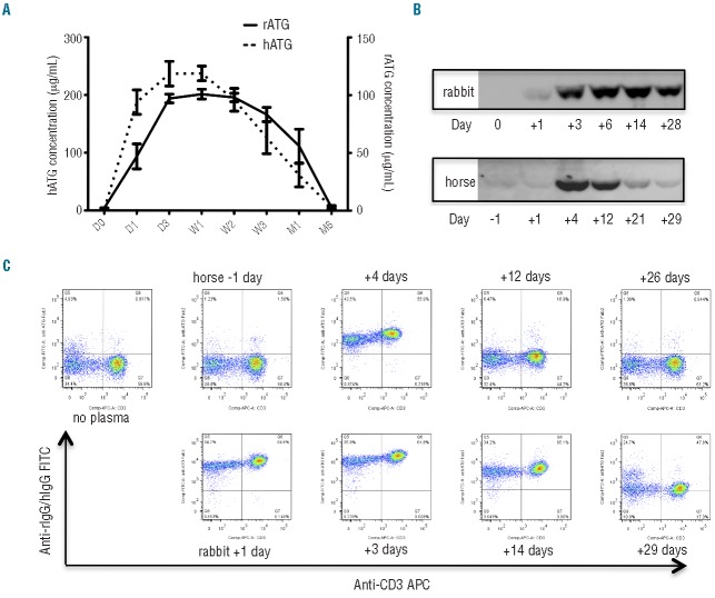
We evaluated the pharmacokinetics of ATG in the patients’ plasma by ELISA. To avoid the possible impact of clinical SS on ATG concentrations in blood, we only included patients without SS in this analysis. In both rATG-treated (n=39) and hATG-treated (n=43) patients, ATG reached high plasma concentrations 2 days after the first infusion, peaking at week 1 (100.8±4.2 μg/mL and 237.6±12.7 μg/mL for rATG and hATG, respectively), and then gradually decreased. The hATG concentration decreased by 18%, 46%, and 74%, while the rATG concentration decreased by 3%, 18%, and 45% from peak values when assessed at week 2, week 3, and 1 month, respectively (Figure 1A). When patients’ serial plasma samples were assayed by western blotting, ATG in the plasma was detected by anti-horse IgG or anti-rabbit IgG-conjugated with peroxidase. The results of the immunoblotting were consistent with those of ELISA, and a representative result is shown in Figure 1B: a weak band was detected in the plasma of a rATG-treated patient on day 1, followed by much more intense bands, sustained until day 28. The kinetics of hATG differed: peak levels occurred on day 4 (similar to rATG) but levels gradually decreased by day 14 and were barely detectable on days 21 and 28. These data indicate that the pharmacokinetics of rATG was different from that of hATG: plasma levels of rATG remained high for longer than those of hATG.
Figure 1.
Pharmacokinetics of rabbit ATG and horse ATG. (A) Concentrations of rATG (n=39) and hATG (n=43) detected by ELISA. ELISA plates were coated with rabbit anti-horse IgG (Fab’2) or chicken anti-rabbit IgG to capture hATG and rATG in the plasma, respectively. The bars represent mean ± standard error (SE). (B) Western blotting for the different types of ATG in the plasma. One microliter of plasma obtained from patients at different time points was used for western blotting. Representative data from at least three experiments with similar results are shown. (C) Binding of rATG and hATG in patients’ plasma to lymphocytes from healthy volunteers. Representative data from at least three experiments with similar results are shown. Cells in the plots were gated from lymphocytes based on forward and side scatter.
We next assessed whether free ATG in patients’ plasma could bind to normal lymphocytes, using flow cytometry to detect cell membrane-bound heterologous protein. rATG in patients’ plasma at day 3, day 14 and 1 month after therapy was found bound to CD3+ T cells and CD3-lymphocytes obtained from healthy controls (lower panel of Figure 1C), while hATG binding to T cells and non-T cells was not detectable 2 weeks after the initial infusion (upper panel of Figure 1C), consistent with a shorter half-life of hATG in the circulation and more binding of rATG to human lymphocytes in vivo. Similarly, rATG in patients’ plasma was also shown to have more sustained binding capacity to CD4+, CD8+ T cells, and CD19+ B cells as compared to hATG in patients’ plasma (data not shown). These differences are likely to account for the more prolonged lymphopenia observed with rATG than with hATG.
Effect of antithymocyte globulins on neutrophils
We compared complete blood cell counts from baseline to 6 months in all patients treated with rATG or hATG. hATG induced a stable increase in absolute neutrophil counts in the first week, distinct from its effects on lymphocytes, while rATG induced a decrease in absolute neutrophil counts similar to its effect on lymphocytes in the first 5 days after treatment. Ten days later, both hATG and rATG resulted in similar changes in absolute neutrophil counts, although the counts were higher in hATG-treated patients than in rATG-treated patients at 3 and 6 months, perhaps because of the higher response rate in the hATG group (Figure 2A). Indeed, when stratifying based on hematologic response, among the responding patients in these two ATG groups, there were no differences in absolute neutrophil counts at 3 and 6 months (Figure 2B). In contrast to the differences in absolute neutrophil counts and lymphocyte counts, no differences were observed in hemoglobin and platelet levels between the two groups (data not shown).
Figure 2.

Changes of absolute neutrophil count (ANC) in patients treated with different types of ATG. (A) Changes of ANC in all hATG (n = 60) and rATG (n = 60)-treated patients (including responders and non-responders). (B) Changes of ANC in responder patients treated with rATG (n = 22) or hATG (n = 41). The bars represent mean ± SE. *P<0.05; **P<0.01; ***P<0.001.
Effect of antithymocyte globulins on lymphocytes
Our previously published flow cytometry data showed that rATG induced more severe and prolonged lymphopenia than did hATG, especially affecting the CD4+ T-cell subset; absolute numbers of regulatory T cells were also lower in rATG-treated patients, both during and after treatment.6 In the current work, we extended flow cytometry to other immune cells. The frequencies of activated CD4+ T cells (CD38+), double-negative (CD3+CD4−CD8−) T cells, and B cells (CD19+) were higher in the peripheral blood of rATG-treated patients from the first week to months after treatment, but these effects were negated by stronger lymphocyte depletion after rATG, and the absolute number of these cells was not higher in rATG-treated patients (Figure 3). Double-negative T cells constitute a distinct regulatory T-cell population that may play an immune regulatory role, but the small total number of these cells may limit their functional effect, as we found for (CD4+CD25highCD127low) regulatory T cells.6
Figure 3.
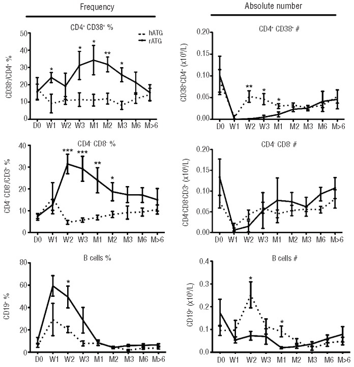
Recovery of different lymphocyte populations after hATG or rATG treatment. CD38+ cells were gated from CD45+CD3+CD4+CD8− T cells; double-negative T cells were gated from CD45+CD3+CD4−CD8− T cells; B cells (CD19+) were gated from CD45+ cells. Left panel shows frequencies and right panel shows absolute numbers of different cell populations. The bars represent mean ± SE. *P<0.05; **P<0.01; ***P< 0.001.
Effect of antithymocyte globulins on cytokines
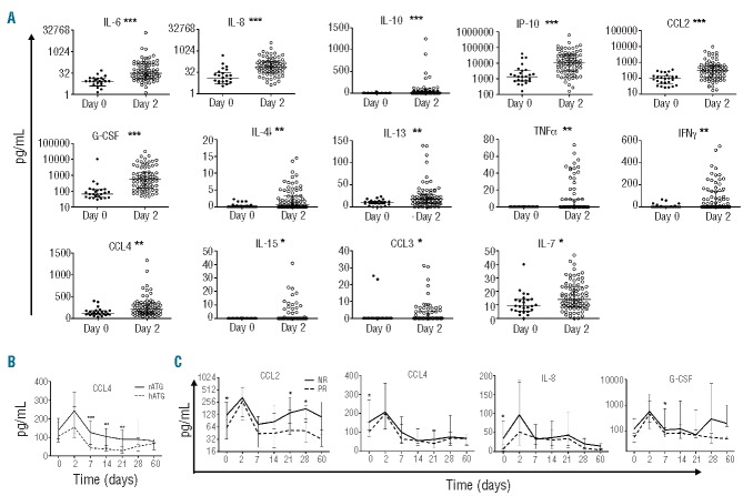
We evaluated changes in plasma cytokine levels using magnetic multiplex assays of 27 cytokines simultaneously. Both types of ATG induced massive, transient release of interleukin (IL)-6, IL-8, chemokine (C-C motif) ligand (CCL)-2, granulocyte colony-stimulating factor (G-CSF), interferon-gamma inducible protein-10 (IP-10), IL-10 (P<0.001), IL-4, IL-13, interferon gamma (IFNγ), CCL4, tumor necrosis factor-alpha (TNFα) (P<0.01), IL-7, IL-15, and CCL3 (P<0.05) on day 2 of administration, likely a direct effect of rATG and hATG (Figure 4A). There were no differences between the two types of ATG except for CCL4 levels (Figure 4B), which were higher in rATG-treated patients from day 7 to 21 than in hATG-treated patients (day 7, P<0.001; days 14 and 21, P<0.01). G-CSF and CCL-2 levels were higher in the rATG cohort than in the hATG patients at day 7 (P<0.05), while eotaxin and IL-5 levels were higher in hATG recipients than rATG recipients at week 2 (P<0.05, data not shown). These results suggest that some inflammatory cytokines (CCL4 and CCL2) may be preferentially increased after rATG. No differences in plasma levels of other cytokines were observed between rATG- and hATG-treated patients.
Figure 4.
Effect of ATG on cytokine levels. (A) “Cytokine storm” on day 2 in patients treated with rATG (n = 36) or hATG (n = 41). (B) Different effects of rATG (n = 36) or hATG (n = 41) on the cytokine CCL4. (C) Different cytokine levels between responders (n = 44) and non-responders (n = 33). The bars represent median and interquartile range. *P<0.05; **P< 0.01; ***P<0.001.
When comparing patients who responded and those who did not (Figure 4C), we found that non-responders had higher levels of CCL2, CCL4, and IL-8 (P<0.05) before treatment, especially of CCL2, the higher levels of which persisted over time in non-responders (days 21 and 28, P<0.05).
Serum sickness during antithymocyte globulin treatment
As foreign proteins, ATG elicit strong immune responses in humans. Foreign antigens and host antibodies form circulating immune complexes that may deposit in tissues and cause SS, usually around day 10 after initiation of ATG therapy. Indeed, ATG administration has provided a unique opportunity for the modern study of a historic syndrome.9 In our cohort, 15 of 60 (25%) rATG-treated patients and 9 of 60 (15%) hATG-treated patients developed clinical SS, with typical manifestations of fever, arthralgia, malaise, and cutaneous eruptions. Surprisingly, we found that ATG concentrations, as measured by ELISA (Figure 5A) and western blotting (Figure 5B), were higher on day 1 in patients with SS than in those without the syndrome, especially in the rATG-treated group. Both rATG and hATG concentrations decreased sharply in patients with SS, compared to those in patients without SS, after week 2, especially for rATG, reaching almost undetectable levels on day 14 in patients with SS treated with rATG (Figure 5B). These observations suggest that the early appearance of rATG in the circulation on day 1 may be predictive of SS, and that rapid disappearance of rATG in the circulation on day 14 is associated with the syndrome.
Figure 5.
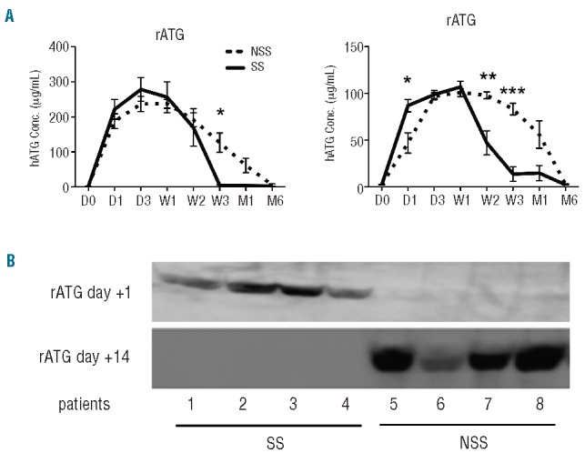
Pharmacokinetics of the two types of ATG in patients with serum sickness (SS) and without serum sickness (NSS). (A) Pharmacokinetics of ATG in SS (hATG n = 5, rATG n = 10) and NSS (hATG n = 49, rATG n = 43) patients detected by ELISA. The bars represent mean ± SE. *P<0.05; **P<0.01; ***P<0.001. (B) Western blotting of rATG in the plasma of SS and NSS patients.
To confirm the relationship of the clearance of ATG and antibody titers against ATG, we examined human anti-ATG antibody titers in the plasma of 42 patients treated with hATG and 38 treated with rATG. When measured by ELISA, human anti-hATG and rATG antibody titers peaked at weeks 2–3 and then declined. All 11 rATG-treated patients who developed SS had significantly higher antibody titers, including IgG, IgA, and IgM isotypes from week 2 to 1 month, than those found in 29 rATG-treated patients without SS. Among the hATG-treated patients, only IgM antibody titers were higher in SS patients (Figure 6A). Human anti-rATG or hATG antibody titers correlated with the occurrence of SS, the IgM subtype appearing more specific, consistent with previous findings.9,10 In addition, we did not find any correlation of SS with response to ATG treatment, or with neutrophil and red blood cell counts, but SS was associated with higher platelet counts from week 2 to 6 months (Figure 6B).
Figure 6.
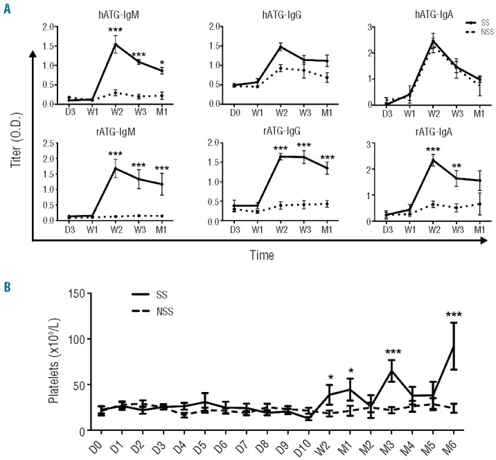
Different immune responses in ATG-treated AA patients with serum sickness (SS) or without serum sickness (NSS). (A) Human anti-ATG antibodies, including IgG, IgA, and IgM, in SS (hATG n = 5, rATG n = 10) and NSS (hATG n = 49, rATG n = 43) patients detected by ELISA. ELISA plates were coated with rATG or hATG for capture anti-rATG and anti-hATG antibodies in the plasma, respectively. (B) Comparison of platelet counts between SS and NSS patients. The bars represent mean ± SE. *P<0.05; **P<0.01; ***P<0.001.
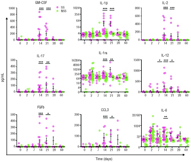
In Luminex assays, patients with SS (whether treated with hATG or rATG) had significantly higher plasma levels of granulocyte-monocyte colony-stimulating factor (GM-CSF), IL-1b, IL-2, IL-17, IL-1 receptor antagonist (IL-1ra), IL-12, fibroblast growth factor-b (FGFb), CCL3, and IL-6 at weeks 2 and 3 than did patients without SS (Figure 7); these increased cytokines may represent a SS cytokine signature, and this late-phase response is likely secondary to immune complex formation. There were no significant differences in SS-associated cytokine release between rATG- and hATG-treated patients.
Figure 7.
Cytokine signature of serum sickness. Red dots represent SS (n = 15), while green dots represent NSS (n = 77). The bars represent mean ± SE. Asterisks indicate differences between SS and NSS at the same time points: *P<0.05; **P<0.01; ***P<0.001.
Discussion
rATG has been widely used as preconditioning for organ transplantation and in preventing graft rejection.11 Direct comparison of rATG and hATG in the treatment of acute graft rejection episodes following renal transplantation showed that rATG is more effective.12 In severe AA, the result was the reverse. The pharmacokinetics and immunological perturbations of both rATG and hATG have not been studied systematically in severe AA.
In the current work, we found that rATG, functionally able to bind human lymphocytes, remained in blood longer than did hATG. Although long-lasting rATG in blood could reduce the numbers of presumably pathogenic T cells, it also decreased likely non-pathogenic T cells (especially CD4+ T cells) and neutrophils, potentially predisposing to infections and delaying evidence of hematologic recovery. It has been reported that renal transplant recipients with persistent CD4+ T-cell lymphopenia induced by rATG had higher rates of mortality and morbidity, including infections and cancer.13 In our ATG-treated patients, the most prevalent complications were infections: one hATG patient and eight rATG patients went “off study” due to recurrent life-threatening systemic infections not adequately controlled with antimicrobials, in the setting of progressive pancytopenia. CD4+ T cell lymphopenia might contribute to this complication, adding to susceptibility from the more severe and prolonged neutropenia with rATG. As we previously reported,14 Epstein-Barr viral loads were observed to be higher, and lymphopenia was also more prolonged, following rATG when it was administered as salvage therapy, as compared to hATG as first-line treatment. The current analysis directly comparing lymphocyte depletion kinetics between types of ATG as first therapy confirms these data, the likely mechanism being depression of both neutrophils and T cells in patients who received rATG, compared to patients who received hATG. The T-cell/B-cell ratio has been reported to be associated with Epstein-Barr virus-associated lymphoproliferative disorders after bone marrow transplantation from matched unrelated donors.15 The EBMT prospective study comparing outcomes between patients treated with rATG or hATG showed increased infectious deaths with rATG;16 in addition, a recent Japanese study on rATG in children also showed an increased infection rate.17 Moreover, imbalanced lymphocyte subsets of activated CD4+ (CD38+), double-negative T cells (CD3+CD4−CD8−), and regulatory T cells in rATG-treated patients may also affect immune system functions.
Guttmann et al. reported that the first dose of ATG induced TNFα and IL-6 secretion in renal transplantation patients but that subsequent doses of ATG did not have the same effect on cytokine production.18 We found that the ATG-induced “cytokine storm” includes not only TNFα and IL-6 but also IL-8, CCL-2, G-CSF, IP-10, IL-10, IL-4, IL-13, IFNγ, CCL4, IL-7, IL-15, and CCL3, and occurs after either rATG or hATG infusion. These cytokines appeared in the blood transiently, likely due to accelerated activation and elimination of T-lymphocytes and other immune cells. The pattern of cytokine release was very similar for rATG and hATG, except that CCL4 levels were much higher in rATG-treated patients than in hATG-treated ones, from 1 to 3 weeks. CCL2 was also inversely correlated with hematologic response. This persistent inflammatory environment might affect the efficacy of rATG and increase its toxicity.
SS is a historically important syndrome, first reported in individuals who received animal antisera, and, as we had observed previously, it occurred in some of our patients after ATG. The pathophysiology of SS is understood to be secondary to host antibody responses to foreign proteins followed by formation and then deposition of immune complexes in tissues (skin, joints, and other organs). Both the dose and regimen of antigen administration and the quality of the immune response would be factors in the production of SS; additionally, certain antigens might stimulate specific patterns of host immune response and cytokine release. The total dose of hATG is much higher (almost 10-fold) than that of rATG. However, rATG is detectable in blood for much longer and at apparently equivalent or higher amounts, as determined by immuneblotting. Within the group of patients who received rATG, ATG concentrations on day 1 were higher than in non-SS patients; we did not see such a difference in hATG-treated patients. Concentrations of both types of ATG decreased at about week 2 when SS occurred, with clearance being associated with the appearance of antibodies against ATG, especially IgM. It is remarkable that, despite marked differences in the amount of antigen infused between hATG and rATG, and presumed differences also in the immune response to horse and rabbit proteins, the clinical pattern of SS, with regards to both the proportion of patients affected and the time of onset of symptoms, was similar with the two biologics.
Patients with SS (following either hATG or rATG) had significantly higher plasma levels of IL-2, IL-12, IL-1b, IL-17, IL-1ra, GM-CSF, FGFb, and CCL3 than did non-SS patients at weeks 2 and 3. This is the first report, to our knowledge, to identify a cytokine signature of SS, concurrent with anti-ATG antibodies, suggesting that the cytokine production was incited by antibody production. SS was not associated with a difference in hematologic response, even if it shortened the duration of rATG in the circulation.
In summary, despite biological similarities in manufacturing, there are many differences in pharmacokinetics and effects on neutrophils, lymphocyte subsets and cytokine release, beyond regulatory T cells, between the two types of ATG. Any one of these differences might be clinically important. Biologics can lead to unpredictable and complex changes in the immune system, and these differences may be related to their efficacy in suppressing the immune system and restoring hematopoiesis in AA patients.
Footnotes
The online version of this article has a Supplementary Appendix.
Funding
This work was supported by the Intramural Research Program of the National Heart, Lung, and Blood Institute, National Institutes of Health
Authorship and Disclosures
Information on authorship, contributions, and financial & other disclosures was provided by the authors and is available with the online version of this article at www.haematologica.org.
References
- 1.Scheinberg P, Young NS. How I treat acquired aplastic anemia. Blood. 2012;120(6):1185–96. [DOI] [PMC free article] [PubMed] [Google Scholar]
- 2.Bacigalupo A, Broccia G, Corda G, Arcese W, Carotenuto M, Gallamini A, et al. Antilymphocyte globulin, cyclosporin, and granulocyte colony-stimulating factor in patients with acquired severe aplastic anemia (SAA): a pilot study of the EBMT SAA Working Party. Blood. 1995;85(5):1348–53. [PubMed] [Google Scholar]
- 3.Rosenfeld S, Follmann D, Nunez O, Young NS. Antithymocyte globulin and cyclosporine for severe aplastic anemia: association between hematologic response and long-term outcome. JAMA. 2003;289(9):1130–5. [DOI] [PubMed] [Google Scholar]
- 4.Frickhofen N, Heimpel H, Kaltwasser JP, Schrezenmeier H. Antithymocyte globulin with or without cyclosporin A: 11-year follow-up of a randomized trial comparing treatments of aplastic anemia. Blood. 2003; 101(4):1236–42. [DOI] [PubMed] [Google Scholar]
- 5.Frickhofen N, Kaltwasser JP, Schrezenmeier H, Raghavachar A, Vogt HG, Herrmann F, et al. Treatment of aplastic anemia with antilymphocyte globulin and methylprednisolone with or without cyclosporine. The German Aplastic Anemia Study Group. N Engl J Med. 1991;324(19):1297–304. [DOI] [PubMed] [Google Scholar]
- 6.Scheinberg P, Nunez O, Weinstein B, Biancotto A, Wu CO, Young NS. Horse versus rabbit antithymocyte globulin in acquired aplastic anemia. N Engl J Med. 2011;365(5):430–8. [DOI] [PMC free article] [PubMed] [Google Scholar]
- 7.Bourdage JS, Hamlin DM. Comparative polyclonal antithymocyte globulin and antilymphocyte/antilymphoblast globulin anti-CD antigen analysis by flow cytometry. Transplantation. 1995;59(8):1194–200. [PubMed] [Google Scholar]
- 8.Feng X, Kajigaya S, Solomou EE, Keyvanfar K, Xu X, Raghavachari N, et al. Rabbit ATG but not horse ATG promotes expansion of functional CD4+CD25highFOXP3+ regulatory T cells in vitro. Blood. 2008;111(7):3675–83. [DOI] [PMC free article] [PubMed] [Google Scholar]
- 9.Bielory L, Gascon P, Lawley TJ, Young NS, Frank MM. Human serum sickness: a prospective analysis of 35 patients treated with equine anti-thymocyte globulin for bone marrow failure. Medicine (Baltimore). 1988;67(1):40–57. [PubMed] [Google Scholar]
- 10.Prin Mathieu C, Renoult E, Kennel De March A, Bene MC, Kessler M, Faure GC. Serum anti-rabbit and anti-horse IgG, IgA, and IgM in kidney transplant recipients. Nephrol Dial Transplant. 1997;12(10):2133–9. [DOI] [PubMed] [Google Scholar]
- 11.Ault BH, Honaker MR, Osama Gaber A, Jones DP, Duhart BT, Jr, Powell SL, et al. Short-term outcomes of Thymoglobulin induction in pediatric renal transplant recipients. Pediatr Nephrol. 2002;17(10):815–8. [DOI] [PubMed] [Google Scholar]
- 12.Schroeder TJ, Moore LW, Gaber LW, Gaber AO, First MR. The US multicenter double-blind, randomized, phase III trial of thymoglobulin versus atgam in the treatment of acute graft rejection episodes following renal transplantation: rationale for study design. Transplant Proc. 1999;31(3B Suppl):1S–6S. [DOI] [PubMed] [Google Scholar]
- 13.Ducloux D, Courivaud C, Bamoulid J, Vivet B, Chabroux A, Deschamps M, et al. Prolonged CD4 T cell lymphopenia increases morbidity and mortality after renal transplantation. J Am Soc Nephrol. 2010;21(5):868–75. [DOI] [PMC free article] [PubMed] [Google Scholar]
- 14.Scheinberg P, Fischer SH, Li L, Nunez O, Wu CO, Sloand EM, et al. Distinct EBV and CMV reactivation patterns following antibody-based immunosuppressive regimens in patients with severe aplastic anemia. Blood. 2007;109(8):3219–24. [DOI] [PMC free article] [PubMed] [Google Scholar]
- 15.Meijer E, Slaper-Cortenbach IC, Thijsen SF, Dekker AW, Verdonck LF. Increased incidence of EBV-associated lymphoproliferative disorders after allogeneic stem cell transplantation from matched unrelated donors due to a change of T cell depletion technique. Bone Marrow Transplant. 2002; 29(4):335–9. [DOI] [PubMed] [Google Scholar]
- 16.Marsh JC, Bacigalupo A, Schrezenmeier H, Tichelli A, Risitano AM, Passweg JR, et al. Prospective study of rabbit antithymocyte globulin and cyclosporine for aplastic anemia from the EBMT Severe Aplastic Anaemia Working Party. Blood. 2012;119(23):5391–6. [DOI] [PubMed] [Google Scholar]
- 17.Jeong DC, Chung NG, Cho B, Zou Y, Ruan M, Takahashi Y, et al. Long-term outcome after immunosuppressive therapy with horse or rabbit antithymocyte globulin and cyclosporine for severe aplastic anemia in children. Haematologica. 2014;99(4):664–71. [DOI] [PMC free article] [PubMed] [Google Scholar]
- 18.Guttmann RD, Caudrelier P, Alberici G, Touraine JL. Pharmacokinetics, foreign protein immune response, cytokine release, and lymphocyte subsets in patients receiving thymoglobuline and immunosuppression. Transplant Proc. 1997;29(7A):24S–6S. [PubMed] [Google Scholar]


