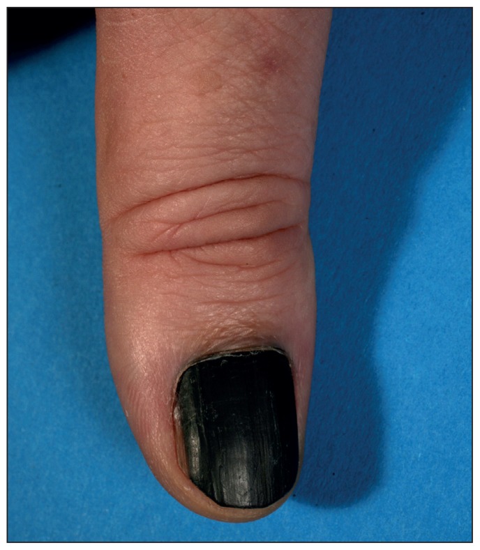
A 53-year-old man presented with a painless black discolouration affecting the complete nail of his right thumb (Figure 1). The discolouration had begun 15 years previously as a thin black line stretching from the lunula to the free edge of the nail, but this line had widened in the past year.
Figure 1:
Melanonychia, or black discolouration of the nail plate, of the right thumb of a 53-year-old man. Histopathological examination showed a subungual melanoma with a Breslow depth of 1.5 mm.
On examination, the nail plate was smooth with a homogenous black colour, consistent with a diagnosis of melanonychia totalis. Histopathologic evaluation showed a subungual melanoma with a Breslow depth of 1.5 mm. We performed a wide excision (margins about 0.5 cm), and all lateral margins and the base of the nail bed were tumour free. Whole-body computed tomography and examination of the sentinel lymph node showed no metastasis. We covered the defect with a full-thickness skin graft, and the patient was given low-dose interferon-α (3 million IU subcutaneously 3 times/wk) for 18 months; he remains free of any metastases three years later.
Subungual melanoma is an uncommon variant of malignant melanoma, representing only 1%–2% of all cutaneous melanoma in the white population and usually affecting the thumb or the big toe.1 Diagnosing subungual melanoma is challenging owing to the diversity of its clinical presentations, including band-like black nail discolourations (melanonychia striata longitudinalis), nail dystrophy, subungual (amelanotic) masses and splits of the nail plate. Differential diagnoses include benign melanocytic nevi, subungual hematoma, vascular tumours (e.g., pyogenic granuloma), discolouration caused by drugs and fungal infections. Signs of malignant disease include extension of the pigmentation onto the proximal or lateral nailfold (Hutchinson sign) and rapid progress of discolouration without any traumatic injury. Signs on dermoscopy may be irregular bands, disruption in parallelism, micro–Hutchinson sign and pigment on the ridges of the hyponychium. More gradual progression, as seen in our patient’s case, has been described2 and may help explain why 20% of cases are not diagnosed on first presentation.1 Thus, if a malignant process cannot be clearly ruled out, histopathologic examination of the nail bed and the germinal matrix should be done.
Clinical images are chosen because they are particularly intriguing, classic or dramatic. Submissions of clear, appropriately labelled high-resolution images must be accompanied by a figure caption and the patient’s written consent for publication. A brief explanation (250 words maximum) of the educational significance of the images with minimal references is required.
Footnotes
Competing interests: None declared.
This article has been peer reviewed.
The authors have obtained patient consent.
References
- 1.Phan A, Touzet S, Dalle S, et al. Acral lentiginous melanoma: a clinicoprognostic study of 126 cases. Br J Dermatol 2006;155:561–9. [DOI] [PubMed] [Google Scholar]
- 2.Kim JY, Choi M, Jo SJ, et al. Acral lentiginous melanoma: indolent subtype with long radial growth phase. Am J Dermatopathol 2014;36:142–7. [DOI] [PubMed] [Google Scholar]


